Susiane Allgayer*, Fernanda Santos Mezzomo**, Waldemar Daudt Polido***, Gabriella Rosenbach****, Carlos Alberto Estevanell Tavares*****
Orthodontic-surgical treatment of skeletal
facial asymmetry: Case report
Introduction: Facial asymmetries consist of an imbalance between the homologous skeletal structures of the face. Most people present some degree of facial asymmetry, since a state of perfect symmetry is rare. This common asymmetry only becomes relevant when it is perceiv-able by the patient. In this situation, either orthodontic surgical correction or orthodontic treatment is normally chosen. Objective: This study, based on literature review, has been illus-trated by a case report comprising Le Fort I orthognathic surgery for maxillary advancement and rotation, with conservative treatment for the mandible. Conclusion: Knowledge of the patient’s chief complaint and expectations, as well as proper diagnostic exams, are important factors to decide the treatment plan and for the final treatment outcome.
Abstract
Keywords: Facial asymmetry. Corrective orthodontics. Tooth extraction. Esthetics.
IntrOduCtIOn
Facial symmetry is a state of balance in which both sides of the face are perfectly related and therefore present the same size, shape and posi-tion. Conversely, the term asymmetry is used when there is imbalance between the homologous parts of the dentofacial complex, thereby affect-ing the proportion between structures.1
The facial asymmetry may often be subclini-cal. In this condition, the skeletal disharmony ex-ists yet it is masked by the overlying soft tissues.2,3
Thus, the soft tissues superimposed to the bone structures, such as the masseter muscle, may minimize or even compensate an existing skeletal deformity. Therefore, when there is discrepancy between the skeletal measurements and facial ap-pearance, the influence of soft tissues on the facial asymmetries should be considered.3
According to some authors,2,3 the clinical
expression of asymmetry only occurs when the bone deviation is at least 4 mm. Below this value, the asymmetry is considered to be subclinical.
* PhD student and MSc in Orthodontics, Pontiical Catholic University of Rio Grande do Sul, Brazil (PUC/RS). Specialist in Orthodontics and Endodontics, ABO/RS.
** Specialist in Orthodontics, ABO/RS.
*** PhD and MSc in Oral and Maxillofacial Surgery, PUC/RS. Head of Implant Specialization Course, ABO/RS.
**** PhD, University of Munich, Germany. MSc and Specialist in Orthodontics, State University of Rio de Janeiro. Professor, Specialization Course in Orthodontics, ABO/RS.
***** PhD and MSc in Orthodontics, Federal University of Rio de Janeiro. Professor, Specialization Course in Orthodontics, ABO/RS. Diplomate of the Brazilian Board of Orthodontics and Facial Orthopedics (BBO).
How to cite this article: Allgayer S, Mezzomo FS, Polido WD, Rosenbach G, Tavares CAE. Orthodontic-surgical treatment of skeletal facial asymmetry: Case report. Dental Press J Orthod. 2011 Nov-Dec;16(6):100-10.
That is to say, human sensitivity to notice facial imbalances occurs more easily if the deformation is closer to or greater than 4 mm. However, the expression of asymmetry or its attenuation de-pend on individual characteristics, such as the soft tissue thickness over the imbalance region.
Within this context, this study conducted a literature review on the skeletal facial asymme-tries, illustrating with a case report of asymmetry involving both the maxilla and the mandible, in which the treatment plan of choice was the as-sociation of surgical treatment on the maxilla and conservative treatment for the mandible.
SKELEtAL FACIAL ASYMMEtrIES
Dentofacial deformities affect approximate-ly 20% of the population, and patients with these discrepancies may present several degrees of functional and esthetic involvement,4 being
classified as isolated mandibular asymmetries or maxillomandibular asymmetries. However, there are no isolated maxillary asymmetries, be-cause a deformed maxilla simultaneously be-causes mandibular disorders.5
The mandibular asymmetries may be caused by excessive or deficient growth of the mandibular body and ramus, or the mandible may be deviated due to asymmetric growth of other structures. These con-ditions cause mandibular laterognathism, i.e., man-dibular deviation to one side of the facial midline.5
Some studies explain that the higher prevalence of mandibular asymmetries may be related to the lon-ger period of mandibular growth when compared to the maxilla, thus increasing the chances of devia-tions. Also, the mandible is a mobile bone, while the maxilla is rigidly connected to other bone structures through sutures and synchondroses.3,6 The condyle
is the main growth center of the mandible; for this reason, injuries to this area during the growth period may cause disturbances in mandibular growth.7,8
Even though the studies conducted by H. Peck and S. Peck9 did not reveal significant difference
in the side of mandibular deviation, in cases of
mandibular skeletal asymmetry, according to Ha-raguchi et al,6 the lateral deviation is more
com-mon on the lower facial third, and 85% of dento-facial deformities present lateral deviations to the left side, a tendency that is corroborated by other studies.2,6,10 In addition, Bell, Proffit and White11
re-lated skeletal asymmetries to Class III malocclusion observing that 40% of these malocclusions cases presented some degree of facial asymmetry.
During the anamnesis, it is important to estab-lish the chief complaint of the patient, identify if the facial imbalance is perceived and if this con-dition causes discomfort and dissatisfaction. The history of traumas, ankylosis or lesions such as osteochondromas affecting the temporomandibu-lar joint, intra-articutemporomandibu-lar disorders, birth by forceps, condylar fractures, ear infections, inadequate use of orthopedic appliances,11,13 besides lesions to facial
nerves, are possible causes of asymmetries.12
During the clinical examination, the extraoral analysis is fundamental in the diagnosis of asym-metries, since analysis of the facial proportions or of the degree of imbalance between the facial thirds and homologous facial structures usually indicates the site of imbalance.11 Analysis of facial proportions
allows evaluation of the harmony between facial thirds, which should have a 1:1 ratio. In the lateral evaluation, if one lip is behind or beyond the Stein-er’s S line, there may be a disproportion between the maxilla and mandible, consequently causing an imbalance between the facial structures.11
In the intraoral examination, dental evaluation in centric relation should include the analysis of dental midlines, their relation to the facial mid-line, existence of crossbite or inclination of the occlusal plane.14 If the dental midlines are
coinci-dent, a deviation of up to 4 mm to one side of the facial midline may not be detected by laypersons; however, if the crowns of incisors are tipped, de-viations above 2 mm may be perceptible.15,16,17
magnitude of asymmetry.13 The posteroanterior
ra-diograph (PA) is a valuable tool to compare struc-tures between the right and left sides of the face.6
If the dental midlines are coincident and deviated up to nearly 2 mm from the facial midline, this will still be considered a harmonious situation, since the aforementioned degree of deviation is not percep-tible to the layperson and may be attenuated by the soft tissues of the face.3,6,18 The lateral
cephalomet-ric radiograph provides information on the antero-posterior component of the deformity.5
When the treatment options are mentioned, it is important to evaluate the efficacy of these. In case of skeletal imbalance, in which the profes-sional must decide between surgical or non-sur-gical treatment, it should be clear the amount of esthetic, dental and facial improvements provided by the selected treatment plan.13 Even though the
surgical correction may be preferable for severe cases and after completion of bone growth, cam-ouflage is a conservative treatment option indicat-ed for correction of mild asymmetries.16,19,20 The
treatment for skeletal asymmetries may comprise an association between conservative treatment and orthognathic surgery.21,22 Thus, the imbalance
may be surgically corrected in one jaw and by den-tal compensation in the other one. Following the alignment and leveling stage, the final objective is to achieve adequate occlusion with coincidence of maxillary and mandibular midlines.20
CASE rEPOrt
A Caucasian female patient, 17 years and 4 months old, searched for orthodontic treatment with the chief complaint of “crossbite”. The pa-tient reported a history without dental and/or skeletal traumas to the facial structures and did not present any systemic alteration or history of previous pathologies.
diagnosis
The extraoral examination revealed fa-cial asymmetry of the lower fafa-cial third, with
mandibular skeletal laterognathism to the left side and mild maxillary skeletal laterognathism to the right side. The deficiency in anteroposte-rior direction in the mid facial third was easily identified by the deep paranasal and infraorbital regions, deep nasogenian grooves, lack of sup-port to the upper lip and thin nasal base. The lower lip was protruded in 4 mm in relation to the S line (Figs 1A-E).
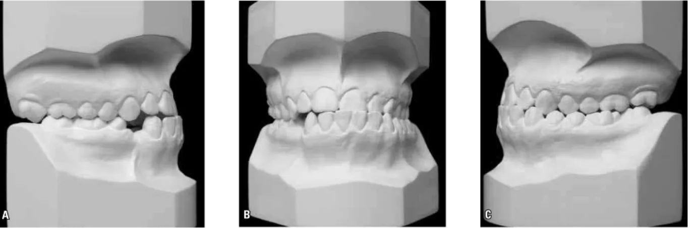
The intraoral examination revealed molar Class III relationship on both sides and canine Class III relationship on the right side and Class I relationship on the left side, as well as absence of third molars and mandibular right first premo-lar. The maxillary dental midline was dislocated 2 mm to the right side, and the mandibular dental midline was deviated 2.5 mm to the left side. The premature contact caused by the maxillary left central incisor led to a forward mandibular devia-tion, generating crossbite at the region of maxil-lary left central and lateral incisors and canine, and left posterior region (Figs 1F-J).
The cephalometric analysis (Figs 3B, 3C and Table 1) presented a skeletal Class III pat-tern with an important vertical component, as displayed by these cephalometric measures: ANB= -2°, WITTS= -6,5 mm, SN.GoGn= 35° and FMA= 29°. Analysis of the posteroanterior radio-graph (Figs 3D, E) revealed mild maxillary devia-tion of 0.5 mm to the right side and mandibular deviation of 3 mm to the left side. The panoramic radiograph (Fig 3A) revealed the presence of im-pacted third molars, except for the maxillary right and left third molars, which were absent.
treatment options
The following treatment options were present-ed to the patient:
I J
H G
F
E
D C
B A
The orthodontic treatment should be finished in Class I molar and canine relationship and dental midlines coincident to each other and to the facial midline.
2) Orthodontic treatment associated to or-thognathic surgery only in the maxilla, with ex-traction of three premolars, followed by maxillary advancement with rotation to the left side. The orthodontic treatment should be finished with
Class I molar and canine relationship and the den-tal midlines coincident to each other, yet deviated to the left side in relation to the facial midline.
C
A B
D E
A B C
FIGURE 3 - A) Initial panoramic radiograph, B) initial lateral cephalometric radiograph, C) initial cephalometric tracing, D) initial posteroanterior radio-graph, E) initial cephalometric tracing.
MEAsurEs NOrMAL INITIAL FINAL
(A) (B)
skeletal pattern
SNA (Steiner) 82° 77° 79°
SNB (Steiner) 80° 75° 79°
ANB (Steiner) 2° - 2° 0°
Facial conv. angle (Downs) 0° - 3° 0°
Y axis (Downs) 59° 62° 60°
Facial angle (Downs) 87° 85° 86°
SN-GoGn (Steiner) 32° 35° 35°
FMA (Tweed) 25° 29° 29°
Dental pattern
IMPA (Tweed) 90° 89° 81°
1–NA (°) (Steiner) 22° 33° 34°
1–NA (mm) (Steiner) 4 mm 9.5 mm 7 mm
1–NB (°) (Steiner) 25° 24° 17°
1–NB (mm) (Steiner) 4 mm 8 mm 4 mm
Interincisal angle (Downs) 130° 125° 129°
LI-to-AP (Ricketts) 1 mm 10 mm 5 mm
Proile S line - Upper lip (Steiner) 0 mm - 2 mm - 2 mm
S line - Lower lip (Steiner) 0 mm 4 mm 1 mm
TABLE 1 - Summary of cephalometric measurements.
treatment stages
In the maxillary arch, a transpalatal bar with Nance button was anchored on the maxillary sec-ond molars, and the mandibular first and secsec-ond molars were banded. The patient was referred to extraction of the maxillary right second premo-lar, maxillary left first premolar and mandibular left second premolar. After bonding of Edgewise standard brackets on the other teeth, alignment and leveling were performed using 0.014-in to 0.020-in up to 0.019 x 0.025-in stainless steel archwires. Elastomeric chains were used to re-tract the premolars and canines mesial to the extraction spaces, for distal movement of the maxillary left central and lateral incisors, and me-sial movement of the maxillary right central and lateral incisors until the midline coincided with the center of the maxilla, as well as for distal movement of the mandibular right canine, cen-tral and lateral incisors, and for mesial movement of mandibular left central and lateral incisors.
The remaining spaces were closed using 0.019 x 0.025-in rectangular archwires with loops. In the immediate preoperative period, the transpalatal bar was removed and a 0.020 x 0.025-in rectan-gular archwire with hooks was installed.
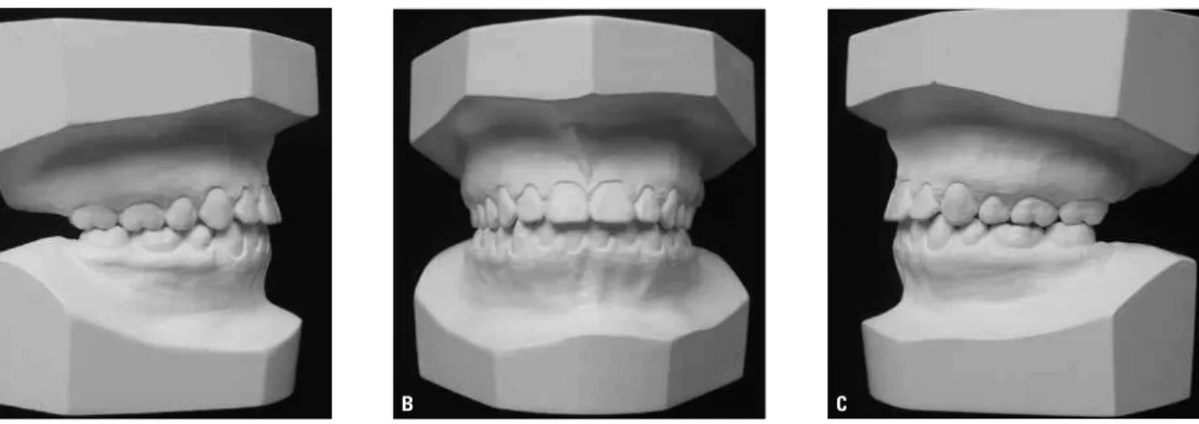
Subsequent impressions were taken to evalu-ate the intercuspation and simulevalu-ate the move-ment for maxillary advancemove-ment and rotation to the left side (Fig 4).
In collaboration with the bucomaxillofacial surgeon, a Le Fort I osteotomy was planned for maxillary advancement and rotation to the left side (Fig 5). One month after surgery, the pa-tient underwent orthodontic detailing and the appliances were removed.
A B C D
G
H I
E F
A B C
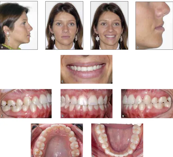
FIGURE 4 - Preoperative extraoral (A, B, C, D) and intraoral (E, F, G, H, I) photographs.
G
I J
H F
E
D C
B A
Regarding function, the patient exhibited lateral and protrusive mandibular movements. Further-more, the facial profile was more harmonious, considering the improved relationship between the lips (Figs 6, 7 and 8).
The final radiographs demonstrate orthodontic finishing with correct root position and absence of root resorptions. The cephalometric analysis re-vealed significant changes, presenting a final skel-etal Class I pattern (ANB= 0°, WITTS= -4mm) (Fig 9 and Table 1).
dISCuSSIOn
In an ideal face, all structures of the craniofacial complex at one side should be perfectly equal to the structures on the opposite side.10 However, even
pleasant faces exhibited mild degrees of asymmetry between the two sides, and total symmetry is not a common condition.13,23,24,25 Even though some
stud-ies report a tendency that the left side of the face is dominant,24,26,27 others state that skeletal facial
struc-tures are larger on the right side compared to the left side, with statistically significant difference.2,6,10,28,29
G F
E D
C B
A
C B
A
FIGURE 7 - Photographs of excursive movements. Canine disocclusion of all teeth during mandibular movements. PROTRUSIVE: A) right side view, B) frontal view, C) left side view. RIGHT LATERAL MOVE-MENT: D) right side view, E) left side view. LEFT LATERAL MOVEMENT: F) right side view, G) left side view.
C B
A
FIGURE 9 - A) Final panoramic radiograph, B) final lateral cephalometric radiograph, C) final cephalo-metric tracing.
The surgical approach for correction of severe facial skeletal asymmetries is usually the treatment of choice.21,30 However, milder or developing cases
may be treated by less invasive techniques.16,19,20,22
In the present case, the extent of improvement of facial appearance from correction by orthognathic surgery on both jaws was carefully considered. Thus, considering that the anteroposterior maxil-lary deficiency had the greatest negative impact on the facial esthetics and that the mandibular lateral deviation was not the main cause of facial imbal-ance — which could be masked by coinciding the dental midlines —, it was decided to perform
Contact address Susiane Allgayer ABO/RS
R. Furriel Luiz Antônio de Vargas, 134 – Mont’Serrat Zip code: 90.470-130 – Porto Alegre/RS, Brazil E-mail: [email protected]
1. Fischer B. Asymmetries of the dentofacial complex. Angle Orthod. 1954;24(4):179-92.
2. Peck S, Peck L, Kataja M. Skeletal asymmetry in esthetically pleasing faces. Angle Orthod. 1991;61(1):43-8.
3. Masuoka N, Momoi Y, Ariji Y, Nawa H, Muramatsu A, Goto S, et al. Can cephalometric indices and subjective evaluation be consistent for facial asymmetry? Angle Orthod.
2005;75(4):651-5.
4. Silva EDO, Laureano Filho JR, Rocha NS, Annes PMR, Tavares PO. Tratamento cirúrgico de assimetria mandibular: relato de caso clínico. Rev Cir Traumat Buco-Maxilo-Fac. 2004;4(1):23-9.
5. Medeiros JP, Medeiros PP. Cirurgia ortognática para o ortodontista. 2nd ed. São Paulo: Ed. Santos; 2004.
6. Haraguchi S, Takada K, Yasuda Y. Facial asymmetry in subjects with skeletal Class III deformity. Angle Orthod. 2002;72(1):28-35.
7. Yamashiro T, Okada T, Takada K. Case report: facial asymmetry and early condylar fracture. Angle Orthod. 1998;68(1):85-90.
8. Duthie J, Bharwani D, Tallents RH, Bellohusen R, Fishman L. A longitudinal study of normal asymmetric mandibular growth and its relationship to skeletal maturation. Am J Orthod Dentofacial Orthop. 2007;132(2):179-84.
9. Peck H, Peck S. A concept of facial esthetics. Angle Orthod. 1970;40(4):284-318.
10. Shah S, Joshi M. An assessment of asymmetry in the normal craniofacial complex. Angle Orthod. 1978;48(2):141-8. 11. Bell WH, Profit WR, White RP. Surgical correction of
dentofacial deformities. Hardcover: Saunders; 1980. v. 1. 12. Legan HL. Surgical correction of patients with asymmetries.
Semin Orthod. 1998;4(3):189-98.
13. Carlini JL, Gomes KU. Diagnóstico e tratamento das assimetrias dentofaciais. Rev Dental Press Ortod Ortop Facial. 2005;10(1):18-29.
14. Burstone CJ. Diagnosis and treatment planning of patients with asymmetries. Semin Orthod. 1998;4(3):153-64. 15. Kokich VO, Kiyak HA, Shapiro PA. Comparing the perception
of dentists and lay people to altered dental esthetics. J Esthet Rest Dent. 1999;11(6):311-24.
16. Anhoury PS. Nonsurgical treatment of an adult with mandibular asymmetry and unilateral posterior crossbite. Am J Orthod Dentofacial Orthop. 2009;135(1):118-26.
17. Janson G, Camardella LT, Freitas MR, Almeida RR, Martins DR. Treatment of a Class II subdivision malocclusion with multiple congenitally missing teeth. Am J Orthod Dentofacial Orthop. 2009;135(5):663-70.
rEFErEnCES
18. Joondeph DR. Mysteries of asymmetries. Am J Orthod Dentofacial Orthop. 2000;117(5):577-9.
19. Shroff B, Siegel SM. Treatment of patients with asymmetries using asymmetric mechanics. Semin Orthod. 1998;4(3):165-79. 20. Mucha JN. As limitações do tratamento ortodôntico
não-cirúrgico. In: Medeiros PJ, Medeiros PP. Cirurgia ortognática para ortodontista. São Paulo: Ed. Santos; 2004. p. 29-56. 21. Rizzatto SM, Menezes LM, Farret MM, Lima EM, Belle R,
Lanes MA. Surgically assisted rapid maxillary expansion combined with maxillary protraction in an adult: a patient report. World J Orthod. 2009;10(4):334-44.
22. Lima EMS, Farret MM, Araújo LL. Tratamento ortodôntico-cirúrgico da má-oclusão de classe III: relato de caso. Rev Clín Ortod Dental Press. 2010 dez-jan 2011;8(6):61-70. 23. Kurt G, Uysal T, Sisman Y, Ramoglu SI. Mandibular
asymmetry in Class II subdivision malocclusion. Angle Orthod. 2008;78(1):32-7.
24. Letzer GM, Kronmam JH. A Posteroanterior cephalometric evaluation of craniofacial asymmetry. Angle Orthod. 1967;37(3):205-11.
25. Sutton PRN. Lateral facial asymmetry: methods of assessment. Angle Orthod. 1968;38(1):82-92.
26. Williamson EH, Simmons MD. Mandibular asymmetry and its relation to pain dysfunction. Am J Orthod. 1979;76(6):612-7. 27. Chebib FS, Chamma AM. Indices of craniofacial asymmetry.
Angle Orthod. 1981;51(3):214-26.
28. Woo TL. On the asymmetry of the human skull. Biometrika 1931;22(3-4):324-41.
29. Lundström A. Some asymmetries of the dental arches, jaws and skull, and their etiological signiicance. Am J Orthod. 1961;47(2):81-106.
30. Villegas C, Uribe F, Sugawara J, Nanda R. Expedited correction of signiicant dentofacial asymmetry using a “surgery irst” approach. J Clin Orthod. 2010;44(2):97-103.
Submitted: August 29, 2010 Revised and accepted: January 20, 2011
COnCLuSIOn
By analysis of the treatment outcomes, it may be concluded that the technique employed in the present case, namely surgical treatment in the maxilla associated to conservative therapy in the mandible, allowed successful results. Even though