Susana Maria Deon Rizzatto1
Class III malocclusion with severe
anteroposterior discrepancy*
This study aims at reporting the clinical case of a patient with Class III skeletal malocclusion with severe maxil-lary deficiency, producing a reduced midface associated with severe mandibular prognathism. The pre-surgical orthodontic preparation was composed mainly by dentoalveolar expansion and repositioning of the incisors in the lower arch. Then, a combined maxillary and mandibular orthognathic surgery was performed. The treatment ob-jectives were achieved, with significant improvement in facial esthetics and occlusion, followed by post-treatment stability. This case was presented to the Brazilian Board of Orthodontics and Facial Orthopedics (BBO), as part of the requirements for obtaining the title of Diplomate by BBO.
Keywords: Class III malocclusion. Corrective orthodontics. Orthognathic surgery.
How to cite this article: Rizzatto SMD. Class III malocclusion with severe anteroposterior discrepancy. Dental Press J Orthod. 2012 Sept-Oct;17(5):178-89.
Submitted: July 19, 2012 - Revised and accepted: August 08, 2012
» The author reports no commercial, proprietary or financial interest in the products or companies described in this article.
» Patients displayed in this article previously approved the use of their facial and in-traoral photographs.
* Case Report, category 4, approved by the Brazilian Board of Orthodontics and Dentofacial Orthopedics (BBO).
1 MSc and Specialist in Orthodontics, PUCRS/UFRGS. Professor of Orthodontics,
PUCRS. Diplomate by the Brazilian Board of Orthodontics and Facial Orthopedics.
O objetivo deste artigo é relatar o caso clínico de um paciente portador de má oclusão de Classe III esquelética com acentuada deficiência maxilar, causando redução do terço médio da face, associada a severo prognatismo mandibu-lar. O preparo ortodôntico pré-cirúrgico foi composto, principalmente, pela expansão dentoalveolar da maxila e o reposicionamento dos incisivos na arcada inferior. Depois, foi realizada a cirurgia ortognática combinada maxilo-mandibular. Os objetivos do tratamento foram atingidos, com significativa melhora da oclusão e da estética facial, seguida de estabilidade pós-tratamento. Esse caso foi apresentado à Diretoria do Board Brasileiro de Ortodontia e Ortopedia Facial (BBO), como parte dos requisitos para obtenção do título de Diplomado pelo BBO.
Rizzatto SMD
INTRODUCTION
Caucasian patient, male, 19 years and 7 months old, with accentuated Class III malocclusion and family history of Class III, being the mother the car-rier. He had poor oral hygiene, caries, marginal gin-givitis and good general health. His chief complaint was difficulty to chew.
DIAGNOSIS
During facial examination, a strongly concave pro-file was observed, with great midface deficiency and accentuated mandibular prognathism, followed by a flat malar region and deep nasolabial sulcus. The face presented symmetry, but with an increased low-er third, prevailing the distance from the lowlow-er lip to
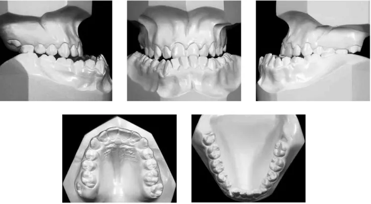
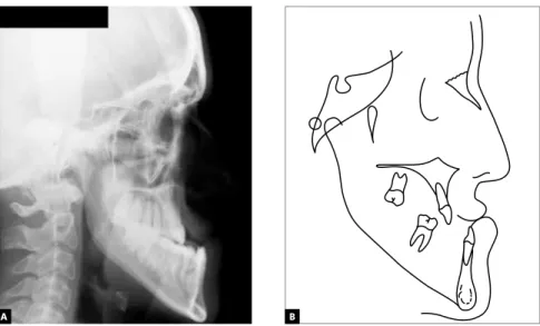
the menton base. The nasolabial angle was obtuse and the mentolabial angle was almost flat, characteristic of excessive compensation by lingual inclination of lower incisors in Class III. The absence of lip volume support was evident (Fig 1). The skeletal pattern was Class III, ANB = -11 ° (SNA = 73° and SNB = 84°); the mandible was elongated with a strongly obtuse go-nial angle; high GoGnSN = 45° and FMA = 38° angles, characterizing a predominance of vertical face devel-opment (Fig 4 and Table 1). From the dental point of view, patients presented Angle Class III malocclusion, with anterior and posterior crossbite and relative maxillary constriction. The upper incisors were well positioned and the lower were retroclined with mod-erate crowding and severe Curve of Spee (Figs 1, 2, 4).
A C B
Figure 2 - Initial dental casts.
Rizzatto SMD
A B
Figure 4 - Initial lateral cephalometric radiograph (A) and cephalometric tracing (B).
TREATMENT OBJECTIVES
The initial treatment objective was to correct the dental compensations in the maxilla and mandible through orthodontic treatment previous to the com-bined orthognathic surgery. In the maxilla, a dentoal-veolar expansion should be performed by increasing the upper intermolar distance to adjust its transverse relation. In the mandible, the severe lingual inclina-tion of the incisors should be corrected, leveling the curve of Spee and obtaining space for the canines and for positioning the incisors in their bone bases. The maxilomandibular sagittal and vertical will be cor-rected by orthognathic surgery with anterior and infe-rior repositioning of the maxilla and mandibular rota-tion and setback, aimed at obtaining the ideal occlusal relationship, function and facial harmony.
TREATMENT PLAN
The treatment plan consisted of three different steps:
First step: Refer the patient to carry out clinical review and preventive care. Following, bonding of up-per and lower brackets and decompensation of the lower arch by leveling the curve of Spee and projection of the incisors. Also, placement of an upper removable plate with expanding screw to promote a mild maxil-lary dentoalveolar expansion and retention with the mechanics advocated by Mulligan.
Second step: At this phase to perform the com-bined orthognathic surgery (for maxillary advance-ment and mandibular setback).
Third step: A post-surgical immobilization of the jaws with rubber bands and intensive elastic physio-therapy was planned. After orthodontic treatment finishing, the appliances would be removed and the retainers would be installed.
TREATMENT PROGRESS
wire hooks. After this first phase, an increase on the profile’s disharmony was observed due to increased sagittal dental discrepancy, mainly due to the buccal inclination of the lower incisors . However, the men-tolabial angle approached normality, as well as the volume of the lower lip (Figs 5 to 8).
The patient was referred to combined orthognathic surgery, consisting of maxillary protrusion with lower replacement and mandibular retrusion with upward rotation. For the following six months, the arches were
stabilized with rubber bands with Class III orientation and elastic physiotherapy was suggested. During this phase, intercuspation refining was performed, and then the appliance was removed. The lower retainer was installed, with a bonded intercanine fixed m re-tainer made with stainless steel 0.032-in archwire, and the upper retainer, wraparound type, was made with stainless steel 0.036-in archwire. This should be used full-time for 24 months followed by overnight use for another 12 months.
Rizzatto SMD
A C
B
Figure 6 - Pre-surgical dental casts.
A B
RESULTS
At the end of treatment, satisfactory occlusion was obtained with good dental intercuspation and pres-ence of anterior and lateral disocclusion guides. The esthetic result was pleasing, harmonious, respecting the individual characteristics of the patient. Orthog-nathic surgery provided maxillary protrusion with an increase in SNA of 6º, plus lower replacement and
Rizzatto SMD
A
B
C
Figure 10 - Final dental casts.
Rizzatto SMD
A B
Figure 13 - Facial and intraoral control photos, four years after retention.
A C B
A B
Figure 15 - Lateral cephalometric radio-graph (A) and cephalometric tracing (B), four years after retention.
Rizzatto SMD
Table 1 -Summary of cephalometric measurements.
Measurements Normal A P B C Difference A/B
Skeletal pattern
SNA (Steiner) 82 73 73 79 81 6
SNB (Steiner) 80 84 84 80 80 4
ANB (Steiner) 2 -11 11 -1 1 10
Angle of convexity (Downs) 0 -23 27 1 0 24
Y axis (Downs) 59 60 63 60 61 0
Facial angle (Downs) 87 91 90 90 90 1
Sn-GoGn (Steiner) 32 45 45 45 45 0
FMA (Tweed) 25 38 39 39 39 1
Dental pattern
IMPA (Tweed) 90 57 77 69 70 12
1–NA (mm) (Steiner) 22 27 26 19 22 8
1.NA (degrees) (Steiner) 4 5 5 5 5 0
1–NB (mm) (Steiner) 25 25 47 20 20 5
1.NB (degrees) (Steiner) 4 3 6 4 4 1
1
1 – interincisal angle (Downs) 130 160 140 145 144 15
1–APo (mm) (Ricketts) 1 -6 -11 3 2 9
Profile Upper lip – S line (Steiner) 0 -3 -6 0 -1 3
Lower lip –S line (Steiner) 0 4 8 1 1 3
3. Vig KD, Ellis E. Diagnosis and treatment planning for the surgical-orthodontic patient.
Dent Clin North Am. 1990;34(2):361-84.
4. Dann JJ, Fonseca R, Bell WH. Soft tissue changes associated with total maxillary
advancement: a preliminary study. J Oral Surg. 1976;34:19-23. CONCLUSION
The Class III malocclusion is considered most chal-lenging for the orthodontist, particularly when there is skeletal involvement.1 In this case, the diagnosis
conclusion was the recommendation of orthognathic surgery due to the dominant etiologic factor - severe maxillary and mandibular malocclusion - this being the primary focus on the treatment strategy. With the surgical approach, psychosocial aspects related to the deformity can also be privileged2 and it is important
that the diagnosis and treatment plan are performed in conjunction with the maxillofacial surgeon in order to maximize results and reduce the time and compli-cations inherent to treatment.3,4 Combined maxillary
and mandibular orthognathic surgery allowed the orthodontic treatment to be efficient, reaching its
goals, with appropriate overjet and overbite relation-ship between canines and molars in key of occlusion. Decompensation of the lower incisor inclination, con-sidered severe, did not cause changes that compro-mised the periodontal support structures. Final and control radiographs showed moderate levels of root resorption, which reveals the low biological cost of the treatment. The change in the mentolabial angle, with the positioning of the lower lip near normality, helped to reduce the subjectivity degree in the analysis of the soft tissue profile in the pre-surgical phase, enhanc-ing the final esthetic result. The improvement in facial esthetics contributed to raising the self-esteem of the patient. The follow-up of four years post-treatment denotes stability in occlusal characteristics, morphol-ogy of the arches and facial harmony.
REFERENCES
1. Stellzig-Eisenhauer A, Lux Cj, Schuster G. Treatment decision in adult patients with Class III malocclusion: orthodontic therapy or orthognathic surgery. Am J Orthod Dentofacial Orthop. 2002;122:27-38.