D e p a rtamento de Neurologia da Faculdade de Medicina da Universidade de São Paulo, São Paulo, Brasil (FMUSP):1P ro f e s s o r Associado;2Doutor;3Acadêmica da FMUSP; 4Professor Titular.
Received 5 January 2005, received in final form 8 April 2005. Accepted 21 May 2005.
Dra. Umbertina Conti Reed - Departamento de Neurologia, Hospital das Clínicas FMUSP - Av. Enéas de Carvalho Aguiar 255/5oa n d a r, sala 5131 - 05403-900 São Paulo SP - Brasil. E-mail: [email protected]
ULLRICH CONGENITAL MUSCULAR DYSTROPHY
AND BETHLEM MYOPATHY
Clinical and genetic heterogeneity
Umbertina Conti Reed
1, Lucio Gobbo Ferreira
2, Enna Cristina Liu
3,
Maria Bernadete Dutra Resende
2, Mary Souza Carvalho
2,
Suely Kazue Marie
1, Milberto Scaff
4ABSTRACT - Ullrich congenital muscular dystrophy (UCMD), due to mutations in the collagen VI genes, is an autosomal recessive form of CMD, commonly associated with distal joints hyperlaxity and severe course. A mild or moderate involvement can be occasionally observ e d . Objective:To evaluate the clinical picture of CMD patients with Ullrich phenotype who presented decreased or absent collagen VI immunoreactivity on muscular biopsy. Results:Among 60 patients with CMD, two had no expression of collagen V and their clinical involvement was essentially diff e rent: the first (3 years of follow-up) has mild motor difficulty ; the second (8 years of follow-up) never acquired walking and depends on ventilatory support. A molecular stu-d y, perf o rmestu-d by Pan et al. at the Thomas Jefferson University, stu-demonstratestu-d in the first a known mutation of Bethlem myopathy in COL6A1 and in the second the first dominantly acting mutation in UCMD and the first in COL6A1, previously associated only to Bethlem myopathy, with benign course and dominant inher-i t a n c e . Conclusion:Bethlem myopathy should be considered in the diff e rential diagnosis of UCMD, even in patients without fingers contractures; overlap between Ullrich and Bethlem phenotypes can be supposed.
KEY WORDS: Ullrich congenital muscular dystrophy, congenital muscular dystrophy, joint hyperlaxity, col-lagen VI, Bethlem myopathy.
D i s t rofia muscular congênita com hiperextensibilidade articular distal (Ullrich) e miopatia de Bethlem: heterogeneidade clínica e genética
RESUMO - A distrofia muscular congênita (DMC) com hiperextensibilidade articular distal (fenótipo Ullrich) a s-socia-se a mutações nos genes do colágeno VI e corresponde a um grave quadro congênito de herança autossômica recessiva e curso pro g ressivo, ocasionalmente mostrando menor gravidade. Objetivo:Av a l i a r o quadro clínico dos pacientes com DMC tipo Ullrich que apresentam imunoexpressão baixa ou ausente d o colágeno VI na biópsia muscular. Resultados:E n t re 60 pacientes com DMC, dois mostravam imunomarc a-ção negativa do colágeno VI. Mostravam-se clinicamente essencialmente diferentes: o primeiro, com 8 anos de idade e três de seguimento mostra leve dificuldade motora; o segundo, com 14 anos de idade e 8 de seguimento, não deambula e apresenta insuficiência respiratória. O estudo molecular, realizado na Thomas Jefferson University por Pan et al., revelou no primeiro, no gene COL6A1, mutação típica da miopatia de Bethlem, que tem curso benigno e herança autossômica dominante; e no segundo a primeira mutação de efeito dominante e do gene COL6A1, previamente associado apenas à miopatia de Bethlem. C o n c l u s ã o :
A miopatia de Bethlem deve constar no diagnóstico diferencial da DMC tipo Ullrich, mesmo na ausência das típicas contraturas dos dedos; pode existir sobreposição dos fenótipos Ullrich e Bethlem.
PA L AV R A S - C H AVE: distrofia muscular congênita; hiperextensibilidade art i c u l a r, colágeno VI, fenótipo Ullrich, miopatia de Bethlem.
Bethlem myopathy is a dominantly inherited dis-o rder caused by mutatidis-ons in the three genes dis-of cdis-ol- col-lagen VI, i.e. COL6A1 (21 q22.3), COL6A2 (21 q22. 3)
and COL6A3 (2 q37)1 - 4. Although Bethlem myopathy
period, childhood or adolescence and contracture s of fingers, elbows and ankles joints re p resent a hall-mark of this phenotype3 - 5. In addition, from 2001,
the deficiency of collagen VI in muscle has been asso-ciated with Ullrich scleroatonic congenital muscular d y s t rophy (UCMD) that is caused by diff e rent types of recessive and dominantly acting mutations in the same three collagen VI genes4 , 6 - 8. UCMD is clinically
less heterogeneous than Bethlem myopathy; howe-v e r, although the majority of patients hahowe-ve the clas-sic severe form that is characterized by neonatal muscle weakness, proximal joint contractures, hyper-laxity of the distal joints and severe course4, milder
patients have now been re p o rt e d9.
Both, Ullrich and Bethlem phenotypes are linked to the COL6A1, COL6A2 or COL6A3 genes, encoding respectively the alpha 1, alpha 2 and alpha 3 chains of collagen VI, and show clinical a n d genetic heterogeneity; there f o re mutation detec-tion in essential in these disorders for allowing the correct diagnosis, the establishment of prognosis and an accurate genetic counseling.
For emphasizing this clinical and genetic hetero-g e n e i t y, we re p o rt on two patients with distal joint h i p e r l a x i t y, the first with a mild to moderate myo-pathic phenotype including joint hyperlaxity and the second with a severe Ullrich phenotype. In both, a molecular analysis was perf o rmed at the Thomas J e fferson University, Philadelphia, by Pan et al.7a n d
revealed in the first a dominantly acting mutat i o n in the COL6A1 gene, that has not been desc r i b e d yet, and in the second a heterozygous in-fram e deletion in the COL6A1 that has been pre v io u s l y described in Bethlem myopathy10-11.
METHOD
Sixty children with clinical and histopathological d i a g-nosis of congenital muscular dystrophy (CMD) had their muscle samples evaluated immunohistochemically by means of immunofluorescency or immunoperoxidase m e-thods, utilizing antibodies for dystrophin (C-term i n a l ) , m e rosin (80 Kda and 300 Kda), sarcoglycans (,, y and
-SGs) and dystroglicans (-DG and- D G )1 2. Among t h e m ,
7 presented marked distal hyperlaxity and had their muscle samples also tested for collagen VI immunore a c-tivity using Hybridoma Bank antibody, code 5C6,1/100. In two patients (Cases 1 and 2) collagen VI immunore a c-tivity was absent.
CASES
Case 1– A 4 year-9 month-old male was born at term following an uneventful pregnancy from non consangui-neous parents who had already two healthy children. A t b i rth, the boy presented bilateral hip dislocation that was treated by the pediatrician and orthopedist. Motor development was mildly delayed: the child acquired sup-ported walking by 15 months of age and unsupsup-ported walking by 21 months of age. From the age of two y e a r s , f requent falls and a difficulty for running and climbing stairs were noted by the parents and other relatives. L a n-guage and mental development were normal. Our first examination at 4 years of age revealed, a mild to mod-erate difficulty for getting up from the floor, a mild pro-ximal weakness of the four limbs (MRC 4), a marked ge-neralized hypotonia, as well as a striking and widespre a d joint hypere x t e n s i b i l i t y. Deep tendon reflexes were hypoactive. On physical examination lumbar lord o s i s and a few small areas of abnormal hypochromic pigmen-tation in the skin of the lower limbs were noted. Seru m c reatine kinase levels were two-fold increased and elec-t romyography denoelec-ted abnormal myopaelec-thic paelec-telec-tern of muscle discharges. A muscle biopsy was perf o rmed at 5 years and 2 months of age and revealed mild to modera-te dystrophic changes re p resenmodera-ted by size fiber variabili-t y, moderavariabili-te perimysial fibrous infilvariabili-travariabili-tion, mild endo-mysial fibrous infiltration, a scarce fatty deposition and some necrotic fibers. Cardiac evaluation was normal. A f t e r a follow-up of 39 months, the course can be considere d slowly pro g ressive as we observed a worsening of pro x-imal muscle weakness (MRC 3 to 4) and the installation of mild distal weakness (MRC 4). The joint hyperlaxity persists and the boy did not develop any joint contractu-re. A molecular study of the patient’s DNA was done at the Department of Dermatology and Cutaneous Biology, Jefferson Institute of Molecular Medicine, Thomas Jef-ferson University, Philadelphia, by Pan et al.7and
demon-strated a Bethlem myopathy heterozygous in-frame dele-tion in the COL6A1 gene, that had been previously des-c r i b e d1 0 , 1 1. The p a t i e n t ’s father has normal posture,
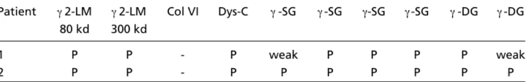
mus-Table 1. Immunohistochemical analysis of the muscle samples of the two patients.
Patient 2-LM 2-LM Col VI Dys-C -SG -SG -SG -SG -DG -DG 80 kd 300 kd
1 P P - P weak P P P P weak
2 P P - P P P P P P P
cular strength and tendon reflexes but presents mode-rate hyperlaxity of both thumbs, the left hand fingers and the left e l b o w. The analysis of his DNA did not re v e a l any mutation.
Case 2– A 6-year-old boy was born at term follow-ing an uneventful pregnancy from non consanguineous p a rents who had already two healthy children. The child presented from birth severe congenital hypotonia and generalized muscle weakness with proximal pre d o m i-nance. Motor development was delayed: he sits with-out support by the age of one year and never acquire d independent walking. Language and mental develop-ment were normal. From the second year of life he grad-ually developed elbows and knees contractures. Our first examination at 6 years of age revealed decre a s e d muscular strength [score of 3 and 2, following the Me-dical Resource Council (MRC) scale, respectively in the distal and proximal segments of the 4 limbs] widespre a d muscular hypotonia and hypotro p h y, distal joints hyper-laxity, as well as absent deep tendon reflexes. We n o t-ed a mild ankles pro t rusion which the parents re f e rre d as more pronounced in the past years. The mother consd e reconsd, after being inquireconsd, that the chilconsd has hyperh i-d rosis when comparei-d to his two normal oli-der siblings. C a rdiac evaluation was normal. Serum creatine k i n a s e levels were normal and electromyography revealed ab-n o rmal myopathic patterab-n of muscle discharges. The f i r s t muscle biopsy, perf o rmed at the age of 6 years, showed moderate size fiber variability, and mild to moderate perimysial as well as endomysial fibrous infiltration. A second biopsy was perf o rmed at 9 years of age and evi-dentiated marked worsening of the former aspects and an additional accentuated fatty deposition, as well as some necrotic fibers. Immunohistochemical analysis with
d i ff e rent antibodies was done (Table 1) and showed no collagen VI immunoreactivity (Fig 1). In a previous stu-d y1 3, the patient’s muscle sample had also been analysed
for laminins 1,1,2 and 1 chains immunore a c t i v i t y. The result was normal, i.e. negative immunomarcation for1 laminin chain, striking immunomarcation for1 and1 laminin chains and a little less pronounced im-m u n o im-m a rcation for2 laminin chain. Along the 8 years of follow-up, the boy manifested a pro g ressive worsen-ing, that was characterized by an accentuation of the h i p o t rophy and of the contractures which became wide-s p read, awide-s well awide-s by the inwide-stallation of wide-scoliowide-siwide-s. Cur-re n t l y, the boy is 14-year-old and depends on ventilato-ry support from 11 years of age. A molecular analysis w a s p e rf o rmed at the Department of Dermatology and Cuta-neous Biology, Jefferson Institute of Molecular Medicine, Thomas Jefferson University, Philadelphia, by Pan et al.7
and revealed a de novo in-frame heterozygous deletion of the COL6A1 gene.
DISCUSSION
Collagen VI is a protein that provides a micro f i-lamentous network in the extracellular matrix of the muscular tissue, as well as in other organs. It i s essential for the correct function of muscle fibers, maintaining its structural integrity. An animal mod-el of human Bethlem myopathy was already des-cribed and the details about the composition and the role of collagen VI have been widely discussed3.
Bethlem myopathy and Ullrich CMD result fro m molecular changes in each one of the three genes encoding collagen VI. The exact mechanism by which collagen VI leads to the myopathy is not
p e rfectly clear. Recently, a possible mitochondria l dysfunction in myofibers has been implicated in this m e c h a n i s m1 4. According to Mercuri et al.1 5,
colla-gen VI involvement is associated to molecular c h a n-ges in collagen VI genes in near to 40% of the C M D patients with Ullrich phenotype. In addition, there a re re p o rts of patients with low collagen VI re a c t i-vity without mutations in collagen VI genes1 5, as
well as of patients with mutations in collagen VI genes without changes in collagen VI re a c t i v i t y1 6,
t h e re f o redocumenting the genetic hetero g e n e i-ty of Ullrich phenoi-type. Although the role of colla-gen VI seems be excluded in a number of cases15,
Ishikawa et al.1 7recently considered that in patients
with Ullrich phenotye who have no mutations in the collagen VI genes and there f o re a normal amo-unt of Collagen VI in the interstitium, a primary a b n o rmality of other not yet identified molecules could cause a failure of collagen VI to anchor the basal lamina to the interstitium.
The first description of Bethlem myopathy was re f e rred by Bethlem & van Wi j n g a a rd e n1 8, who in
1976 re p o rted 28 patients from three families with an autosomal dominant, benign and slowly pro-g ressive myopathy. The most characteristic aspect of Bethlem myopathy is the occurrence of early c o n-t r a c n-t u res of n-the inn-terphalangeal joinn-ts and n-the el-bows. Merlini et al.1 9considered that the fingers
c o n t r a c t u resare the hallmark of Bethlem myopa-t h y. Clinical presenmyopa-tamyopa-tion and age of onsemyopa-t are hi-ghly variable3 , 5and although the clinical course of
the disease is thought to be benign, some re p o rt s emphasize that Bethlem myopathy can be slowly p ro g ressive and can culminate in wheelchair use2 0 -2 -2. Histopathological findings were either
nonspeci-fic or compatible with dystrophic changes and cre a-tine kinase levels can be normal or mildly elevate d3.
Collagen VI can be norm a l3. As diff e rent kinds of
mutations have been found in Bethlem myopathy, t h e reare attempts of establishing genotype/phe-notype correlation in Bethlem patients and some data indicate that large deletions and mutations inside the triple-helical collagen VI monomer helix f o rmed by the three collagenous polypeptides1 ,
2 and3 are associated with a more severe phe-notype than those occurring in the N-terminal glo-bular region4.
The first report of Ullrich phenotype occurred in 1930 by Ullrich2 3who named it scleroatonic form
of CMD and until 2002 only recessive mutations had been described in patients with UCMD3 , 6. In
2003, the first dominantly acting mutation in the
COL6A1 gene was found in one of our Brazilian p a-tients who we are now re p o rt i n g7and re c e n t l y
m o re three patients with a dominantly acting mu-tation in the COL6A1 gene were published8. Bes i d e
the genetic hetero g e n e i t y, UCMD also exhibits cli-nical hetero g e n e i t y4 , 9 , 1 5 - 1 6 , 2 4that is not related to
each of the 3 loci, but can be associated to the d e g ree of the deficiency of collagen VI in muscle o r c u l t u red fibro b l a s t s1 6. A complete deficiency has
been observed in the severe cases while the milder ones show a partial deficiency1 6. However, the
ma-jority of patients have a severe involvement that includes scoliosis, failure to thrive, and early and s e v e re re s p i r a t o ry impairment by the end of the first decade of life15. Mildly affected patients can
be related to mutations leading to a partial defi-ciency of collagen VI9,16.
The present re p o rt intends to emphasize the wide spectrum of phenotypes that can be associa-ted to collagen VI deficiency. Both patients have marked distal hyperlaxity, and histopathological d y s t rophic pattern, but clinical involvement was essentially diff e rent: the first (with 3 years and 4 months of follow-up) acquired independent walk-ing and shows a mild difficulty for runnwalk-ing and climbing; the second (with 8 years of follow-up) never acquired independent walking and needs i n t e rmittent ventilatory support from the begin-ning of the second decade of life. A molecular stu-dy of both patients was perf o rmed by Pan et al.7
at the Thomas Jefferson University and demonstra-ted in each one a diff e rent type of deletion of COL6A1 gene: in the first a heterozygous in-frame deletion in the COL6A1 that has been pre v i o u s l y described in Bethlem myopathy1 0 , 1 1and in the
sec-ond a dominantly acting mutation in the COL6A1 gene, that has not been described yet. This gene had been previously associated only to Bethlem myopathy and from 2003 is associated also to UCMD, as well as with a particular aspect of ossifcation of the posterior longitudinal ligament of s p i-ne in some subjects25.
In Patient 1, the absence of contractures, the marked joint hyperlaxity, and the dystrophic pat-t e rnfound on muscle biopsy had been supposed b y us as suggestive of a non specific mero s i n - p o s i t i v e CMD diagnosis. The result of the molecular analy-sis denoting a previously described Bethlem myo-pathy heterozygous in-frame deletion in the C O L 6 A 1 gene10,11indicates that the boy, currently young,
compatible with Bethlem myopathy. However, as his foll o w -up is now completing three years, he can be consi-d e reconsi-d an atypical case. During the 100th Euro p e a n N e u romuscular Center (ENMC) international work-s h o p4, Muntoni considered that joint laxity, aff e c
t-ing especially the knees and elbows, can be a com-mon finding at presentation and diseappears along the years. In the same opport u n i t y4, this author
re p o rted a case particularly coincidental to ours including by the presence of bilateral hip disloca-tion. His patient, currently aged 28, has also con-genital torticollis, a finding that has been com-monly described3. In addition, the dystrophic
chan-ges on the muscular biopsy, previously considere d non compatible with Bethlem myopathy2 6, have
been found so frequently as the non specific chan-g e s3. However, even considering that the lack of
c o n t r a c t u res and the dystrophic changes on muscu-lar biopsy, as observed in our Patient 1, have been re p o rted in patients with confirmed molecular diagnosis of Bethlem myopathy3 , 4such findings at
p resentation can not be considered typical. In fact, Jobsis et al.2 0followed-up 23 children and 36 adult
patients with Bethlem myopathy and found that nearly all children exhibit weakness or contractu-res during the first two years of life. In addition, a c c o rding to Bertini and Pepe5, muscle biopsy fro m
Bethlem cases shows non specific changes and an i n c rease of endomysial connective tissue is rare l y o b s e rved. Merc u ry et al.2 1re p o rted that the degre e
of muscle involvement varies according to the de-g ree of motor impairment. There f o re in Patient 1 a less amount of muscle changes would be expect-ed. In addition, as in our patient was found a het-e rozygous in-framhet-e dhet-elhet-etion of 18 aminoacids so-mewhat downstream in the triple-helical domain, a result of exon 14 skipping in the COL6A1 gene7,
we could theoretically expect a more severe clini-cal involvement. The patients already re p o rted wi-th a type of mutation similar to wi-that observed in o u r patient have either a typical clinical picture with finger contracture s1 0or a severe course when comp
a-red to that of other reported Bethlem myopathy f a m i l i e s1 1. In one familiy one of the affected
mem-bers had lost the deambulation at the age of 35 years and another member had developed Achilles’ tendons bilateral shortening and finger contrac-t u res from 7 years of age1 1. Finally, as muscle
immu-n o h i s t o c h e m i s t ry with Col VI aimmu-ntibodies caimmu-n be normal in the muscle3, being detected only by
fi-b rofi-blast culture, that is not a routine pro c e d u re, t h e description of the clinical findings of our Patient
1 intends to emphasize that Bethlem myopathy should be included among the diff e rential diagno-sis of merosin-positive CMD. Besides this, in spora-dic patients with clinical and histopathological fin-dings suggestive of merosin-positive CMD, who al-so manifest joint hyperlaxity, a molecular analysis looking for Bethlem mutations is recommended. Patient 2 has a classic severe form of UCMD and re p resented the first example of UCMD with a het-e rozygous in-framhet-e dhet-elhet-etion in COL6A17, there f o re
i n c reasing the already marked genetic hetero g e n e-ity observed in this form of CMD. Ve ry recently more t h ree patients with dominant mutations have been described, all manifesting severe phenotype char-acterized by marked restriction of re s p i r a t o ry func-tion, scoliosis and lack of independent ambulation in one of them8. According to Baker et al.8, these
new genetic data in UCMD7 - 8highlighted the
neces-sity of a careful mutation investigation for pro v i d-ing an accurate genetic counseld-ing advice. Patient 2 was the only who presented a typical severe Ul-lrich phenotype among around 80 childrens with cli-nical and histopathological diagnosis of CMD, in-cluding 34 typical MD-CMD cases, who we have at-tended and followed-up since 1990 at our institu-tion. There f o re, although Muntoni and Vo i t9h a v e
re f e rred that UCMD is probably the second most fre-quent variant of CMD, it is our impression that UCMD is not so common among Brazilian patients. In conclusion, the new molecular data seem suggest that new phenotypes linked to collagen VI unit and particularly to COL6A1 gene can be identified in a next future, so defining if Ullrich and Bethlem phenotypes are independent enti-ties or, as re p o rted by Bertini and Pepe5, re p re s e n t
an overlap between the clinical phenotypes and the molecular defects. A probable overlap between UCMD, Bethlem myopathy and Ehlers-Danlos syn-d romes has been the focus of recent re s e rc h e s4 , 9 , 2 7.
A c k n o w l e d g e m e n t s– We are grateful to Dr. Cars-ten G. Bonnemann who kindly provided the molecular analysis of the patients and to Dr. Stephan Kroger for his generous gift of the antibody to collagen VI.
REFERENCES
1. Jobsis GJ, Keizers H, Vreijling JP, et al. Type VI collagen mutations in Bethlem myopathy, an autosomal dominant myopathy with contractu-res. Nat Genet 1996;14:113-115.
2. Speer MC, Tandan R, Rao PN, et al. Evidence for locus hetero g e n e i t y in the Bethlem myopathy and linkage to 2q37. Hum Mol Genet 1996; 5:1043-1046.
3. Pepe G, de Visser M, Bertini E, et al. Bethlem myopathy (BETHLEM) 86th ENMC international workshop, 10-11 November 2000, Naarden, The Netherlands. Neuromuscul Disord 2002;12:296-305.
4. Pepe G, Bertini E, Bonaldo P, et al. Bethlem myopathy (BETHLEM) and Ullrich scleroatonic muscular dystrophy: 100th ENMC international workshop, 23-24 November 2001, Naarden, The Netherlands. Neuro muscul Disord 2002;12:984-993.
5. Bertini E, Pepe G. Collagen type VI and related disorders: Bethlem myopathy and Ullrich scleroatonic muscular dystro p h y. Eur J Paediatr Neurol 2002;6:193-198.
6. Camacho Vanegas O, Bertini E, Zhang RZ, et al. Ullrich sclero a t o n i c muscular dystrophy is caused by recessive mutations in collagen type VI. Proc Natl Acad Sci USA 2001;98:7516-7521.
7. Pan TC, Zhang RZ, Sudano DG, Marie SK, Bonnemann CG, Chu ML. New molecular mechanism for Ullrich congenital muscular dystro p h y : a heterozygous in-frame deletion in the COL6A1 gene causes a severe phenotype. Am J Hum Genet 2003;73:355-369.
8. Baker NL, Morgelin M, Peat R, et al. Dominant collagen VI mutations a re a common cause of Ullrich congenital muscular dystro p h y. Hum Mol Genet 2005;14:279-293.
9. Muntoni F, Voit T. The congenital muscular dystrophies in 2004: a centu-ry of exciting progress. Neuromuscul Disord 2004;14:635-649. 10. Lamande SR, Shields KA, Kornberg AJ, Shield LK, Bateman JF. Bethlem
myopathy and engineered collagen VI triple helical deletions pre v e n t intracellular multimer assembly and protein secretion. J Biol Chem 1999;274:21817-21822.
11. Pepe G, Giusti B, Bertini E, et al. A h e t e rozygous splice site mutation in COL6A1 leading to an in-frame deletion of the alpha1(VI) collagen chain in an Italian family affected by Bethlem myopathy. Biochem Bio-phys Res Commun 1999;258:802-807.
12. F e r reira LG. Análise imunohistoquímica das proteínas do complexo
d i s t ro f i n a - g l i c o p roteínas associadas em pacientes com distrofia muscu-lar congênita. Tese., São Paulo, 2002.
13. Reed UC. Distrofia muscular congênita: estudo da variabilidade feno-típica e análise da correlação clínico-imunohistoquímica. Tese. São Paulo, 1999.
14. Irwin WA, Bergamin N, Sabatelli P, et al. Mitochondrial dysfunction and apoptosis in myopathic mice with collagen VI deficiency. Nat Genet 2003;35:367-371.
15. M e rcuri E, Yuva Y, Brown SC, et al. Collagen VI involvement in Ullrich s y n d rome: a clinical, genetic, and immunohistochemical study. Neuro-logy 2002;58:1354-1359.
1 6 . Demir E, Ferre i ro A, Sabatelli P, et al. Collagen VI status and clinical severity in Ullrich congenital muscular dystrophy: phenotype analysis of 11 families linked to the COL6 loci. Neuropediatrics 2004;35:103-11 2 . 17. Ishikawa H, Sugie K, Murayama K, et al. Ullrich disease due to defi-ciency of collagen VI in the sarcolemma. Neurology 2004;62:620-623. 18. Bethlem J, Wijngaarden GK. Benign myopathy, with autosomal
dom-inant inheritance. A report on three pedigrees. Brain 1976;99:91-100. 19. Merlini L, Morandi L, Granata C, Ballestrazzi A. Bethlem myopathy:
early-onset benign autosomal dominant myopathy with contracture s : description of two new families. Neuromusc Disord 1994;4:503-511. 20. Jobsis GJ, Boers JM, Barth PG, de Visser M. Bethlem myopathy: a
slow-ly pro g ressive congenital muscular dystrophy with contractures. Brain 1999;122:649-655.
21. M e rcuri E, Cini C, Counsell S, et al. Muscle MRI findings in a thre e -generation family affected by Bethlem myopathy. Eur J Paediatr Neuro l 2002;6:309-314.
22. Haq RU, Speer MC, Chu M-L, Tandan R. Respiratory muscle involve-ment in Bethlem myopathy. Neurology 1999;52:174-176.
23. Ullrich O. Kongenitale atonisch-sklerotische Muskeldystrophie, ein w e i t e rer Typus der heredodegeneration Erkrankungen des nuero m u s-kularen Systems. Z Ges Neurol Psychiat1930;126:171-201.
24. Demir E, Sabatelli P, Allamand V, et al. Mutations in COL6A3 cause s e v e re and mild phenotypes of Ullrich congenital muscular dystro p h y. Am J Hum Genet 2002;70:1446-1458.
25. Tanaka T, Ikari K, Furushima K, et al. Genomewide linkage and link-age disequilibrium analyses identify COL6A1, on chromosome 21, as the locus for ossification of the posterior longitudinal ligament of the spine. Am J Hum Genet 2003; 73: 812-822.
26. Tohyama J, Inagaki M, Nonaka I. Early onset muscular dystrophy with autosomal dominant here d i t y. Report of a family and CT findings of skeletal muscle. Brain Dev 1994;16:402-406.