https://doi.org/10.1590/0004-282X20170109
ARTICLE
Bereitschaftspotential preceding eyelid blinks
in Parkinson’s disease
Potencial de Bereitschafts precedendo o piscamento ocular na doença de Parkinson
Isabella Araújo Mota¹, Maria das Graças Coriolano2, Otávio Gomes Lins3
he increasing prevalence and incidence of Parkinson’s disease (PD), along with the increase in the population’s life expectancy, has made it the second most frequent neurode
-generative disorder. his has a signiicant socio-economic impact on the quality of life of PD patients and caregivers due to progressive physical dependence in activities of daily living, in addition to subsequent cognitive impairment.
Hypomimia, or masked facies, in PD is character
-ized by reduced automatic and voluntary facial muscle expressiveness from the early stages, progressing with advancing disease1 and generating social implications due
to the expressionless, apathetic, depressed and introspec
-tive appearance2
. Among the hypomimia findings, there is a decrease of the frequency of eyelid blink by different pathways related to the planning and execution of this
motor act. This signal is one component of the extensive bradykinesia presentation.
Bradykinesia results from neuronal damage to pathways related to motor planning3
. Voids in the dopaminergic and nondopaminergic circuits have been involved in this patho
-physiology, generating an imbalance between inhibition and excitation, deicits and compensation4,5,6.Changes in the
dopaminergic pathways explain, at least in part, disturbances in the kinematics of spontaneous eyelid blink7,8,9, and the vol
-untary one10
and in the blink relex habituation of PD11
. Pharmacological treatment with dopaminergic medi
-cations, considered the gold standard12,13, results in partial
problem-solving of bradykinesia as it improves the speed of movement7,8,10, but not its rhythm and amplitude14
, although the damage regarding the amplitude is very evident1
.
1Hospital Universitário Lauro Wanderley, Ambulatório de Neurologia, João Pessoa PB, Brasil; 2Universidade Federal de Pernambuco, Departamento de Anatomia, Recife PE, Brasil; 3Universidade Federal de Pernambuco, Departamento de Neuropsiquiatria, Recife PE, Brasil.
Correspondence: Isabella Araújo Mota Fernandes; BR 230, Km 10, Condomínio Villas do Atlântico, casa 3B; 58310-000 Cabedelo PB, Brasil; E-mail: [email protected]
Conflict of interest: There is no conlict of interest to declare. Received 10 February 2017; Accepted 04 May 2017. ABSTRACT
The Bereitschaftspotential (BP) is a negative wave observed in EEG retrograde averaging, preceding a motor act. The objective was to study the BP preceding voluntary eyelid blinks in Parkinson’s disease (PD) patients during off and on phases of levodopa. Methods: Ten PD patients in stages 1 and 2 of the Hoehn & Yahr classiication were compared to 18 healthy controls. Artifact-free EEG segments of two seconds preceding the onset of the blink potential were averaged and analyzed, and the statistical signiicance of the measured amplitudes were evaluated by analysis of variance models. Results: The presence of a BP in the PD patients was demonstrated. The mean amplitudes at 0 ms were respectively 0.6 µV and 3.3 µV for the BP patients and the normal controls, respectively. Conclusions: The BP amplitudes were signiicantly smaller in PD patients than normal participants. The amplitudes of the BP were not modiied by levodopa.
Keywords: contingent negative variation; Parkinson disease; levodopa
RESUMO
O Potencial de Bereitschafts (PB) é uma onda negativa observada retrogradamente no EEG precedendo um ato motor. Objetivo: Estudar o PB precedendo o piscamento palpebral voluntário em pacientes com doença de Parkinson (DP) durante as fases off e on da levodopa. Foram comparados dez pacientes com DP nos estágios 1 e 2 de Hoehn & Yahr com 18 controles saudáveis. Os segmentos de EEG livres de artefatos 2 segundos antes do início do potencial foram calculados e analisados e a signiicância estatística das amplitudes foi medida por modelos de análise de variância. Resultados: A presença de PB nos pacientes com DP foi demonstrada. As amplitudes médias a 0 ms foram respectivamente 0,6 μV e 3,3 μV para os pacientes com DP e controles respectivamente. Conclusões: As amplitudes do PB foram signiicativamente menores nos pacientes com DP do que controles. As amplitudes do PB não foram modiicadas pela levodopa.
Bereitschaftspotential (BP), or readiness potential, described by Kornhuber and Deecke in 1964 corresponds to the negative gradient starting one-and-a-half to two sec
-onds before the motor potential, seen in electroencephalo
-gram (EEG) retrograde averaging. his potential is divided into an early component related to planning and a late poten
-tial related to motor execution15,16,17. his study aimed to eval
-uate the ocular blink in patients with PD in the of (without levodopa) and on phases (with levodopa) by measuring the BP.
METHODS
Subjects
he study was approved by the University Human Research Ethics Committee (statement 90208). All partici
-pants signed an informed consent form.
Ten PD patients (nine males) aged 43 to 78 years were randomly recruited in the Parkinsonian Patient Care Service (Pro-Parkinson) at the Clinic Hospital of the Federal University of Pernambuco, Brazil. All patients were diagnosed by a neurologist trained in movement disorders. Eighteen normal individuals (eight males) aged 17 to 60 years served as a comparison group. he analysis of this normal data was recently accepted for publication18.
Procedures
he participants sat comfortably on a chair and were instructed to relax, restrain moving the muscles of the neck, head or face and ix their gaze on a spot placed two meters away at eye level, blinking as naturally as possible, once every ten sec
-onds. here were no auditory or visual clues for the time of blink
-ing and the rhythm was rehearsed before start-ing the record-ings. he PD patients who met the inclusion criteria were invited to return the next morning. he patients were instructed not to take their morning anti-Parkinson’s med
-ication, before coming to the laboratory. hey were also advised to avoid the intake of protein foods.
he PD patients were initially recorded at least 12 hours after the last anti-Parkinson’s medication (of phase). hen levodopa/benserazide 100/25 mg was ofered and, after 40 minutes, a second recording section (on phase) was made.
he normal controls were recorded only once.
he EEG was recorded from 11 EEG electrodes placed in positions F3, FZ, F4, C3, Cz, C4, P3, Pz, P4, O1, O2 of the inter
-national 10-20 system of electrode placement, referenced to two electrodes placed on the mastoids and linked together. he vertical electrooculogram was recorded from two electrodes placed one centimeter above and below the right eye. he ground electrode was placed on the skin over the right clavicle.
he recordings were made using a Neuron-Spectrum NET polygraph (Neurosoft). he bandpass of the ilters were 0.1 Hz to 35 Hz. Inter-electrode impedances (10 Hz) were kept below 3kΩ.
Data analysis
Blinks were visually identiied as large positive delections on the vertical electrooculogram channel and the beginning of the blink potential was marked. he two-second EEG seg
-ments preceding the mark were back-averaged together. Blinks that presented artifacts during or close to this period were eliminated from the analysis.
he averages were saved in text iles and analyzed in a speciic program written in Matlab software. Initially, the waves obtained by the general averaging of each group of participants were observed. he measurement points were established at -1800 ms (at the beginning of the BP), -500 ms (at the start of the late component of the BP) and 0ms (at the onset of the blink potential). To compensate for background noise, the amplitudes were averaged in a 100 ms segment centered on the latency to be measured.
Statistical analysis of the BP amplitudes was performed on the C3, C4 and Cz electrodes, using the STATISTICA ver
-sion 10 statistical package (StatSoft). he statistical analysis was performed using analysis of variance (ANOVA) models. A three-way repeated measures ANOVA model, condition (of
and on), time (-1800 ms, -500 ms, 0s) and electrodes (C3, Cz, C4) was used to compare the PD group during the of phase and the on phase. A mixed-model ANOVA, group (PD and con
-trols) and two repeated-measures, time (-1800 ms, -500 ms and 0 ms) and electrode (C3, Cz and C4), was used to com
-pare the PD patients with the normal controls. he Geisser-Greenhouse correction was applied for violation of sphericity. he Newman-Keuls test was used for post hoc analysis, when
necessary. he critical p-value was 0.05.
RESULTS
Table 1 lists some demographic and clinical characteris
-tics of the studied PD patients.
Table 1. Some characteristics of the studied Parkinson’s disease patients.
Initials Sex Age TD H&Y UPDRS II
UPDRS III
UPDRS T MMSE
AC M 43 8 3 9 14 23 29
EL M 44 3 1 10 29 39 25
DA M 48 6 1 10 15 25 30
SF F 52 2 1 10 24 34 26
JF M 58 4 1 12 30 42 27
CE M 53 5 2 19 90 109 30
MA M 61 3 1 11 29 40 30
AJ M 61 5 1 5 20 25 30
EV M 68 1 2 8 35 43 24
JQ M 78 4 1 5 7 12 30
A total of 230 blinks during the of phase and 250 blinks during the on phase were averaged from the 10 PD patients,
giving a mean of 23 blinks per patient during the of phase and 25 blinks per patient during the on phase.
Figure 1 shows the grand average of the EEG recorded at all electrodes from the PD patients in the of (dashed line) and
on (solid line) phases.
In both conditions, a negative potential of increasing amplitude was observed starting around -1,500 ms before the blink onset. his potential had a broad scalp distribution, but was most evident at the central regions. Around -500ms before the blink potential, a change in the wave coniguration was observed. No obvious diferences were seen between the potentials obtained during the of phase and the on phase.
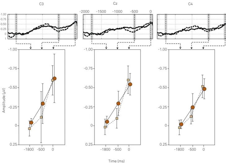
Figure 2 shows the analysis of the amplitudes of the potentials at C3, Cz and C4 electrodes measured -1800 ms, -500 ms and 0 ms before the onset of the blink potential. he waveforms and measurement times are shown at the top of the igure. Amplitudes were averaged in a segment of 100ms centered on the latency to be measured (vertical gray shadows). he means and standard deviations of the amplitudes at each electrode, condition and time-point are illustrated at the bottom of the igure. Table 2 shows the ANOVA of the data.
he ANOVA showed a signiicant efect for time. No inter
-actions were signiicant. he post hoc analysis
(Newman-Keuls) showed that the amplitudes at -500ms (onset of the late component) and 0ms (end of the BP) were both signii
-cantly larger than the amplitude at -1800 ms (just before
the onset of the early BP component). he amplitudes at -500 and 0ms were not diferent. his is consistent with the presence of a BP preceding the voluntary blink of PD patients, during both of and on phases. No signiicant dif
-ferences between the amplitudes during the of phase and the on phase were observed.
A total of 900 blinks were averaged from the 18 normal controls, giving a mean of 50 blinks per individual. As there were no signiicant diferences between the measured ampli
-tudes of the BP during of and on phases, all 580 blinks from
the 10 PD patients were averaged, giving a mean of 58 blinks per participant.
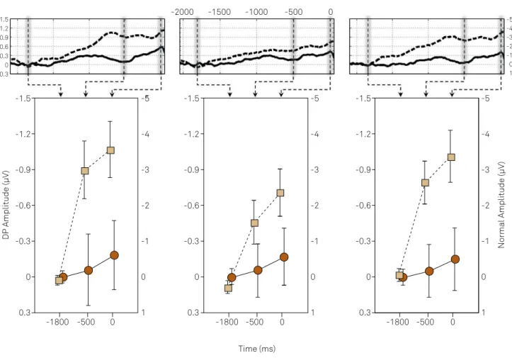
Figure 3 shows the analysis of the amplitudes of the poten
-tials at C3, Cz and C4 measured -1800, -500 and 0 ms before the onset of the blink potentials from the PD patients (solid line) and the normal controls (dashed line). he waveforms and measurement times are shown at the top of the igure. he means and standard deviations of the amplitudes at each electrode, condition and time-point are illustrated at the bottom of the igure. Diferent scales were used for the PD patients (right side) and for the control group (left side). Table 3 shows the ANOVA of the data.
he ANOVA showed a signiicant interaction between the time and the group. he post hoc analysis showed that the
amplitudes at -500 ms and 0ms were both signiicantly larger in the control group than in the PD group. At -1800 ms the amplitudes were not diferent between groups. herefore, the amplitude of the BP of PD patients is signiicantly smaller than the BP of normal controls at all analyzed electrode positions.
On
Off
F3 Fz F4
C3 Cz C4
P3 Pz P4
O1 O2
-1 -.75 -.5 -.25 0 .25
-1 -.75 -.5 -.25 0 .25
-1 -.75 -.5 -.25 0 .25
-1 -.75 -.5 -.25 0 .25
-1 -.75 -.5 -.25 0 .25
-1 -.75 -.5 -.25 0 .25
-1 -.75 -.5 -.25 0 .25
-1 -.75 -.5 -.25 0 .25
-1 -.75 -.5 -.25 0 .25
-1 -.75 -.5 -.25 0 .25
-1 -.75 -.50 -.25 0 -0.25 -2000 -1500 -1000 -500 0
-2000 -1500 -1000 -500 0
-2000 -1500 -1000 -500 0
Figure 1. Grand average of the bereitschaftspotential of the Parkinson’s disease patients during the off phase (dashed line) and on
C3 Cz C4
-0.75
-0.25 -1.00
-0.50
1 0
-0.75 -1.00
-0.50
-0.25
0.25 0
-1800 -500 0 -1800 -500 0 -1800 -500 0
Time (ms)
Amplitude (µV)
-2000 -1500 -1000 -500 0
-0.75 -1.00
-0.50
-0.25
0.25 0
-0.75 -1.00
-0.50
-0.25
0.25 0
Figure 2. Analysis of the amplitudes of the bereitschaftspotential of the Parkinson’s disease patients at C3, Cz and C4 electrodes measured -1800, -500 and 0 ms before the onset of the blink potentials. The waveforms and measurement times are shown on the top of the igure. Amplitudes were averaged within a segment of 100 ms centered on the latency to be measured (vertical gray shadows). The means and standard deviations of the amplitudes at each electrode, condition and time-point are illustrated at the bottom of the igure.
Table 2. Factorial repeated-measurement analysis of variance of the amplitudes of the bereitschaftspotential of the Parkinson’s disease patients, with three repeated measurements: condition (off and on medication), time (-1800, -500 and 0ms, relative to the onset of the blink potential) and electrode (C3, Cz and C4).
Variable SS df MS F p
Intercept 11.706 1 11.706 12.145 0.007
Error 8.675 9 0.964
Condition 0.212 1 0.212 0.284 0.607
Error 6.716 9 0.746
Time 9.552 2 4.776 7.072 0.005*
Error 12.157 18 0.676
Electrode 0.063 2 0.032 0.099 0.906
Error 5.730 18 0.318
Condition × time 0.260 2 0.130 0.275 0.762
Error 8.494 18 0.472
Condition × electrode 0.021 2 0.010 0.207 0.815
Error 0.8900 18 0.049
Time × electrode 0.064 4 0.016 0.176 0.950
Error 3.293 36 0.092
Condition × time × electrode 0.018 4 0.005 0.063 0.992
Error 2.534 36 0.070
Newman-Keuls test of the main effect TIME
-1800 ms × 0 ms 0.005*
-1800 ms × -500 ms 0.024*
-500 ms × 0 ms 0.233
C3 Cz C4
-1.2
-0.6 -1.5
-0.9
-0.3
0.3 0
-1.2
-0.6 -1.5
-0.9
-0.3
0.3 0
-1.2
-0.6 -1.5
-0.9
-0.3
0.3 0
-1.2
-0.6 -1.5
-0.9
-0.3
0.3 0 -4
-2 -5
-3
-1
1 0
-4
-2 -5
-3
-1
1 0
-4
-2 -5
-3
-1
1 0
-1800 -500 0 -1800 -500 0 -1800 -500 0
Time (ms)
DP Amplitude (µV)
Normal Amplitude (µV)
-4
-2 -5
-3
-1
1 0
-2000 -1500 -1000 -500 0
Figure 3. Analysis of the amplitudes of the bereitschaftspotential of the Parkinson’s disease patients (grand average of all recordings during the off phase and the on phase) and the normal controls at C3, Cz and C4 electrodes measured -1800, -500 and 0 ms before the onset of the blink potentials. The waveforms and measurement times are shown on the top of the igure. Amplitudes were averaged within a segment of 100ms centered on the latency to be measured (vertical gray shadows). The means and standard deviations of the amplitudes at each electrode, condition and time-point are illustrated at the bottom of the igure. Different scales were used for the PD patients (right side) and the normal controls (left side).
Table 3. Factorial mixed-model analysis of variance of the amplitudes of the bereitschaftspotential of the Parkinson’s disease patients and normal controls: Group (PD patients and normal controls) and two repeated measures: time (-1800, -500 e 0 ms, relative to the onset of the blink potential) and electrode (C3, Cz and C4).
Variable SS Df MS F p*
Intercept 227.770 1 227.770 10.379 0.004*
Group 128.687 1 128.687 5.864 0.024*
Error 504.716 23 21.944
Time 134.349 2 67.175 7.847 0.001*
Time × group 74.475 2 37.238 4.350 0.019*
Error 393.785 46 8.561
Electrode 9.394 2 4.697 2.296 0.112*
Electrode × group 9.731 2 4.866 2.379 0.104*
Error 94.087 46 2.045
Time × electrode 2.860 4 0.715 1.244 0.298*
Time × electrode × group 2.665 4 0.666 1.159 0.334*
Error 52.878 92 0.5748
Newman-Keuls test of the interaction time × group
Parkinson × Control at -1800 ms 0.902
Parkinson × Control at -500 ms 0.033*
Parkinson × Control at 0 ms 0.012*
DISCUSSION
his study demonstrated that there is a BP preceding vol
-untary blinks in PD patients. his BP does not difer signii
-cantly during the of phase and the on phase of medication.
To our knowledge, there have been no studies on BP preced
-ing blinks in PD patients. In a previous recently-accepted study18, we reported the presence of a BP preceding voluntary
(but not spontaneous blinks) in normal participants. he BP of DP patients had a broad scalp distribution, most clearly identiied at the central regions, similar to the BP of normal controls and to the BP preceding other move
-ments in PD and normal controls19,20,21.
he amplitude of the BP of the PD patients was much smaller than the BP of the control group, even though the PD patients were at a relatively mild stage of the disease. At its largest negativity (just preceding the blink) the mean ampli
-tudes of the BP of the PD patients and the normal controls were respectively 0.6 µV and 3.3 µV. his suggests a signiicant dysfunction of the motor neural networks, even in patients with few symptoms. his inding is consistent with previ
-ous descriptions of reduced amplitude of BP preceding other movements in PD patients, even in the early stages12,22,23,24,25,26.
It is also in accordance with the early clinical inding of pau
-city of facial expression and reduced blink rate in PD patients, suggesting that neural systems related to motor planning, especially those related to facial expression and blinks, are afected early in PD1
.
Most PD patients were at stage 1 of the H&Y scale and showed low scores on the motor and daily living activities of the Uniied Parkinson’s Disease Rating Scale. Although the disease has a progressive symptomatology, it is known that damage to neuronal pathways begins 8–17 years before the appearance of the irst symptoms, with diferent progression characteristics depending on the compensatory mechanisms and the age of onset. herefore, even patients who are diag
-nosed early may present with signiicant neural dysfunction. Although most studies have reported smaller BP ampli
-tudes in PD patients compared to normal controls, some stud
-ies have reported no diferences21,24,26 or even larger ampli
-tudes27,28. he reasons for these discrepancies are not clear.
In our study, the dopaminergic drug did not signiicantly afect the BP. A larger amplitude during the on phase has been
described in BP preceding the movement of the ingers22,23.
he absence of an inluence by levodopa on the BP preceding the eye blink may result from an earlier, and more severe, dys
-function of the dopaminergic networks involved in the plan
-ning of eye blinks, than inger movements.
Although levodopa is considered the gold standard drug for PD therapy, it does not appear to act on all the mechanisms that cause bradykinesia14
. Nondopaminergic pathways play a role in the clinical improvement of motor planning and BP amplitude in PD patients, as observed after unilateral postero
-ventral pallidotomy28 and after neurofeedback techniques25.
hese studies corroborate the presence and importance of nondopaminergic or dopaminergic pathways resistant to modulation mechanisms of the motor planning in PD6.
he amplitudes at C3, Cz and C4 were not signiicantly diferent in PD patients. In our previous study with normal controls18, we found larger amplitudes at C3 and C4 in rela
-tion with Cz. his was also observed by Yamamoto29, but not
Shimizu and Okiyama20
, in their study with saccadic move
-ments. A lateral component of the BP source complex, per
-haps the face motor cortex, is less active in PD patients than in normal subjects. A source analysis study may shed some light on this issue.
he main objective of this study was to determine if a BP preceding voluntary blinks could be demonstrated in PD patients. We believe this was accomplished. he poten
-tials we obtained had very low amplitudes in comparison with the potentials obtained from normal participants. Although the normal participants we used were not perfectly age-matched with the PD patients, we believe the diferences in amplitudes are obvious. Furthermore, it has been shown that there is no signiicant change in BP amplitudes with increasing age, within certain limits30
.
In conclusion, this is the irst study demonstrating a BP preceding eyelid blinks in PD patients. he BP of the PD patients had much smaller amplitudes than the BP of normal subjects, even though the PD patients were in the early stages of the disease. hese potentials could be used in the future as early diagnostic and evolutionary markers of Parkinson’s dis
-ease. Further investigations should be conducted. Currently considered the gold standard drug in Parkinson’s disease, levodopa did not obviously modify the BP preceding blinks, suggesting that the resistant nondopaminergic or dopami
-nergic accessory pathways related to motor planning should be investigated in search of more efective medications.
References
1. Korosec M, Zidar I, Reits D, Evinger C, Vanderwerf F. Eyelid
movements during blinking in patients with Parkinson’s disease. Mov Disord. 2006;21(8):1248-51. https://doi.org/10.1002/mds.20930
2. Tickle-Degnen L, Zebrowitz LA, Ma HI. Culture, gender and health care stigma: practitioners’ response to facial masking experienced by people with Parkinson’s disease. Soc Sci Med. 2011;73:95-102. https://doi.org/10.1016/j.socscimed.2011.05.008
3. Berardelli A, Rothwell JC, Thompson PD, Hallett M. Pathophysiology of bradykinesia in Parkinson’s disease. Review Article. Brain. 2001;124(11): 2131-46. https://doi.org/10.1093/brain/124.11.2131
5. Yu R, Liu B, Wang L, Chen J, Liu X. Enhanced functional connectivity between putamen and supplementary motor area in Parkinson’s disease patients. PLoS One. 2013;8:59717. https://doi.org/10.1371/journal.pone.0059717
6. Gröger A, Kolb R, Schäfer R, Klose U. Dopamine reduction in the substantia nigra of Parkinson’s disease patients conirmed by in vivo magnetic resonance spectroscopic imaging. PLoS One. 2014;9(1):84081. https://doi.org/10.1371/journal.pone.0084081
7. Deuschl G, Goddemeier C. Spontaneous and relex activity of facial muscles in dystonia, Parkinson’s disease, and in normal subjects. J Neurol Neurosurg Psychiatry. 1998;64(3):320-4. https://doi.org/10.1136/jnnp.64.3.320
8. Taylor JR, Elsworth JD, Lawrence MS, Sladek JR Jr, Roth RH, Redmond DE Jr. Spontaneous blink rates correlate with dopamine levels in the caudate nucleus of MPTP-treated monkeys. Exp Neurol. 1999;158(1):214-20. https://doi.org/10.1006/exnr.1999.7093
9. Colzato LS, Wildenberg, WPM, Hommel, B. Reduced spontaneous eye blink rates in recreational cocaine users: evidence for dopaminergic hypoactivity. PLoS One. 2008;3(10):3461. https://doi.org/10.1371/journal.pone.0003461
10. Agostino R, Bologna M, Dinapoli L, Gregori B, Fabbrini G, Accornero N et al. Voluntary, spontaneous, and relex blinking in parkinson’s disease. Mov Disord. 2008;23(5):669-75. https://doi.org/10.1002/mds.21887
11. Penders CA, Delwaide PJ. Blink relex studies in patients with Parkinsonism before and during therapy. J Neurol Neurosurg Psychiatry. 1971;34(6):674-8. https://doi.org/10.1136/jnnp.34.6.674
12. Tolosa E, Katzenschlager R. Pharmacological management of Parkinson`s disease. In: Jankovic J, Tolosa E. Parkinson´s disease and movement disorders. Philadelphia: Lippincott Williams and Wilkins; 2006. p. 113-6.
13. Porras G, De Deurwaerdere P, Li Q, Marti M, Morgenstern R, Sohr R et al. L-dopa-induced dyskinesia: beyond an excessive dopamine tone in the striatum. Sci Rep. 2014;4(1):3730. https://doi.org/10.1038/srep03730
14. Espay AJ, Giuffrida JP, Chen R, Payne M, Mazzella F,
Dunn E et al. Differential response of speed, amplitude, and rhythm to dopaminergic medications in Parkinson’s disease. Mov Disord. 2011;26(14):2504-8. https://doi.org/10.1002/mds.23893
15. Caviness JN, Evidente VG, Joshi N. An investigation on the reproducibility of the Bereitschaftspotential. Neurology. 1998;50:A224-5.
16. Shibasaki H, Hallet M. What is the Bereitschaftspotential? Clin Neurophysiol. 2006;117(11):2341-56.
https://doi.org/10.1016/j.clinph.2006.04.025
17. Colebatch JG. Bereitschaftspotential and movement-related potentials: origin, signiicance, and application in disorders of human movement. Mov Disord. 2007;22(5):601-60. https://doi.org/10.1002/mds.21323
18. Mota IA, Lins OG. Bereitschaftspotential preceding spontaneous and voluntary eyelid blinks in normal individuals. Clin Neurophysiol. 2017;128(1):100-5. https://doi.org/10.1016/j.clinph.2016.10.010
19. Shimizu N, Okiyama R. Bereitschaftspotential preceding voluntary saccades is abnormal in patients with Parkinson’s disease. Adv Neurol. 1993;60:398-402.
20. Jahanshahi M, Jenkins IH, Brown RG, Marsden CD, Passingham RE, Brooks DJ. Self-initiated versus externally triggered movements. I. An investigation using measurement of regional cerebral blood low with PET and movement-related potentials in normal and Parkinson´s disease subjects. Brain. 1995;118(4):913-33. https://doi.org/10.1093/brain/118.4.913
21. Praamstra P, Cools AR, Stegeman DF, Horstink MW. Movement-related potential measures of different modes of movement selection in Parkinson’s disease. J Neurol Sci. 1996;140(1-2):67-74. https://doi.org/10.1016/0022-510X(96)00076-7
22. Dick JP, Rothwell JC, Day BL, Cantello R, Buruma O, Gioux M et al. The Bereitschaftspotential is abnormal in Parkinson’s disease. Brain. 1989;112(1):233-44. https://doi.org/10.1093/brain/112.1.233
23. Cunnington R, Iansek R, Johnson KA, Bradshaw JL. Movement-related potentials in Parkinson’s disease. Motor imagery and movement preparation. Brain. 1997;120(8):1339-53. https://doi.org/10.1093/brain/120.8.1339
24. Filipović SR, Sternić N, Svetel M, Dragasević N, Lecic D, Kostić VS. Bereitschaftspotential in depressed and non-depressed patients with Parkinson’s disease. Mov Disord. 2001;16(2):294-300. https://doi.org/10.1002/mds.1059
25. Fumuro T, Matsuhashi M, Mitsueda T, Inouchi M, Hitomi T, Nakagawa T et al. Bereitschaftspotential augmentation by neuro-feedback training in Parkinson’s disease. Clin Neurophysiol. 2013;124(7):1398-405. https://doi.org/10.1016/j.clinph.2013.01.026
26. Touge T, Werhahn KJ, Rothwell JC, Marsden CD. Movement-related cortical potentials preceding repetitive and random-choice hand movements in Parkinson’s disease. Ann Neurol. 1995;37(6):791-9. https://doi.org/10.1002/ana.410370613
27. Fattapposta F, Pierelli F, My F, Mostarda M, Del Monte S, Parisi L et al. L-dopa effects on preprogramming and control activity in a skilled motor act in Parkinson’s disease. Clin Neurophysiol. 2002;113(2):243-53. https://doi.org/10.1016/S1388-2457(01)00723-4
28. Gironell A, Rodríguez-Fornells A, Kulisevsky J, Pascual B, Barbanoj M, Otermin P. Motor circuitry re-organization after pallidotomy in Parkinson disease: a neurophysiological study of the bereitschaftspotential, contingent negative variation, and N30. J Clin Neurophysiol. 2002;19(6):553-61. https://doi.org/10.1097/00004691-200212000-00009
29. Yamamoto J, Ikeda A, Satow T, Matsuhashi M, Baba K, Yamane F et al. Human eye ields in the frontal lobe as studied by epicortical recording of movement-related cortical potentials. Brain. 2004;127(4):873-87. https://doi.org/10.1093/brain/awh110