ELECTROMYOGRAPHIC ASSESSMENT OF SWALLOWING
DIFFERENT TYPES OF CLINICAL DISEASE, PARKINSON’S
ON AND OFF PHASES
Avaliação eletromiográica da deglutição dos diferentes
tipos clínicos da doença de Parkinson nas fases on e off
Douglas Monteiro(1), Maria das Graças Wanderley de Sales Coriolano(2), Luciana Rodrigues Belo(3),
Etenildo Dantas Cabral(2), Amdore Guescel Asano(4), Otávio Gomes Lins(3)
(1) Centro Universitário Maurício de Nassau (UNINASSAU,
Recife, PE, Brasil.
(2) Departamento de Anatomia da Universidade Federal de
Pernambuco - UFPE, Recife, PE, Brasil.
(3) Pós-Graduação em Neuropsiquiatria e Ciências do
Com-portamento da Universidade Federal de Pernambuco -
resting tremor, rigidity, bradykinesia and postural
problems1.
The classic clinical picture of PD can be divided into two basic types (clinical): the akinetic-rigid, characterized by the presence of akinesia and/ or rigidity, and the hyperkinetic, present only in
tremors2.
Besides the motor symptoms, different studies showed the frequent existence of dysphagia during
the PD evolution not always associated to it3-5.
Dysphagia is a symptom related to any change when swallowing, hampering secure, eficient and
. Its exact prevalence in
INTRODUCTION
First described by James Parkinson and then characterized by Jean-Martin Charcot, the Parkinson Disease (PD) has several symptoms as
ABSTRACT
Purpose: to evaluate the electromyographic parameters of swallowing different types of clinical stages of idiopathic Parkinson disease on and off. Methods: the study was performed at the Clinic of Neurology, Hospital of the Federal University of Pernambuco. The population of the study were 20 patients with Parkinson disease, divided by Uniied Scale for Assessment of Parkinson’s disease in three groups: lickering, akinetic-rigid and mixed. The surface electromyography examination was collected on suprahyoid muscles during swallowing and 3 ml water and 10 ml of yoghurt, which was repeated 5 times for each volume and consistency. This protocol was carried out before and after the medication period off and on. Results: it was found that the phase off, akinetic-rigid group had the highest average in parts of swallows and duration of surface electromyography, while the mixed group had the lowest average amplitude. In phase on the three groups tended to improve or maintain the averages of the variables, but there was no signiicant difference between the clinical types, before or after Levodopa. Conclusion: drug therapy through Levodopa shows no consistent differences in surface electromyography of swallowing the clinical types of Parkinson disease.
The study was performed in the Pro-Parkinson Program of the Clinic Hospital of the Federal University of Pernambuco (HC/UFPE) together with the Pro-Parkinson Extension Project: Speech Therapy linked to Pro-Parkinson Program. The program is multidisciplinary and attend patients with PD arriving at the hospital with a doctor routine follow up. Twenty-six patients with clinical idiopathic PD diagnosis were chosen, proven by the Neurologist of the Pro-Parkinson Program.
In the research, subjects with craniofacial abnor-malities or injuries in speech articulation organs; associated neurological disorder; decompensated systemic diseases; total absence of teeth; without the use of dental prosthesis, poorly itting prosthesis; compromised cognitive level (identiied by the Mini Mental State Examination - MMSE); in use of alternative ways to diet; subject to imminent risk of aspiration, demonstrating weak and ineffective cough and classiied in stages 4 and 5 according to
the scale original version of “Hoehn & Yarh” (HY)16
and who did not take Levodopa.
The study population were 20 patients with idiopathic PD. There were 6 losses because of the bad itting prosthesis, parkinsonism induced by drugs, other neurological disorders associated with cognitive retardation patient identiied by the MMSE, strong dysarthria with exaggerated tongue movement and not able to complete the exam.
The stage classiication of the disease according to the HY scale observed four patients in stage 1, eight in stage 2 and eight in stage 3.
In PD, patients have predominantly tremor have higher average of age, disease duration, duration of medication and daily dose of Levodopa, following then by rigid-akinetic and mixed group respectively (Table 1).
authors associate it with rigidity7. These symptoms
are result of degeneration of black substance of the
midbrain and consequently diminution of dopamine8.
Dopamine reposition is the main treatment of PD and it is done through the use of Levodopa, a therapy very eficiency for the improvement of symptoms and it is considered a pattern when compared to
other medicine9-11. However, the speciic neural
changes caused for this medicine improving the
motor function are not clear12. This brings
contro-versies in the swallowing abnormality in PD13.
Within the instrumental examinations used in the swallowing evaluation, there is the Surface Electromyography (EMGs) as a simple, repro-ducible, not invasive technic of low discomfort level during the examination giving important data for the evaluation of swallowing parameters. It enables to measure the muscle activity collected through
surface electrodes put in the muscle skin14,15.
In this way, the objective of this study is to evaluate the swallowing electromyography param-eters of different clinical PD types in on and off phases.
METHODS
It is an analytic study, longitudinal type, observing Levodopa action compared between swallowing on and off phases of clinical PD types.
This study was approved by the Ethic Committee in Research with Human Beings of the Health Science Center of the Federal University of Pernambuco (UFPE), nº368/2010 – CEP/CCS. All participants were informed of the objective signing the free and clarify consent term.
Table 1 – Characterization of sample
CLINICAL
TYPE N
AGE (Years
old)
GENDER (M/F)
TIME OF THE DISEASE
(years)
MEDICATION TIME (Years)
LEVODOPA (mg / day)
Tremor 6 68±10 3/3 8±3,1 7±4,3 688±190
Rigid/akinetic 9 57±11 7/2 4±2,8 4±2,2 611±176
Mixed 5 59±7* 5/0 3±1,2 3±1,4 340±114
Total 20 60±10 15/5 5±3,3 4±3,3 566±211
500 Hz) and digitized (8 kHz, 2 kHz per channel) for a surface electromyograph 4 channel EMG System of Brazil, EMG model 400c.
The swallowing start was considered when the EMG activity increased activity clearly above the previous base. The end of swallowing was scored when the EMG activity levels returned to base activity. The difference between the beginning and the end of swallowing determines the duration of the EMG activity during swallowing.
The records were saved as text iles (.txt) so they
could be read by EMG BioanalyzerBR (version 1.0)
to perform the data analysis obtained through the
EMG19.
The studied variables were: number of swallows in parts (or multiple), duration (in seconds) and amplitude (through the root mean square - RMS average) of swallowing, which are continuous quanti-tative variables. Data were tabulated in Microsoft Excel spreadsheets and the results were presented as mean (±) patterns deviation and percentages.
Following the prerequisites, the Shapiro-Wilk and Kolmogorov-Smirnov test showed that the amplitude and number of swallows in parts variables opposed to duration variables were not normally distributed, so the comparison of these variables between the on and off phases of the types clinical PD was performed using the Kruskal Wallis test.
The comparison of the duration of swallowing variable was performed using ANOVA test. As level of statistical signiicance p <0.05 was considered. Data were analyzed through the statistical program Statistical Package for TM Social Sciences, version 19.0 (SPSS).
RESULTS
Swallowing in parts:
In off codiication, the presence of swallowing in parts in at least one of the volumes and consis-tencies offered, was high in the three groups evaluated, being a little bigger in the group of rigid-akinetic patients (56% to 67%), followed by tremors (33% to 67%) and mixed (40% to 60%).
After the use of Levodopa, phase on, percentages of rigid-akinetic patients with swallowing in parts decreases for all the volumes and consistencies. In the group of tremors there was also decrease the swallowing in parts with water, being similar For data collection, the research was divided into
two stages: irst the PD patients conirming clinical diagnosis, disease stage (HY) and they answered the questions of the data record where the eligibility criteria were observed.
Item III of the Uniied Parkinson’s Disease
Rating Scale - UPDRS)17 on the motor examination
was used to verify the predominant symptom. With three groups patients with tremor, patients with predominant rigidity (or bradykinesia) and patients who had the symptoms of tremor and rigidity (or bradykinesia) similarly.
The scores of the UPDRS were considered as following: the scores for questions 20 (resting tremor) and 22 (rigidity) were compared. If the highest score was observed in question 20, the subject was included in the group of tremor, if the higher score was noted in question 22, the patient was included in the group of rigid-akinetic, but if the scores of both questions were equal or if the difference was 1 point, the subject was included in the “mixed” group. Then it was scheduled another day more convenient for the patient come back for the second stage, to do the sEMG.
On sEMG examination the patients arrived in the Service in the off period, i.e., without making use of Levodopa for at least 12 hours, according to previous guidance, but taking their medication.
For the sEMG examination volumes 3 and 10 ml were used for the liquid (water) and paste (yogurt) consistencies. The patient was requested to swallow each volume ive times. This protocol was performed before and after medication, in off and on period, respectively.
Each volume was measured with a syringe, the yogurt in the syringe placed directly in the patient’s mouth, while water was placed in a disposable cup led to the patient´s mouth. The patient kept the volume in the mouth and waited until the command to swallow given after 2 seconds of record. Each volume offered was recorded during a maximum time of 10 seconds.
For the record disposable self-adhesive electrodes (Meditrace 200) were used ixing in the suprahyoid region. Before ixing the electrodes, the skin was cleaned with gauze with alcohol at 70 ° and slightly scorched with Nuprep (gel abrasion). The ground electrode was ixed on the right collarbone.
Table 2 - Percentage of the subjects of each clinical type with swallowing in parts in the on and off phases
Consistency Volume (ml) Tremor Rigid/akinetic Mixed
(off) (on) (off) (on) (off) (on)
Water 3 67% 33% 56% 11% 40% 20%
10 50% 33% 56% 44% 40% 40%
Yogurt 3 33% 33% 56% 22% 40% 40%
10 50% 50% 67% 56% 60% 40%
ml: milliliter.
When the average swallowing in parts for water is analyzed, in off phase, despite the rigid-akinetic patients present higher average in swallowing 10 ml of water, there was not signiicant difference among the groups for any of the volumes.
In the on phase of clinical types, the group of rigid-akinetic patients presented higher decrease in the average of swallowing in parts, in the group of tremors there was a small decrease and in the mixed group were the same average for swallowing water volume. However, there were not signiicant
differences between phase on and off in none of the groups.
In the off phase, the average of the number of swallowing in parts of each clinical type, there were again higher in the rigid-akinetic group, however there was no signiicant difference among the groups.
After the medication, only the rigid-akinetic group showed reduction in the average of swallowing in parts. The other groups of clinical type kept similar average before and after the medication (Figure 1).
PD: Parkinson disease; ml: milliliters. *p ≤0,05 (Teste Kruskal Wallis).
Figure 1 – Average of number of swallowing in parts of 3 ml and 10 ml of water and yogurt in the clinical type of Parkinson disease in on and off phase.
sw
a
llo
w
in
g
i
n
p
a
rt
s
water yogurt
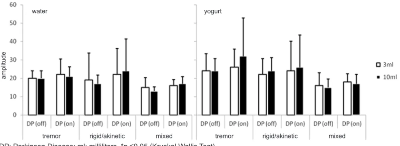
As well as in the swallowing of water, in off phase, sEMG amplitudes of swallowing of yogurt were less in the mixed group. The other group has equal amplitudes.
In the on phase, all group presented small elevation of the amplitudes, however the mixed group continue presenting lower values. These differences were not signiicant statistically (Figure 2).
sEMG Amplitude:
In the off phase of PD clinical types, the average of the sEMG amplitudes of swallowing of 3 ml and 10 ml of water was lower in the mixed patients group, the rigid-akinetic and tremors groups showed similar averages.
In the medication effect, all groups showed small elevation of the sEMG amplitudes, especially the rigid-akinetc group. However, there was no signif-icant difference among the groups.
Duration of sEMG:
When checking the duration of sEMG of swallowing water, both the off and on condition, the rigid-akinetic group present electrophysiological activity slightly longer than the tremor and mixed.
After the medication, time of sEMG did not suffer big changes in none of the groups, with no signiicant differences.
The duration of the sEMG in swallowing yogurt was also higher in the rigid-akinetic group, followed by the mixed group, before and after Levodopa.
In the on phase, the mixed group showed little decrease in the duration of sEMG of volumes of 3 ml and 10 ml, while tremor and rigid-akinetic reduced the time in swallowing of 3 ml of yogurt and increased the time swallowing 10 ml . However, all the differ-ences were small and not signiicant (Figure 3). DP: Parkinson Disease; ml: milliliters. *p ≤0,05 (Kruskal Wallis Test).
Figure 2 – Average of amplitude of the electromyography surface of swallowing and 3 ml water and 10 ml of yogurt in clinical type of Parkinson’s disease
a
mp
lit
u
d
e
water yogurt
groups. That is, when considering the average of swallowings in parts and the duration of the EMGs, tremors have values similar to those observed in the mixed group. When the scope is analyzed, tremor
group shows similar values to those observed in
rigid-akinetic group.
After using the medication, changes og the tremor group were minimal, which agrees with other authors when they state that nigrostriatal degeneration is very related to bradykinesia and rigidity than tremor. The mechanism of subthalamic stimulation and pallidotomy show a better response to the treatment of tremor than for the other two
symptoms25.
However, there are reports that surgical inter-ventions such as pallidotomy, thalamotomy, and deep brain stimulation have no positive effects on swallowing function, with worsening dysphagia often
cited as a complication of surgical interventions26.
It is noteworthy that the average age and duration of disease higher in predominantly tremulous group may indicate a longer life expectancy, thus strength
-ening claims of some authors16,27,28 that the PD would
present earlier with slower progression and better prognosis in this clinical type of disease. However, despite the rigid-akinetic group presents a slightly bigger loss than tremor in some of the electrophysi-ological parameters, there were no signiicant differ-ences between groups.
The mixed group was highlighted because it had lower EMGs amplitudes than the other groups, which may indicate that the association of rigidity/ bradykinesia, more tremor symptoms can bring greater harm to the activation of muscles, but no signiicant difference. After the use of Levodopa,
DISCUSSION
The rigid-akinetic group in off phase, showed the highest number of patients with swallowing in parts, as well as higher average in swallowing in parts. The duration of EMGs of this clinical type was also a bit longer than the other groups. These indings indicate an impairment in swallowing of these patients because any duplication or swallowing multiplication with less than or equal to 20 ml of water (dysphagia limit) volumes, is considered
pathological20.
In this way, the higher duration of EMGs may be related with bradykinesia, and/or incoordination of the muscles involved in swallowing process of
PD patients15. Adaptive lack of mechanisms to
create space in the oral cavity was observed in patients with PD, and it is interpreted as a form
of hypokinesia21. The prolongation of the transit
time of oro-pharyngeal is a data that relects most consistently the dysfunction caused by the rigidity
and hypokinesia22.
On the other hand, in this study although not signiicant, the rigid-akinetic group seem to have a better response to therapy with Levodopa, since in the on phase, little changes occurred as: reduction in the percentage of patients with swallowing in parts, lower average in swallowing in parts, increased amplitudes and reduction duration of EMGs.
When bradykinesia is predominant, treatment with Levodopa is useful and the symptom disap-pears in a short time because this symptom would be directly related to symptom reduction of
dopamine23. The administration of single doses of
Levodopa reduces the rigidity of patients with DP24.
PD: Parkinson disease; ml: milliliters. *p ≤0,05 (Kruskal Wallis Test).
Figure 3 - Average duration (in seconds) the surface electromyography of swallowing 3 milliliters and 10 milliliters of water and yogurt in clinical types of Parkinson’s disease
se
co
n
d
s
water yogurt
are fed by clinical heterogeneity, suggesting at least two different clinical forms: PD tremor and PD rigid-akinetic.
CONCLUSIONS
Medicine therapy through Levodopa has not shown consistent differences in electromyographic parameters of swallowing between clinical types of PD.
However, due to the large interindividual variability, it is suggested to conduct further studies with larger sample.
ACKNOWLEDGEMENT
To Conselho Nacional de Desenvolvimento Cientíico e Tecnológico – CNPq
To Núcleo de Atenção ao Idoso – NAI/UFPE To Programa Pró-Parkinson do HC/UFPE which remained lower than the other groups,
although not signiicant, too.
The scarcity of electrophysiological studies that address the effect of Levodopa in the types of clinical swallowing in PD makes dificult comparison of results.
In a study using videoluoroscopy (VF) as an evaluation tool, patients were separated into tremor and non-tremor. The author states not founding signiicant differences between the groups and that generally half of patients improved on the on phase, the author assuming that occurred due to decreased
bradykinesia and rigidity of the tongue29.
Another author also used the VF, to verify the differences between dyskinetic and non-dyskinetic patients, concluding that dyskinetic patients have a higher eficiency of oropharyngeal swallowing, which can be explained by higher doses of Levodopa. The author also suggests that other neurotransmitter systems in addition to dopamine are involved in swallowing disorders in PD 13.
Other studies28,30 also raise questions about PD
constitute a single disease entity. These doubts
RESUMO
Objetivo: avaliar os parâmetros eletromiográicos da deglutição dos diferentes tipos clínicos da
doença de Parkinson idiopática nas fases on e off. Métodos: foram estudados 20 pacientes com
doença de Parkinson, divididos através da Escala Uniicada de Avaliação da Doença de Parkinson em três grupos: tremulantes, rígido-acinético e misto. O exame de eletromiograia de superfície foi coletado sobre a musculatura supra-hióidea durante a deglutição de 3 ml e 10 ml de água e iogurte, que foi repetida 5 vezes para cada volume e consistência. Este protocolo foi realizado no antes e após
a medicação, período off e on. Resultados: veriicou-se que na fase off, o grupo rígido-acinético
apre-sentou as maiores médias de deglutições em partes e duração das eletromiograias de superfície,
enquanto que o grupo misto apresentou as menores médias de amplitude. Na fase on, os três grupos
tenderam a melhorar ou manter as médias das variáveis estudadas, porém não houve diferença sig-niicante entre os tipos clínicos, antes ou depois da Levodopa. Conclusão: a terapia medicamentosa através da Levodopa não apresenta diferenças consistentes nas eletromiograias de superfície da deglutição dos tipos clínicos da doença de Parkinson.
A Surface Electromyography Study. Dysphagia. 2012;27:550-5.
16. Hoehn MM, Yahr MD: Parkinsonism: onset, progression, and mortality. Neurology. 1967;17(5):427-42.
17. Fahn S, Elton RL,Committee. AMoutUD. Uniied Parkinson’s disease rating scale. In: Fahn S, Marsden CD, Goldstein M, Calne DB, editors. Recent developments in Parkinson’s disease. New Jersey: Macmillan Healthcare Information. 1987. P.;153-63.
18. SENIAN: Surface ElectroMyoGraphy for the Non-Invasive Assessment of Muscles. Disponível em: <http://www.seniam.org/> Acesso em 26 jan. 2013.
19. Feodrippe P, Belo LR, Coriolano MGWS, Carneiro D, Lins OG. EMG BioanalyzerBR para a análise de sinais eletromiográicos na deglutição. Rev CEFAC. 2011;14(3):498-505.
20. Ertekin C, Aydogdu I, Yuceyar N, Tarlaci S, Kiylioglu N, Pehlivan M. et al. Electrodiagnostic methods for neurogenic dysphagia. Electroencephalography and clinical neurophysiology. 1998;109:331-40. 21. Wintzen AR, Badrising UA, Roos RAC, Vielvoye J, Liauw L. Inluence of bolus volume on hyoid movements in normal individuals and patients with Parkinson’s disease. Canadian Journal of Neurological Sciences. 1994;21:57-9.
22. Nilsson H, Ekberg O, Olsson R, Hindfelt B. Quantitative assessment of oral and pharyngeal function in Parkinson’s disease. Dysphagia. 1996;11:144-50.
23. Aviles-Olmos I, Martinez-Fernandez R, Foltynie T. L-dopa-induced dyskinesias in Parkinson’s disease. European neurological journal. 2010;2(2):91-100.
24. Pezzoli G, Zini M, Amrein R. L-dopa: the drug that changer the history of Parkinson’s disease. Focus Parkinson ’s Disease. 2011;22(1):7-11.
25. Loureiro F. Alterações da deglutição em pacientes com doença de Parkinson: associação com a clínica e estudo eletroisiológico simultâneo com a respiração [Tese]. Porto Alegre (RS): Universidade Católica do Rio Grande do Sul; 2011. 26. Gross RD, Atwood JR, Ross SB, Eichhorn KA, Olszewski JW, Doyle PJ. The coordination of breathing and swallowing in Parkinson’s disease. Dysphagia. 2008;23:136-45.
27. Graham JM, Sagar HJ. A Data-Driven Approach to the Study of Heterogeneity in Idiopathic Parkinson’s Disease: Identiication of Three Distinct Subtypes. Movement Disorders. 1999;14(1):10-20. 28. Lewis SJG, Foltynie T, Blackwell AD, Robbins
REFERENCES
1. Kansara S, Trivedi A, Chen S, Jankovic J, Le W. Early diagnosis and therapy of Parkinson’s disease: can disease progression be curbed? Journal of Neural Transmission. 2012;120(1):197-210.
2. Barbosa ER, Sallem FAS. Doença de Parkinson – Diagnóstico (artigo de revisão). Neurociências. 2005;13(3):158-65.
3. Yamada EK, Siqueira KO, Xerez D, Koch HA, Costa MMB. A inluência das fases oral e faríngea na dinâmica da deglutição. Arquivos de Gastroenterologia. 2004;41(1):18-23.
4. Gasparim AZ, Jurkiewicz AL, Marques JM, Santos RS, Marcelino PCO, Herrero-Junior F. Deglutição e tosse nos diferentes graus da doença de Parkinson. Arquivos Internacionais de Otorrinolaringologia. 2011;15(2):181-8.
5. Walker RW, Dunn JR, Gray WK. Self-reported dysphagia and its correlates within a prevalent population of people with Parkinson’s disease. Dysphagia. 2011;26:92-6.
6. Ertekin C, Pehlivan M, Aydogdu I, Ertas LM, Uludag B, Çlelebi G et al. An electrophysiological investigation of deglutition in man. Muscle&Nerve. 1995;18:1177-86.
7. Bushmann M, Dobmeyer SM, Leeker L, Perlmutter JS. Swallowing abnormalities and their response to treatment in Parkinson’s disease. Neurology. 1989;39:1309-14.
8. Steidl EMS, Ziegler JR, Ferreira FV. Doença de Parkinson revisão bibliográica. Disc. Scientia. Série: Ciências da Saúde. 2007;8(1):115-29. 9. THE PARKINSON STUDY GROUP. Levodopa and progression of Parkinson’s disease. New England Journal Medicine. 2004;351:2498-508. 10. Poewe W. The natural history of Parkinson’s disease. Journal Neurology. 2006;253(2):2-6. 11. Azevedo LL, Cardoso F. Ação da levodopa e sua inluência na voz e na fala de indivíduos com doença de Parkinson. Rev Soc Bras Fonoaudiol. 2009;14(1):136-41.
12. Robichaud JA, Kerstin D, Comella CL, Corcos DM. Effect of Medication on EMG Patterns in Individuals with Parkinson’s Disease. Movement disorders. 2002;17(5):950-60.
13. Monte FS, Silva-Júnior FP, Braga-Neto P, Souza MAN, Bruin VMS. Swallowing abnormalities and dyskinesia in Parkinson’s disease. Movement disorders. 2005;20:457-62.
14. Pullman SL, Goodin SD, Marquinez AI, Tabbal S, Rubin M. Clinical utility of surface EMG. Neurology. 2000;55:171-7.
30. Uitti RJ, Baba Y, Wszolek ZK, Putzke DJ. Deining the Parkinson disease phenotype: initial symptoms and baseline characteristics in a clinical
cohort. Journal Parkinsonism Relatated Disorders.
2005;11:139-45. using a data driven approach. Journal Neurology
Neurosurgery Psychiatry. 2005;76:343-8.
29. Fuh J, Lee R, Wang S, Lin C, Wang P, Chiang J, Liu H. Swallowing dificulty in Parkinson’s disease. Clinical neurology and neurosurgery. 1997;99:106-12.