Case Report
1 1 8 Arq Bras Oftalmol. 2015;78(2):118-9 http://dx.doi.org/10.5935/0004-2749.20150030
INTRODUCTION
The dexamethasone implant (Ozurdex®; Allergan Inc., Irvine, CA, USA) is a novel treatment modality mainly used to treat macular ede-ma associated with retinal vein occlusions and to treat noninfectious posterior uveitis(1). However, there are quite satisfactory data about
its efficacy in multiple clinical situations associated with refractory macular edema, including diabetic macular edema, macular edema asso ciated with uveitis or Irvine-Gass syndrome, and retinitis pigmen-tosa(2,3). Major concerns with intravitreal dexamethasone (IV-DEX) are
increased intraocular pressure and cataract progression(1,2). Here we
report an unusual case of IV-DEX-related acute retinal necrosis (ARN) in a rheumatoid arthritis patient with refractory posterior uveitis.
CASE REPORT
A 52-year-old woman presented with floaters and decreased vision in the left eye. She had a 7-year history of rheumatoid arthritis. For the year prior to presentation, she had been receiving azathiopri-ne 150 mg daily to treat posterior uveitis in the left eye. Oazathiopri-ne month prior to presentation, intravitreal dexamethasone (IV-DEX) implanta-tion was performed for resistant macular edema in the left eye.
On examination, visual acuity was 20/20 in the right eye and 20/40 in the left eye. There were no pathological findings in the right eye. Slit-lamp examination of the left eye revealed 3+ anterior cham-ber cells. Fundus examination revealed vitreous haze and debris, disc edema, vessel attenuation, scattered retinal hemorrhages, exudation, and peripheral confluent areas of retinal whitening. The implant was ABSTRACT
A 52-year-old woman undergoing azathioprine treatment for rheumatoid arthritis developed acute retinal necrosis a month after intravitreal dexamethasone (Ozur-dex®) implantation for posterior uveitis in the left eye. Varicella zoster virus (VZV) DNA was detected in the anterior chamber and vitreous samples on polymerase chain reaction (PCR) analysis. Retinal detachment occurred despite systemic and intravitreal antiviral therapy. Favorable structural and functional outcomes were achieved after retinal surgery with silicone oil. To the authors’ knowledge, this is the first reported case of acute retinal necrosis following placement of an Ozurdex®
implant. Physicians practicing Ozurdex® implantations should be aware of this unusual but devastating complication. Extra caution and frequent follow-up are required in all immunocompromised patients receiving Ozurdex® implantation.
Keywords: Retinal necrosis syndrome, acute; Immunosuppression; Intravitreal in -jections/methods; Dexamethasone/administration & dosage; Case reports
RESUMO
Uma mulher de idade de 52 anos em tratamento azatioprina para a artrite reumatóide desenvolveu necrose aguda de retina um mês após implantação Ozurdex® para uveíte posterior do olho esquerdo. DNA de varicela zoster (VZV) foi detectado em amostras de câmara anterior e vítreo por análise de PCR. Apesar da terapia antiviral sistêmica e intravítrea, o paciente apresentou descolamento de retina. Desfecho favorável estrutural e funcional foi obtida após a cirurgia retiniana com óleo de silicone. Pelo conhecimento dos autores, este é o primeiro caso relatado de necrose aguda de retina após a colocação de um implante Ozurdex®. Os médicos que implantam Ozurdex® devem estar cientes desta complicação incomum, mas devastadora. É necessário cuidado extra e acompanhamento frequente dos pacientes que recebam o implante Ozurdex® e apresentem qualquer condição imunocomprometedora.
Descritores: Síndrome de necrose retiniana aguda; Imunossupressão; Injeções in tra-vítreas/métodos; Dexametasona/administração & dosagem; Relatos de casos
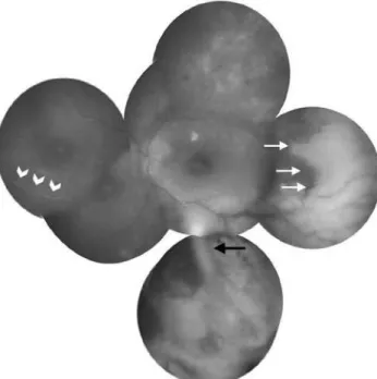
easily observed in the inferior vitreous (Figure 1). Fluorescein an-giography demonstrated staining of the optic disc and vessels, and clear-cut zones of retinal ischemia (Figure 2). Immune suppression caused by the azathioprine treatment was ruled out by a normal white cell count.
The presumed diagnosis was acute retinal necrosis. Anterior chamber paracentesis and vitreous tap samples were analyzed using viral polymerase chain reaction (PCR). Varicella zoster virus (VZV) DNA was identified, but no herpes simplex virus, cytomegalovirus, or Epstein-Barr virus DNA was detected. Systemic and laboratory inves-tigations for syphilis, AIDS, and hepatitis were also negative, and there was no evidence of any systemic azathioprine-related adverse effect. The patient was commenced on intravenous acyclovir (3 × 750 mg/day) treatment for 10 days to manage ARN, in addition to to pical prednisolone acetate 1% drops every hour and cyclopen-tolate 1% drops thrice a day. The patient was followed as inpatient. However, this treatment regimen did not resolve the areas of retinal whitening or the anterior chamber inflammation. Therefore, she received 4 intravitreal ganciclovir injections a week apart, and oral valacyclovir 1 g thrice daily. One month after the first ganciclovir in-jection, the patient developed retinal detachment from the necrotic retinal area in the left eye, and visual acuity decreased to counting fingers at 1 m. She underwent repair of the retinal detachment with vitrectomy, laser retinopexy, and silicone oil. She was discharged after surgery and maintained on oral valacyclovir therapy. Four months after surgery, her visual acuity was 20/60 in the left eye (Figure 3). The right eye re mains unaffected.
Acute retinal necrosis following intravitreal dexamethasone (Ozurdex
®) implant
Necrose aguda de retina após implante de dexametasona intravítrea (Ozurdex
®)
Murat KucuKevcilioglu1, Mustafa eren1, uMit Yolcu2, gungor sobaci3
Submitted for publication: April 22, 2014 Accepted for publication: August 29, 2014
1 Department of Ophthalmology, Gulhane Military Academy of Medicine, Ankara, Turkey. 2 Department of Ophthalmology, Siirt Military Hospital, Siirt, Turkey.
3 Department of Ophthalmology, Hacettepe University Faculty of Medicine, Ankara, Turkey.
Funding: No specific financial support was available for this study.
Disclosure of potential conflicts of interest: None of the authors have any potential conflict of interest to disclose.
Kucukevcilioglu M, et al.
119 Arq Bras Oftalmol. 2015;78(2):118-9 DISCUSSION
The clinical presentation and laboratory workup of the presented case lead to a diagnosis of ARN. To the best of our knowledge, there is no previous report of ARN after an IV-DEX implantation (Pubmed and Medline search). However, ARN has been described following intravitreal injections of triamcinolone(4,5). Recently, Ramaiya et al.
reported a case of ARN 1 year after fluocinolone acetonide (Retisert®; Bausch and Lomb, Irvine, CA, USA) implantation in a young patient with intractable uveitis(6). Prior to implantation, the authors had
tried different immunotherapeutic agents, including azathioprine with high dose oral steroid, to control the inflammation. Shah et al. retrospectively analyzed viral retinitis after triamcinolone injections and found increased number of injections or long duration of drug activity as a risk factor(5). Systemic immunosuppression is another risk
factor, and its incidence was found to be 2 times higher in the immu-nocompromised subset than in the rest of the subjects(7). The current
Figure 1. Fundus photography of the left eye at presentation showing mild optic disc edema, macular striations, occlusive vasculitis (white arrow heads), scattered retinal hemorrhages, clear cut whitening of peripheral retina (white arrows), and the IV-DEX implant in the inferior vitreous (black arrow).
Figure 2. Fluorescein angiography images showing annular (A) retinal ischemia in the periphery and (B) optic disc leakage.
A B
Figure 3. Fundus photography of the left eye 4 months after surgery showing attached retina under silicone oil tamponade, mild obscuration of disc edges, and some retinal scarring.
patient was on azathioprine for rheumatoid arthritis, and despite the normal white cell count, the combined effect of local and systemic immunosuppression could have played a role in the development of ARN. However, further studies are needed to confirm this possibility.
In the literature, early vitrectomy with laser demarcation is recommended for severe ARN(8,9). In this case, we initiated medical
treatment because the macula was unaffected and visual acuity was good at presentation. After progression to retinal detachment, vitrectomy with laser retinopexy and silicone oil tamponade limited the disease progression and provided a favorable visual outcome in the short term. However, long-term follow up is needed to determine whether this unusual gain in vision is sustained.
In conclusion, physicians practicing IV-DEX implantations should be aware of this unusual but devastating complication. Extra caution and frequent follow-up is required in immunocompromised patients receiving IV-DEX implantation.
REFERENCES
1. Haller JA, Bandello F, Belfort R Jr, Blumenkranz MS, Gillies M, Heier J, et al. Randomized, sham-controlled trial of dexamethasone intravitreal implant in patients with macular edema due to retinal vein occlusion. Ophthalmology. 2010;117(6):1134-1146.e3. 2. Dutra Medeiros M, Postorino M, Navarro R, Garcia-Arumí J, Mateo C, Corcóstegui B.
Dexamethasone intravitreal implant for treatment of patients with persistent diabetic macular edema. Ophthalmologica. 2014;231(3):141-6.
3. Srour M, Querques G, Leveziel N, Zerbib J, Tilleul J, Boulanger-Scemama E, et al. Intra-vitreal dexamethasone implant (Ozurdex) for macular edema secondary to retinitis pigmentosa. Graefes Arch Clin Exp Ophthalmol. 2013;251(6):1501-6.
4. Han JM, Ahn J, Park KH, Woo SJ. Presumed necrotizing viral retinitis after intravitreal triamcinolone injection: case report. Korean J Ophthalmol 2011;25(6):451-4. 5. Shah AM, Oster SF, Freeman WR. Viral retinitis after intravitreal triamcinolone injection
in patients with predisposing medical comorbidities. Am J Ophthalmol. 2010;149(3): 433-40 e1.
6. Ramaiya KJ, Rao PK. Herpetic necrotizing retinitis following flucinolone acetonide in travitreal implant. Ocul Immunol Inflamm. 2011;19(1):72-4.
7. Haller JA, Bandello F, Belfort R Jr, Blumenkranz MS, Gillies M, Heier J, et al. Dexametha-sone intravitreal implant in patients with macular edema related to branch or central retinal vein occlusion twelve-month study results. Ophthalmology. 2011;118(12): 2453-60
8. Hillenkamp J, Nolle B, Bruns C, Rautenberg P, Fickenscher H, Roider J. Acute retinal necrosis: clinical features, early vitrectomy, and outcomes. Ophthalmology. 2009;116(10): 1971-5 e2.