Original Article
7 3 Arq Bras Oftalmol. 2015;78(2):73-5 http://dx.doi.org/10.5935/0004-2749.20150020
INTRODUCTION
With the increasing popularity of phacoemulsification as a me-thod of choice for cataract surgery, the incidence of complications such as inadvertent posterior capsule tear, nuclear fragments, and intraocular lens (IOL) loss into the vitreous cavity has increased greatly(1-6).
These complications are directly related to the surgeon’s ex-pertise and tend to increase in specific cases that represent major challenges. These cases include inadequate zonular support (pseu-doexfoliation, trauma, and previous vitrectomy), mature and hyper-mature cataracts, high axial myopia, insuficient mydriasis, patient movements during the perioperative period, among others(2,6,7-12).
Nuclear and IOL fragmentation in the vitreous may trigger serious consequences, including permanent visual loss, if not treated pro-perly. These complications induce a severe inflammatory response
that is proportional to the excessive intraocular manipulation trauma and the nuclear fragment size or IOL model and material. These pa-tients may also develop chronic uveitis, secondary glaucoma, corneal edema, and retinal detachment. One study showed a 52% incidence of glaucoma in such a condition(3).
To determine the best clinical or surgical treatment strategy for such conditions, ophthalmologists mainly consider fragment size and the presence or absence of corneal edema and glaucoma(13-20).
Studies on the removal time of vitrectomy nuclear fragments indicated that it is not necessary for this procedure to be performed on the same day and that these fragments can be removed up till 1-2 weeks after surgery(3,6,13-20).
This study aims to identify the causes and results of pars plana vitrectomy (PPV) in patients who underwent phacoemulsification with intraoperative complications and to analyze whether the surgical
Results of pars plana vitrectomy after complicated phacoemulsification surgery
Resultados pós-vitrectomia via pars plana em pacientes submetidos à cirurgia de facoemulsiicação
com complicação intraoperatória
AlexAndre GrAndinetti1, dioGo SuenAGA2, FernAndA M. oliveirA3, KriSSiS SAlibA uliAnA Cruz2, lAurindA MeneGuette2, luCiAne buGMAn MoreirA4
Submitted for publication: June 9, 2014 Accepted for publication: December 11, 2014
1 Department of Surgery, Federal University of Parana (UFPR), Curitiba, PR, Brazil. 2 Anterior Segment Sector, Paraná Eye Hospital, Curitiba, PR, Brazil.
3 Retina and Vitreous Sector, Paraná Eye Hospital, Curitiba, PR, Brazil.
4 Department of Ophthalmology, Medical School, Positivo University, Curitiba, PR, Brazil.
Funding: No specific financial support was available for this study.
Disclosure of potential conflicts of interest: None of the authors have any potential conflict of interest to disclose.
Corresponding author: Krissis Saliba Uliana Cruz. Rua Frei Caneca, 3.159 - Guarapuava, PR - 85015-220 - Brazil - E-mail: [email protected]
Approved by the following research ethics committee: Positivo University (CAAE 27185914. 6.0000.0093).
ABSTRACT
Purpose: To identify the causes and outcomes of pars plana vitrectomy (PPV) in patients undergoing phacoemulsification with intraoperative complication and to analyze whether the interval between phacoemulsification and PPV interferes with best-corrected final visual acuity.
Methods: This descriptive and retrospective analytical study was conducted in Paraná Eye Hospital in 2013. Data were collected from medical records of 38 patients who underwent complicated phacoemulsification and also required PPV.
Results: The most frequent complication as a result of phacoemulsification was posterior capsule rupture, observed in 35 patients (92.10%), followed by capsular bag detachment, in three patients (7.89%). Twenty-eight patients (73.68%) had cortical fragments that were removed during PPV. Twelve patients (31.57%) had their intraocular lens repositioned. PPV was performed on the same day of pha-coemulsification in one patient (2.63%), within 1 week in 15 patients (39.47%), between 1 week and 1 month in 13 patients (34.21%), and 1 month after phacoe-mulsification in 9 patients (23.68%).
Conclusion: This study is in agreement with worldwide literature, asserting that major complications of phacoemulsification are posterior capsule rupture and capsular bag detachment, and in addition, there is an improvement in the final visual acuity in almost half the cases, even when there are complications during modern cataract surgery, when complementary appropriate treatment is provided.
Keywords: Phacoemulsification/complications; Vitrectomy/methods; Cataract ex-traction
RESUMO
Objetivos: Identificar as causas e os resultados da vitrectomia via pars plana (VPP) em pacientes submetidos à cirurgia de facoemulsificação com complicação intrao-peratória, analisando se o tempo cirúrgico entre a facoemulsificação e a VPP interfere na melhor acuidade visual corrigida final.
Métodos: Estudo analítico descritivo e retrospectivo realizado no Hospital de Olhos do Paraná em 2013. Os dados foram coletados de prontuários de 38 pacientes que foram submetidos à cirurgia de facoemulsificação complicada e que também preci-saram de VPP.
Resultados: A complicação intraoperatória mais frequente na cirurgia de fa-coemulsificação, nos pacientes estudados, foi à ruptura de cápsula posterior, que ocorreu em 35 pacientes (92,10%), seguido de desinserção zonular em 3 pacientes (7,89%). Em 28 pacientes (73,68%) foram encontrados restos corticais, que foram removidos durante a VPP. Em 12 pacientes (31,57%) foi realizado o reposicionamento da lente intraocular. A cirurgia de VPP foi realizada no mesmo dia da facoemul-sificação em 1 paciente (2,63%), dentro de 7 dias em 15 pacientes (39,47%), entre 1 semana e 1 mês em 13 pacientes (34,21%) e após 1 mês da facoemulsificação em 9 pacientes (23,68%).
Conclusão: O presente estudo encontrou dados semelhantes aos descritos na li teratura mundial, que afirmam que as principais complicações da facoemulsificação são a ruptura de cápsula posterior e desinserção zonular; e que a acuidade visual final melhora, em aproximadamente metade dos casos, mesmo após ocorrer complica-ções na cirurgia de catarata moderna, quando instituído tratamento complementar adequado.
Results of pars plana vitrectomy after complicated phacoemulsification surgery
7 4 Arq Bras Oftalmol. 2015;78(2):73-5
interval between phacoemulsification and PPV interferes with the best-corrected final visual acuity (VA).
METHODS
A descriptive and retrospective analytical study was conducted at Paraná Eye Hospital by reviewing 38 patient records.
The inclusion criteria were as follows: patients who underwent cataract surgery by phacoemulsification with intraoperative compli-cations and required postoperative PPV immediately after cataract surgery. Considered surgeries were performed between January and December 2013.
The exclusion criteria were as follows: patients with incomplete follow-up during the study period or those with incomplete medical records.
The following data were collected for this study: gender, age, ori-gin, best-corrected visual acuity (BCVA) prior to phacoemulsification and a month after PPV, and complications or procedures required during the last surgery.
The ophthalmologic examination included the following pro-cedures: average VA with best correction according to the Snellen chart, biomicroscopy with Zeiss slit lamp, intraocular pressure measu-rement by Perkins tonometer, and retinal mapping by EyeTech indi-rect ophthalmoscopy. Legacy 20000™ (Alcon) and Infiniti®
(Alcon) were used for phacoemulsification, and Stellaris (Bausch & Lomb) was used for vitrectomy.
Third-year ophthalmology residents and fellows of the cataract and anterior segment department of Paraná Eye Hospital performed the phacoemulsification surgeries. Experienced physicians expertise in retinal procedures performed the PPV procedures.
The study design was submitted to and approved by the Ethics Committee of Positivo University. Patient identities were not collec-ted, ensuring patient anonymity. Because the aim of the study was the collection of medical appointment data, there were no interven-tions in the physician conduct, which occurred independent of the study.
RESULTS
Between January and December 2013, 42 patients underwent complicated cataract surgery followed by PPV. Four of these patients were excluded from the study because of incomplete records or follow-up. Of the 38 remaining patients, 12 were male (31.5%) and 26 female (68.4%). The average age was 69.42 ± 13.89 years (minimum of 5 years, maximum of 84 years). Half of the patients were from Curitiba, Paraná and the other half were from other locations.
Prior to phacoemulsification, eye comorbidities (besides cataract) were found in six patients (15.78%): glaucoma (five patients) and age- related macular degeneration (one patient). Of the 38 patients, 13 (34.21%) had diabetes mellitus type II and 11 (28.94%) had systemic arterial hypertension without fundoscopic changes.
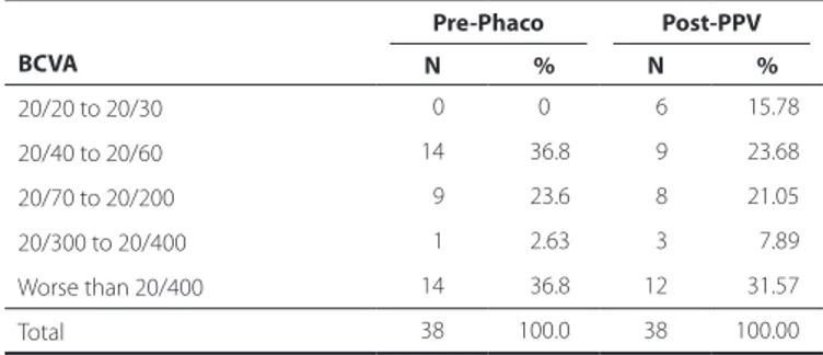
Table 1 shows the general view of the BCVA values prior to pha-coemulsification (BCVA pre-surgery) and 30 days after vitrectomy (BCVA post-surgery).
When analyzed individually, 18 (47.36%) patients presented an improvement in the final BCVA after PPV compared with BCVA prior to phacoemulsification, whereas 11 (28.94%) had worsening of BCVA. BCVA remained unchanged in nine (23.68%) patients. Of the 18 pa-tients with improvement in the final BCVA, 15 (83.33%) underwent PPV within the first month after phacoemulsification.
Eighteen (47.37%) phacoemulsification procedures were perfor-med within the first half of 2013 and 20 (52.43%) in the second half. Vitrectomy (PPV) was performed on the same day as phacoemulsifi-cation in one patient (2.63%), within 7 days in 15 patients (39.47%), between 1 week and 1 month in 13 patients (34.21%), and 1 month after phacoemulsification in nine patients (23.68%)(Table 2).
Posterior capsule tear was the most frequent intraoperative com-plication (35 patients, 92.10%), followed by detachment of the cap-sular bag in three patients (7.89%). Cortical fragments were found in 28 patients (73.68%) and were removed during PPV. Twelve patients (31.57%) required intraocular lens repositioning in the ciliary sulcus.
DISCUSSION
Cortical fragments were observed and surgically removed in 73.68% of the patients who underwent PPV. The remaining patients underwent PPV for removal of the nucleus from the vitreous. Gilliland showed a similar occurrence of cortical fragments in patients who underwent PPV(3).
Lavinski et al. revealed that the surgical interval between cataract surgery and PPV was more than 15 days for most patients. In the present study, this surgical interval ranged from up to 15 days (16 patients; 42.10%) to more than 15 days (22 patients; 57.89%)(21). On the other hand, the authors suggested that a shorter surgical interval between the two procedures results in an improvement of the final VA. This finding is consistent with that of the present study, given that of the 18 patients that showed improvement in the final BCVA after PPV, 13 (72.22%) had undergone this procedure within the first month after phacoemulsification(21,22).
The average patient age in the present study was 69.42 years, similar to that obtained by Santacruz I in his RCP and final VA study,
Table 1. Best-corrected visual acuity (BCVA) prior to phacoemulsiica-tion (Pre-Phaco) and after pars plana vitrectomy (Post-PPV), n=38
BCVA
Pre-Phaco Post-PPV
N % N %
20/20 to 20/30 00 0 06 015.78
20/40 to 20/60 14 036.8 09 023.68
20/70 to 20/200 09 023.6 08 021.05
20/300 to 20/400 01 02.63 03 007.89
Worse than 20/400 14 036.8 12 031.57
Total 38 100.0 38 100.00
BCVA= best-corrected visual acuity.
Table 2. Best-corrected visual acuity (BCVA) prior to phacoemulsiication (Pre-Phaco) and post pars plana vitrectomy (Post-PPV), according to the interval between surgeries, n=38
Surgical interval between phaco and PPV
Pre-Phaco Post-PPV
<20/400 ≥20/400
Average of the eyes
≥20/400 <20/400 ≥20/400
Average of the eyes ≥20/400
Up to 7 days 7 09 20/70 5 11 20/50
Between 8 and 30 days 3 10 20/60 2 11 20/60
Grandinetti A, et al.
75
Arq Bras Oftalmol. 2015;78(2):73-5 and by Falavarjani et al. in their study of PPV for the removal of the
nucleus from the vitreous (23).
Tavares et al. observed ocular comorbidities in 35% of patients, with glaucoma being the most frequent condition. These findings are consistent with those of in the present study(24).
It is important to emphasize that RCP is a complication that can occur with any surgeon and its proper management requires expe-rience. Its management includes vitrectomy in order to minimize final VA losses(25).
As demonstrated, the results obtained in the present study are consistent with existing worldwide literature statistics.
It is crucial to point out the importance of an integrated and readily available team of retina specialists in order to manage such complications, provide the best prognosis, and consequently, increa-se quality of life.
CONCLUSION
In the present study, the most frequent intraoperative compli-cation during phacoemulsificompli-cation was the posterior capsule tear, followed by capsular bag detachment.
Almost half of the patients presented improvement in the final BCVA after PPV compared with BCVA prior to phacoemulsification.
Most of the patients who presented improvement in the final BCVA underwent PPV within the first month after phacoemulsifica-tion, suggesting that a shorter surgical interval between phacoemul-sification with complication and PPV improves the final BCVA.
REFERENCES
1. Emery JM, Wilhelmus KA, Rosenberg S. Complications of facoemulsification. Ophthal-mology. 1978;85(2):141-50.
2. Monshizadeh RSN, Haimovici R. Management of retained intravitreal lens fragments after cataract surgery. Surv Ophthalmol. 1999;43(5):397-404.
3. Gilliland GD, Hutton WL, Fuller DG. Retained intravitreal lens fragments after cataract surgery. Ophthalmology. 1922;99(8):1263-9.
4. Irvine WD, Flynn HW, Murray TG. Retained lens fragment after phacoemulsification manifesting as marked intraocular inflammation with hypopyon. Am J Ophthalmol. 1922;114(5):610-4.
5. Pande M, Dabbs, TR. Incidence of lens matter dislocation during phacoemulsification. J Cataract Refract Surg. 1996;22(6):737-42.
6. Tommila P, Immonen I. Dislocated nuclear fragments after cataract surgery. Eye (Lond). 1995;9(Pt 4):437-41.
7. Allinson RW, Metrikin DC, Fante RG. Incidence of vitreous loss and dislocated lens fragments during phacoemulsification among third-year residents performing pha-coemulsification. Ophthalmology. 1992;99(5):726-30.
8. Gonvers M. New approach to managing vitreous loss and dislocated lens fragments during phacoemulsification. J Cataract Refract Surg. 1994;20(3):346-9.
9. Leaming DV. Practice styles and preferences of ASCRS members: 1994 survey. J Ca-taract Refract Surg. 1995;21(4):378-85.
10. Gusek JP, Holm M, Cotter JB, Cameron JA, Rademaker WJ, Wissinger DH, Tonjum AM, Sleeper LA. Risk Factors for intraoperative complications in 1000 extra-capsular cases. Ophthalmology. 1987;94(5):461-6.
11. Margherio RR, Margherio AR, Pendergas SD, Williams GA, Garretson BR, Strong LE, et al. Vitrectomy for retained lens fragments after phacoemulsification. Ophthalmology. 1997;104(9):1426-32.
12. Streeten BW. Pathology of the lens. In: Albert DM, Jakobiec FA, editors. Principles and practices of ophthalmology: clinical practice. Philadelphia: WB Saunders; 1994. p.2180-239.
13. Hutton WL, Snyder WB, Vaiser A. Management of surgically dislocated intravitreal lens fragments by pars plana vitrectomy. Ophthalmology. 1978;85(2):176-89.
14. Fastenberg DM, Schwarzt PL, Shakin JL, Golub BM. Management of dislocated nu clear fragments after phacoemulsification. Am J Ophthalmology. 1991;112(5):535-9. 15. Lambrou FH Jr, Stewart MW. Management of dislocated lens fragments during
phacoemulsification. Ophthalmology. 1992 Aug;99(8):1260-2; Discussion 1268-9. 16. Kim JE, Flynn Jr HW, Smiddy WE, Murray TG, Rubsamen PE, Davis JL, Nicholson
DH. Retained lens fragments after phacoemulsification. Ophthalmology. 1994; 101(11):1827-32.
17. Blodi BA, Flynn Jr HW, Blody CF, Folk JC, Daily MJ. Retained nuclei after cataract sur-gery. Ophthalmology. 1992;99(1):41-4.
18. Wong D, Briggs MC, Hicley-Dwyer MU, McGalliard JN. Removal of lens fragments from the vitreous cavity. Eye (Lond). 1997;11(Pt 1):37-42.
19. Stewart MW. Management of retained lens fragments: can we improve? Am J Oph-thalmol. 2007;144(3):445-6.
20. Mittra RA, Connor TB, Han DP, Koenig SB, Mieler WF, Pulido JS. Removal of dislocated intraocular lenses using pars plana vitrectomy with placement of an open-loop, flexible anterior chamber lens. Ophthalmology. 1998;105(6):1011-4.
21. Lavinsky J, Fior O, Goldhardt R, Dei Ricardi LM. Complications of lens displacement into the vitreous cavity. Arq Bras Oftalmol. 2002;65(4):435-9.
22. Falavarjani KG. Pars plana vitrectomy and intravitreal phacoemulsification for dropped nuclei. J Ophthalmic Vis Res. 2012;7(2):125-9.
23. Santacruz I. Posterior rupture in cataract surgery: frequency, management and visual result. Mem Inst Invest Cienc Salud. 2011;7(1):43-8.
24. Tavares VN, Colossi CG, Saalfeld V, Vilela MA. Phacoemulsification under topical anes-thesia: series of cases. Rev Bras Oftalmol. 2013;72(3):178-80.