ARTICLE
Neuroendoscopic surgery in children:
an analysis of 200 consecutive procedures
Neuroendoscopia em crianças: uma análise de 200 procedimentos consecutivos
Luciano Lopes Furlanetti1,2,Marcelo Volpon Santos1,Ricardo Santos de Oliveira1
1Division of Pediatric Neurosurgery of the Department of Surgery and Anatomy, University Hospital, Ribeirão Preto Medical School, University of São Paulo
(USP), Ribeirão Preto SP, Brazil;
2Division of Stereotactic and Functional Neurosurgery of the Department of Neurosurgery, University Hospital Freiburg, Freiburg im Breisgau, Germany.
Correspondence: Ricardo Santos de Oliveira; Department of Surgery and Anatomy, University Hospital, Ribeirão Preto Medical School, University of São Paulo; 14049-900 Ribeirão Preto SP - Brasil; E-mail: [email protected]
Conflict of interest: There is no conlict of interest to declare.
Received 05 September 2012; Received in inal form 02 October 2012; Accepted 09 October 2012.
ABSTRACT
Objective: Neuroendoscopic surgery in children has particular features and is associated with different success rates (SR). The aim of this study was to identify putative factors that could inluence the outcome in pediatric patients. Methods: Clinical data of 177 patients under 18 years of age submitted to 200 consecutive neuroendoscopic procedures from January 2000 to January 2010 were reviewed. Results: The overall success rate was 77%. Out of the patients with successful outcomes, 46% were under six months, 68% were between six months and one year of age, and 85% older than one year. Neuroendoscopic techniques provide very good results for a wide number of indications in children. Tumor-related cerebrospinal luid (CSF) circulation problems and aqueductal stenosis seem to be particularly well suited to neuroendoscopic treatment regardless of the patient’s age. Conclusion: Patients’ age and etiology of hydrocephalus were associated with a different outcome. In all cases, surgical experience is extremely important to reduce complications.
Key words: endoscopic third ventriculostomy, hydrocephalus, neuroendoscopy, pediatric neurosurgery.
RESUMO
Objetivo: A cirurgia neuroendoscópica em crianças apresenta particularidades e está associada a diferentes taxas de sucesso (TS). O objetivo deste estudo consistiu em identiicar fatores que pudessem inluir no resultado do tratamento em pacientes pediátricos. Métodos: Dados clíni-cos de 177 pacientes com idade inferior a 18 anos submetidos a 200 procedimentos neuroendoscópiclíni-cos consecutivos entre janeiro de 2000 e janeiro de 2010 foram revisados. Resultados: A taxa de sucesso global foi de 77%. Os pacientes com idade inferior a seis meses apresentaram taxa de sucesso de 46%; pacientes entre seis meses e um ano de vida obtiveram êxito em 68% dos casos; dentre os maiores de um ano, 85% dos procedimentos foram bem-sucedidos. Técnicas neuroendoscópicas proporcionam muito bons resultados para uma grande variedade de indica-ções em crianças. Independentemente da faixa etária, o tratamento endoscópico apresenta-se particularmente adequado para problemas da circulação liquórica relacionados a tumores e à estenose aquedutal. Conclusão: A faixa etária dos pacientes e a etiologia da hidrocefalia estão associadas a diferentes resultados. Em todos os casos, experiência neurocirúrgica é extremamente importante para a redução das complicações.
Palavras-Chave: terceiro ventriculostomia endoscópica, hidrocefalia, neuroendoscopia, neurocirurgia pediátrica.
he use of an endoscope to treat hydrocephalus has be-come a well-established technique that emerged in the ear-ly 20th century when Sir Walter Dandy began treating
hy-drocephalus by endoscopically cauterizing or removing the choroid plexus1. In the past two decades, introduction of new
instruments including rod lenses, Hopkins optic devices and high-resolution cameras has led to a huge increase in the number of neuroendoscopic procedures performed in spe-cialized neurosurgical centers2,3.
Neuroendoscopy is particularly useful as an adjunct tool in the treatment of hydrocephalus. It is an attractive method
owing to its simplicity, durability and because it does not re-quire lifelong implanted hardware4,5. Historically, endoscopic
third ventriculostomy (ETV) always seemed to be a promising technique and can be considered nowadays a standard proce-dure for obstructive hydrocephalus6,7. However, data published
in the medical literature is both extensive and conlicting when they come to the role of patients’ age and etiology of the hydro-cephalus in the success rate (SR) of endoscopic procedures6,8-18.
METHODS
Between 2000 and 2010, 200 neuroendoscopic procedures were performed in 177 patients in the same institution by the senior author (R.S.O) for the treatment of hydrocephalus in patients under 18 years old. A rigid GAAB Karl Storz®
neu-roendoscope (Tuttlingen, Germany) equipped with a nº 8 French diameter Hopkins rod lens system, a 0º iber optic, a nº 3 French working channel and an irrigation channel was used. All procedures were performed free hand after the in-duction of general anesthesia.
Routine postoperative outpatient follow-up appoint-ments were scheduled within one week and, then, one, three and every six months. Success was deined by the following criteria: when no further intervention was required to treat hydrocephalus and the absence of signs or symptoms of raised intracranial pressure.
Data were analyzed with the Fisher test or a chi-square test to determine whether each factor was correlated with the success of the endoscopic procedure for categorical data; p<0.05 values were considered signiicant. he ra-tios of children requiring permanent postoperative shunts or further surgical interventions in the diferent subgroups were compared. he factors were grouped by patient age at surgery, underlying pathology, type of endoscopic proce-dure and postoperative complications.
he length of hospitalization and the learning curve re-lated to the endoscopic approach were also analyzed.
Group classification
hese patients fell into three groups. In Group A, 26 pa-tients (mean age 4.8± standard deviation (SD) 0.86 months) were under six months of age; in Group B, 25 patients were between six months and one year of age (mean age 7.4±1.02 months); and in Group C, 115 patients were older than one year of age (mean age 5.8±1.09 years).
RESULTS
A total of 177 patients were studied. There were 78 male patients (44%) and 99 female patients (56%) ranging in age from 11 days to 18 years (mean age 5.1±1.06 years). The mean follow-up period was 65 months (ranging from 10 months to 9 years).
he etiology of hydrocephalus was as follows: out of the 177 patients, cystic malformations were found in 45 (25%), tumors in 40 (23%), aqueductal stenosis (AS) in 33 (19%), ce-rebral malformation in 30 (17%), meningitis or ventriculitis in 8 (5%), intraventricular hemorrhage in 6 (3%), isolated ventri-cle in 3 (2%) and other causes in 12 out of 177 (7%) patients. In 114 patients (64%), ETV was performed as a single and straightforward procedure. In 29, (16%) endoscopic cyst
fenestration was performed. In 18 (10%), two procedures were associated (i.e. ETV + cyst fenestration), in 11 (6%) a ventricular catheter was placed guided by endoscopy and ive patients (2%) underwent ETV + tumor biopsy. he basic indi-cation for endoscopic-assisted catheter placement was com-plex multiloculate hydrocephalus. Six of these patients were younger than six months and seven were pre-term children.
Sixty-six procedures (33%) were performed in patients under one year of age. In 166 patients, the main goal of the endoscopic intervention was to restore the cerebrospinal lu-id (CSF) low pathways.
Age group and SR
he overall SR for CSF circulation restoration was 77% (127/166). According to the age group, we observed a 46% (12/26) SR in Group A; 68% (17/25) in Group B, and 85% (98/115) in Group C (p=0.001) (Fig 1). Table 1 shows the distri-bution of patients with respect to their hydrocephalus etiol-ogy and SR per group of age according to the CSF restoration.
Hydrocephalus etiology and SR
In Group A, the etiology of hydrocephalus was related to complex cystic lesions or arachnoid cysts in 15 out of 26 (58%) cases, whereas brain or spinal malformations (such as spinal dysraphism, Dandy Walker, Chiari malformation) were noted in four (15%), and hemorrhage and ventriculitis in two cases (3.8%). AS was observed in ive patients (19%).
Out of the 25 patients in Group B, cystic lesions were found in 6 (24%), malformations in 5 (20%), AS in 4 (16%), posterior fossa tumors in 3 (12%), hemorrhage in 2 (8%), and infection in only one case. In Group C, an obvious predominance of pure obstructive hydrocephalus (i.e. posterior fossa tumors and AS) was observed in comparison to the other groups (19% (5/26), 28% (7/25) and 53% (61/115) respectively (p=0.002) (Fig 2).
he overall analysis according to etiology showed a suc-cess rate of 88.1% (29/33) in AS, 83% (33/40) in hydrocepha-lus associated to posterior fossa tumors and 74% (32/43) in cystic lesions. Lower success rates were observed in cases of
Fig 1. Overall success rates according to age group. Line graph showing the correlation between age and success rate.
90% 80% 70% 60% 50% 40% 30% 20% 10% 0%
Success Shunt
A B C Age Group
myelomeningocele, intraventricular hemorrhage and ven-triculitis (p=0.001) (Fig 3).
Poorer outcomes were more frequent in premature in-fants compared to their full-term counterparts (56 and 77% of SR respectively, p=0.042).
he overall success rate of ETV ranged from 33 to 86.4%. ETV alone showed the best overall outcome, with 80% of good results (91/114), followed by 69% (20/29) success rate with cyst fenestrations. Among patients with AS, there was no statisti-cal diference between the age groups: Group A – 60% (3/5), Group B – 75% (3/4) and Group C – 92% (22/24) (p=0.104).
he overall outcome of ETV in patients with previous in-traventricular hemorrhage or infection was 44%. here was no statistical signiicance between age groups (p=0.709). he mean length of time between ETV and failure was four months (ranging from 15 days to 9 months).
Learning curve and complication rate
he overall complication rate in this series was 11% (22/200). hey included: intraventricular hemorrhage in 9/22, infection in 7/22 (three cases of meningitis, three of ventriculi-tis and one wound infection), CSF leakage in three cases, tran-sient disfasia in two, and hypertensive pneumocephalus in one case. he mortality rate was 1%. One patient developed severe
ventriculitis and another patient died due to respiratory com-plications postoperatively. he complication rates in Groups A, B and C were 11.5, 12 and 13%, respectively (p=0.973).
Analyzing the outcome in two diferent periods of time (between 2000–2004 and 2005–2010), an improvement in the success rate of neuroendoscopic procedures can be clearly seen ( from 66 (43/65) to 83% (84/101) (p=0.012)). We ob-served a signiicant complication rate reduction in the same period (21 and 7%, p=0.006) (Fig 4).
he overall length of hospitalization was less than three days in 56% of the patients.
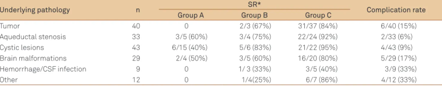
Table 1. Cerebrospinal luid circulation restoration procedures in 166 patients and success rate calculated according to the number of patients and procedures.
Underlying pathology n SR* Complication rate
Group A Group B Group C
Tumor 40 0 2/3 (67%) 31/37 (84%) 6/40 (15%) Aqueductal stenosis 33 3/5 (60%) 3/4 (75%) 22/24 (92%) 2/33 (6%) Cystic lesions 43 6/15 (40%) 5/6 (83%) 21/22 (95%) 4/43 (9%) Brain malformations 29 2/4 (50%) 3/5 (60%) 16/20 (80%) 5/29 (17%) Hemorrhage/CSF infection 9 0 1/ 3 (33%) 3/5 (40%) 3/9 (33%) Other 12 0 1/4(25%) 6/7 (86%) 4/12 (33%)
n: Values represent the number of patients (%) unless otherwise stated; SR: success rate; CSF: cerebrospinal luid; *assortment of the SR by “underlying pathology” did not reveal any statistically signiicant differences among these groups.
Fig 2. Etiology of hydrocephalus according to age group. Line graph showing the etiology of hydrocephalus in each age group. The etiology was grouped as pure obstructive (aqueductal stenosis and posterior fossa tumors) and other.
90% 80% 70% 60% 50% 40% 30% 20% 10% 0%
Pure Obstructive Other
A B C Age Group
Fig 3. Overall success rates according to the etiology of hydrocephalus. Bar graph demonstrating the correlation between SR and the underlying pathology (p=0.001).
100% 90% 80% 70% 60% 50% 40% 30% 20% 10% 0%
88%
83%
74%
69%
60%
20%
Aqueductal stenosis
Tumor Cystic lesion
Malformation Hemorrhage Infection
Fig 4. Learning curve. Line graph showing a synchronous improvement of the success rates and a remarkable decrease in the number of complications in two different time periods (p=0.012).
90% 80% 70% 60% 50% 40% 30% 20% 10% 0%
Success Shunt Complication
2000–2004 2005–2009
83%
66%
34%
17% 7% 22%
Reoperations due to failure on the irst attempt of the en-doscopic procedure were needed in 23 patients. he SR in this group was 74% after a second procedure. Only 6 out of 23 pa-tients required further operations (either endoscopy or shunt). hirty-seven procedures (18%) were performed in children that had been previously shunted. Previous shunt surgery was strong-ly correlated to failure of the endoscopic procedure (p=0.009).
DISCUSSION
Over the last few years, intracranial neuroendoscopy has found its place in pediatric neurosurgery. Current experience throughout the world shows that this treatment is a good alternative to shunts in many cases of cerebral disease, and particularly in obstructive hydrocephalus. ETV is considered to be a simple, fast and safe procedure in children15.
In this paper, we present a single-center experience with 200 consecutive endoscopic intracranial procedures performed in children. he overall success rate to restore CSF circulation was 77%, and the absolute complication rate was 11%. hese data are in accordance with those reported in the literature3,19-21.
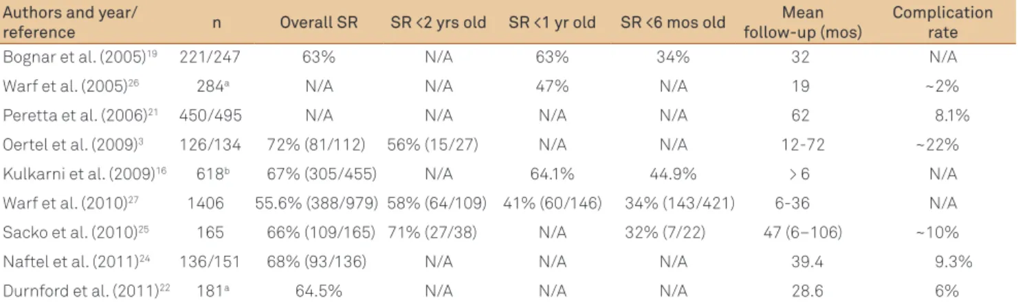
We reviewed only neuroendoscopic pediatric series pub-lished in the literature that had more than 100 cases (Table 2). he overall SR in those series ranged from 55.6 to 72%3,19,21-27.
Despite technological advances in the last century con-cerning the management of hydrocephalus, the issue about timing of neuroendoscopy and its efectiveness in younger patients is still controversial5,12,15,16,20,28-31.
Also, there is no consensus regarding the appropri-ate age of patients to be treappropri-ated with ETV. In most series, younger children were not included. hus, some authors who have defended ETV suggest that it should be attempt-ed only in children older than one year15,32. his may be
ex-plained by the poor CSF resorption capacity of newborn infants due to immaturity of arachnoid granulations. In ad-dition, the anterior fontanel is widely open and the sutures
can become splayed in infants, contributing to the mainte-nance of low intracranial pressure17,32-35.
On the other hand, some authors advocated that ETV has the same long-term results in children younger than six months when compared to older children and, therefore, patient age should no longer be considered a contraindica-tion to using the technique; and in the event of delayed fail-ure (usually, secondary to obstruction of the stoma) this can be often managed by repeating the procedure31,36.
he literature review showed that SR in children under six months of age ranged from 32 to 44.9%, and in children older than one-year age it ranged from 56 to 71% (Table 2).
In our series, the etiology of hydrocephalus and patients’ age group were both relevant factors predicting success. hese results are in accordance with other authors15,36-38.
ETV success rate among patients under one year with AS was 78%, similar to the results among patients older than one year (90%).
Moreover, intraventricular hemorrhage, myelomenin-gocele and previous CSF infection or shunt infection were strongly associated with failure of ETV in our series. Some authors reported similar results analyzing outcome and un-derlying pathology15,28,30,33,39.
Another factor that influenced the outcome in this series was surgical experience21. We observed a
remark-able reduction in the number of complications related to neuroendoscopic procedures in two subsequent time periods analyzed ( from 20 to 11%). Recently, Bouras and Sgouros40 published an extensive review of complications
associated with ETV. Their analysis included 2.985 ETVs performed in 2.884 patients and they concluded that ETV can be regarded as a low-complication procedure, with an overall complication rate of 8.5%, permanent morbidity rate of 2.4%, mortality rate of 0.21%, and delayed “sudden death” rate of 0.07%. According to the literature, the over-all complication rate ranged, in individual series, from 2 to 44.9% (Table 2).
Table 2. Summary of data of the neuroendoscopic pediatric series published in the medical literature*.
Authors and year/
reference n Overall SR SR <2 yrs old SR <1 yr old SR <6 mos old
Mean follow-up (mos)
Complication rate
Bognar et al. (2005)19 221/247 63% N/A 63% 34% 32 N/A
Warf et al. (2005)26 284a N/A N/A 47% N/A 19 ~2%
Peretta et al. (2006)21 450/495 N/A N/A N/A N/A 62 8.1%
Oertel et al. (2009)3 126/134 72% (81/112) 56% (15/27) N/A N/A 12-72 ~22%
Kulkarni et al. (2009)16 618b 67% (305/455) N/A 64.1% 44.9% > 6 N/A
Warf et al. (2010)27 1406 55.6% (388/979) 58% (64/109) 41% (60/146) 34% (143/421) 6-36 N/A
Sacko et al. (2010)25 165 66% (109/165) 71% (27/38) N/A 32% (7/22) 47 (6–106) ~10%
Naftel et al. (2011)24 136/151 68% (93/136) N/A N/A N/A 39.4 9.3%
Durnford et al. (2011)22 181a 64.5% N/A N/A N/A 28.6 6%
1. Dandy WE, Blackfan KD. An experimental and clinical study on internal hydrocephalus. JAMA 1913;61:2216-2217.
2. Cipri S, Gambardella G. Neuroendoscopic approach to complex hydrocephalus. Personal experience and preliminary report. J Neurosurg Sci 2001;45:92-96.
3. Oertel JMK, Gaab M, Schroeder HW, Baldauf J. Endoscopic options in children: experience with 134 procedures. J Neurosurg Pediatr 2009;3:81-89.
4. Enchev Y, Oi S. Historical trends of neuroendoscopic surgical techniques in the treatment of hydrocephalus. Neurosurg Rev 2008;31:249-262.
5. Rekate HL. Selecting patients for endoscopic third ventriculostomy. Neurosurg Clin N Am 2004;15:39-49.
6. O’Brien DF, Javadpour M, Collins DR, Spennato P, Mallucci CL. Endoscopic third ventriculostomy: an outcome analysis of primary cases and procedures performed after ventriculoperitoneal shunt malfunction. J Neurosurg 2005;103:393-400.
7. Oertel JKM, Schroeder H, Gaab MR. Third Ventriculostomy for treatment of hydrocephalus: results of 271 procedures. Neurosurg Quarterly 2006;16:24-31.
8. Balthasar AJ, Kort H, Cornips EM, Beuls EA, Weber JW, Vles JS. Analysis of the success and failure of endoscopic third ventriculostomy in infants less than 1 year of age. Childs Nerv Syst 2007;23:151-55.
9. Cohen AR. Prediction, with restriction. J Neurosurg Pediatr 2010;6:307-309.
10. Di Rocco C, Massimi L, Tamburrini G. Shunts vs endoscopic third ventriculostomy in infants: are there different types and/or rates of complications? A review. Childs Nerv Syst 2006;22:1573-1589.
11. Drake JM. Endoscopic third ventriculostomy in pediatric patients: the Canadian experience. Neurosurgery 2007;60:881-886.
12. Etus V, Ceylan S. Success of endoscopic third ventriculostomy in children less than 2 years of age. Neurosurg Rev 2005;28:284-288.
13. Hellwig D, Grotenhuis JA, Tirakotai W, et al. Endoscopic third ventriculostomy for obstructive hydrocephalus. Neurosurg Rev 2005;28:1-38.
14. Kadrian D, van Gelder J, Florida D, et al. Long-term reliability of endoscopic third ventriculostomy. Neurosurgery 2005;56:1271-1278.
15. Koch D, Wagner W. Endoscopic third ventriculostomy in infants of less than 1 year of age: which factors inluence the outcome? Childs Nerv Syst 2004;20:405-441.
16. Kulkarni AV, Drake JM, Mallucci CL, Sgouros S, Roth J, Constantini S, Canadian Pediatric Neurosurgery Study Group. Endoscopic third ventriculostomy in the treatment of childhood hydrocephalus. J Pediatr 2009;155:254-259.
17. Wagner W, Koch D. Mechanisms of failure after endoscopic third ventriculostomy in young infants. J Neurosurg 2005;103:43-49.
18. Yadav YR, Jaiswal S, Adam N, Basoor A, Jain G. Endoscopic third ventriculostomy in infants. Neurol India 2006;54:161-163.
19. Bognar L, Markia B, Novak L. Retrospective analysis of 400 neuroendoscopic interventions: the Hungarian experience. Neurosurg Focus 2005;19:E10.
20. Gorayeb RP, Cavalheiro S, Zymberg ST. Endoscopic third ventriculostomy in children younger than 1 year of age. J Neurosurg 2004;100:427-429.
21. Peretta P, Ragazzi P, Galarza M, et al. Complications and pitfalls of neuroendoscopic surgery in children. J Neurosurg Pediatr 2006;105:187-193.
22. Durnford AJ, Kirkham FJ, Mathad N, Sparrow OC. Endoscopic third ventriculostomy in the treatment of childhood hydrocephalus: validation of a success score that predicts long-term outcome. J Neurosurg Pediatr 2011;8:489-493.
23. Kulkarni AV, Drake JM, Kestle JR, Mallucci CL, Sgouros S, Constantini S; Canadian Pediatric Neurosurgery Study Group. Predicting who will beneit from endoscopic third ventriculostomy compared with shunt insertion in childhood hydrocephalus using the ETV Success Score. J Neurosurg Pediatr 2010;6:310-315.
24. Naftel RP, Reed GT, Kulkarni AV, Wellons JC. Evaluating the Children’s Hospital Alabama endoscopic third ventriculostomy experience using the endoscopic third ventriculostomy success score. J Neurosurg Pediatr 2011;8:494-501.
25. Sacko O, Boetto S, Lauwers-Cances V, Dupuy M, Roux FE. Endoscopic third ventriculostomy: outcome analysis in 368 procedures. J Neurosurg Pediatr 2010;5:68-74.
26. Warf BC. Comparison of endoscopic third ventriculostomy alone and combined with choroid plexus cauterization in infants younger than 1 year of age: a prospective study in 550 African children. J Neurosurg Pediatr 2005;103:475-481.
27. Warf BC, Mugamba J, Kulkarni AV. Endoscopic third ventriculostomy in the treatment of childhood hydrocephalus in Uganda: report of a scoring system that predicts success. J Neurosurg Pediatr 2010;5:143-148.
28. Elgamal E, El-Dawlatly AA, Murshid WR, El-Watidy SM, Jamjoom ZA. Endoscopic third ventriculostomy for hydrocephalus in children younger than 1 year of age. Childs Nerv Syst 2011;27:111-116.
29. Fritsch MJ, Kienke S, Ankermann T, Padoin M, Mehdorn HM. Endoscopic third ventriculostomy in infants. J Neurosurg 2005; 103:50-53.
30. Kim SK, Wang KC, Cho BK. Surgical outcome of pediatric hydrocephalus treated by endoscopic III ventriculostomy: prognostic factors and interpretation of postoperative neuroimaging. Childs Nerv Syst 2000;16:161-169.
31. Siomin V, Weiner H, Wisoff J, et al. Repeat endoscopic third ventriculostomy: is it worth trying? Childs Nerv Syst 2001;17:551-555.
32. Hopf NJ, Grunert P, Fries G, Resch KD, Perneczky A. Endoscopic third ventriculostomy: outcome analysis of 100 consecutive procedures. Neurosurgery 1999;44:795-806.
References
In the present series, reoperations due to failure of the irst attempt of endoscopic procedure were observed in 11.5% of cases. he success rate was 74% after a second pro-cedure. hese results are similar to other series31.
herefore, despite the fact that some patients sufering from reocclusion of the stoma might have to undergo shunting, several authors consider well worth trying to repeat ETV31,36.
In conclusion, neuroendoscopic techniques provide very good results for a wide number of indications in children.
33. Jones RF, Kwok BC, Stening WA, Vonau M. Third ventriculostomy for hydrocephalus associated with spinal dysraphism: indications and contraindications. Eur J Pediatr Surg 1996;6:5-6.
34. Oi S, Abbott R. Loculated ventricles and isolated compartments in hydrocephalus: their pathophysiology and the eficacy of neuroendoscopic surgery. Neurosurg Clin N Am 2004;15:77-87.
35. Oi S, Di Rocco C. Proposal of “evolution theory in cerebrospinal luid dynamics” and minor pathway hydrocephalus in developing immature brain. Childs Nerv Syst 2006;22:662-669.
36. Cinalli G, Sainte-Rose C, Chumas P, et al. Failure of third ventriculostomy in the treatment of aqueductal stenosis in children. J Neurosurg 199;90:448-454.
37. Baldauf J, Gaab MR, Schroeder H. Endoscopic third ventriculostomy in children younger than 2 years of age. Childs Nerv Syst 2007;23:623-626.
38. Beems T, Grotenhuis JA. Is the success of endoscopic third ventriculostomy age-dependent? An analysis of the results of endoscopic third ventriculostomy in children. Childs Nerv Syst 200;218:605-608.
39. Elbabaa S, Steinmetz M, Ross J, Moon D, Luciano M. Endoscopic third ventriculostomy for obstructive hydrocephalus in the pediatric population: evaluation of outcome. Eur J Pediatr Surg 2001;11:S52-S54.