270 270 270 270 270
Brito Jr. et al
Brachytherapy for in-stent restenosis
Arq Bras Cardiol 2001; 77: 270-3.
Hospital Israelita Albert Einstein – São Paulo
Mailing address: Fábio S. Brito Jr. – Hospital Israelita Albert Einstein - Av. Albert Eistein, 627/701 – 05651-901 – São Paulo, SP, Brazil - E-mail: [email protected] English version by Stela Maris C. e Gandour
Fábi o Sândol i de Br i t o Jr, Rodr i go Hanr i ot , Br eno Ol i vei r a Al m ei da, Mi guel Ant oni o Neves Rat i , Nadi a Suel i de Medei ros, Môni ca Lagatta, José Carl os da Cruz, João Vi ctor Sal vaj ol i , Marco Antoni o Peri n
São Paulo, SP - Brazil
Intracoronary Brachytherapy in the Treatment of In-stent
Restenosis. Initial Experience in Brazil
Brief Report
Intracoronary brachytherapy using beta or gamma radiation is currently the most efficient type of therapy for preventing the recurrence of coronary in-stent restenosis. Its implementation depends on the interaction among inter-ventionists, radiotherapists, and physicists to assure the safety and quality of the method. The authors report the pio-neering experience in Brazil of the treatment of 2 patients with coronary in-stent restenosis, in whom beta radiation was used as part of the international multicenter randomi-zed PREVENT study (Proliferation REduction with Vascu-lar ENergy Trial). The procedures were performed rapidly and did not require significant modifications in the tradi-tional techniques used for conventradi-tional angioplasty. Altera-tion in the radiological protecAltera-tion devices of the hemodyna-mic laboratory were also not required, showing that intra-coronary brachytherapy using beta radiation can be in-corporated into the interventional tools of cardiology in our environment.
As a direct consequence of the impressive growth of the number of stents implanted, problems related to reste-nosis of these metallic endoprostheses have arisen. The in-cidence of restenosis ranges from 10% to 35% 1, depending
on the population studied, resulting fundamentally from exacerbation of neointimal hyperplasia2. Independent of the
device used, the treatment of coronary in-stent restenosis is associated with high rates of recurrence (>30%), which may even exceed 50% when the restenosis is long and diffu-se 1-3. More recently, encouraging results from clinical
stu-dies using coronary radiotherapy (beta or gamma rays) 4-7
have introduced new perspectives for the treatment of this problem. The authors report the 2 pioneering cases that in-troduced this new type of therapy into Brazil.
Case report
Case 1 – The patient is a 53-year-old diabetic, hyper-tensive, dyslipidemic, male ex-smoker who sought our Ser-vice complaining of precordial pain on moderate efforts. He reported using acetylsalicylic acid (200 mg/day), enalapril (20 mg/day), diltiazem (180 mg/day), simvastatin (20 mg/ day), glipizide (20 mg/day), and metformin (2 g/day). He also reported having undergone 2 angioplasties in the proximal 1/3 of the circumflex artery, with stent (Multilink Duet 3.5x18 mm) implantation in the latter angioplasty performed in June ‘99. The physical examination and the electrocardiography performed at the time of hospital admission were normal; a new coronary angiography showed an 80%-obstructing lesion in the initial 1/3 of the circumflex artery at the site of stent implant. The anterior descending and circumflex arteries had only parietal irregularities, and the left ventricle had preserved volume and contractility. In May ’00, based on these findings and after signing the written consent, the patient underwent a new angioplasty in the circumflex artery (fig. 1), which was complemented by intracoronary brachytherapy (beta radiation) as part of the international multicenter PREVENT study. The procedure was guided by intracoronary ultrasonography, which showed a reference diameter of the vascular lumen of 3.6 mm, with a large amount of neointimal tissue between the metallic rods of the stent (fig. 1). After progressive dilations with balloon catheters (3.5 and 4.0 x 20 mm), a good angiographic aspect was ob-tained, with a residual lesion of 20%. The intracoronary echo-graphy confirmed the good result of the intervention, and the smallest area of the lumen obtained was 8 mm 2. Then, the
centralizing perfusion balloon catheter (3.5 x 27 mm) was positioned and inflated (4 atm) in the treated site (figs. 1 and 2), with connection of its distal extremity to the afterloader, which stored the source of beta radiation (32P) (fig. 2). Then
the guidewire (0.018 inches of thickness, and with a ra-dioactive extremity of 27 mm of length) was advanced and positioned (fig. 2) for the application of the dose of 20 Gy at 1mm from the luminal surface. The total duration of the
Arq Bras Cardiol 2001; 77: 270-3.
Brito Jr. et al Brachytherapy for in-stent restenosis
271 271 271 271 271 application of radiation was 2.5 min. After the intervention,
the patient had a good clinical evolution with no electro-cardiographic or enzymatic (CKMB) alteration, being discharged on the following day with the same pres-criptions as that on admission. On clinical control, 1 month after the procedure, the patient remained free of any symp-toms or adverse events. The patient will undergo a late angiographic study (6 months) to assess the efficacy of the treatment used.
Case 2 - The patient is a 74-year-old diabetic hyperten-sive female referred to our service due to angina at rest. She was using acetylsalicylic acid (200 mg), propatyl nitrate (30
mg), diltiazem (90 mg), chlorpropamide (250 mg), and capto-pril (37.5 mg) on a daily basis. The patient reported a previ-ous angioplasty with stent implantation in the right corona-ry and anterior descending coronacorona-ry arteries, which was performed in January ‘00. The physical examination did not show significant alterations, but the electrocardiogram sho-wed alterations in ventricular repolarization in the anterior wall (inversion of the T-wave from V1 to V6). Based on these findings, the patient underwent new coronary angiography, which depicted a good angiographic aspect of the stent im-planted in the right coronary artery and a 90%-obstructive lesion in the middle 1/3 of the anterior descending coronary artery at the site of the previous implantation of the Multi-link Duet 3.0 x 23 mm stent (in-stent restenosis). After sig-ning the written consent for inclusion in the PREVENT stu-dy, the patient underwent a new angioplasty of the anterior descending artery (balloon catheters 3.0x20 and 3.5x15 mm) guided by intracoronary ultrasonography in May ‘00 (fig. 3). The procedure was successfully performed; a residual lesion of 20% was shown on angiography, and a smaller area of the vascular lumen of 5 mm2 was evidenced on
intra-vascular ultrasonography (fig. 3). Then, intracoronary bra-chytherapy (beta radiation) was performed by positioning and inflation (4 atm) of the centralizing perfusion balloon catheter (3.0 x 27 mm) and progression of the guidewire with a radioactive distal extremity (27 mm) up to the site of the lesion (fig. 2). The prescribed dose of radiation was 20 Gy at 1 mm of the surface of the vascular lumen, and the duration of application (5 min) was based on the measurements of the vas-cular lumen assessed by intracoronary ultrasonography (diameter of reference of 3.0 mm). The patient had a good Fig. 1 - A) 70%-obstructive lesion of the proximal 1/3 of the circumflex artery, in a
site of previous stent implantation; B) the arrow indicates the point where the echographic image was obtained and shows the significant narrowing of the vascular lumen due to a large amount of neointimal tissue between the metallic rods of the stent; C) after angioplasty, note the good angiographic aspect with minimum residual stenosis; D) intracoronary echography confirms the good result of the procedure, with widening of the vascular lumen. Note in the detail in figura C, the spiral centralizing perfusion balloon catheter positioned in the site of the lesion, for radiation application. In B and D, the details show the same echographic images in a reduced size, which serve as a refernce for identifying the echographic catheter (in black), the area of the vascular lumen (in red), the neointimal area inside the stent (light gray), and the area of the atheroma plaque + media layer of the vessel (dark gray), compressed behind the stent rods. The stent is depicted as a white line.
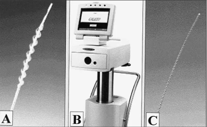
Fig. 2 - Spiral centralizing perfusion balloon catheter (A). Computerized afterload (B) that stores the guidewire with 0.018 inches of thickness and radioactive distal extremity (32P) of 27mm of length (C).
272 272 272 272 272
Brito Jr. et al
Brachytherapy for in-stent restenosis
Arq Bras Cardiol 2001; 77: 270-3.
clinical evolution, with no electrocardiographic or enzy-matic (CKMB) alteration, and was discharged 2 days after the intervention with the following prescription: ace-tylsalicylic acid (200 mg/day), diltiazem (90 mg/day), and chlorpropamide (250 mg/day). On clinical control, 1 month after the procedure, the patient was free from anginal symptoms. The patient will undergo a late angiographic study (6 months).
Discussion
Intracoronary brachytherapy is currently the most effi-cient therapy for preventing recurrence of in-stent resteno-sis 4-7. Three prospective randomized studies (SCRIPPS,
WRIST, and GAMMA-I) using systems based on catheters with gamma radiation showed a 41% to 69% reduction in the rates of angiographic restenosis, associated with a signifi-cant reduction in the need for new revascularization of the vessel treated, as compared with isolated angioplasty or an-gioplasty associated with atheroablative methods 4,6,7.
More recently, with the results of the beta-WRIST5 and
START* studies, the efficacy of beta radiation in the treatment of in-stent restenosis has been shown. In the randomized START* study, a significant reduction occurred in the incidence of angiographic restenosis in patients ceiving radiation (28.8% vs 45.2%), with a corresponding re-duction in the need for target vessel revascularization (16% vs 24.1%).
Recently, intracoronary brachytherapy was introdu-ced into Brazil as part of a double-blind multicenter pros-pective and randomized study named PREVENT (Prolifera-tion REduc(Prolifera-tion with Vascular ENergy Trial). Using the pro-totype of the equipment GalileoTM (Guidant Corporation)
(fig. 2), this study assessed the efficacy of brachytherapy (beta radiation; 32P) in the treatment of primary or restenotic
lesions, including in-stent restenosis, in native coronary arteries (2.4 to 3.7 mm of diameter). As an inclusion criterion, the authors selected lesions up to 15 mm in length, which allowed that, with a source (guidewired) of 27 mm, their pro-ximal and distal margins could also be irradiated. This thera-peutic strategy aims at avoiding the occurrence of reste-nosis in the margins of the segment treated, which occurs in 8% to 18% of the irradiated cases, and in less than 4% of the control groups. Restenosis in the margins of the segment treated accounts for 33% to 75% of all restenoses following brachytherapy in recent clinical trials 4-7. This phenomenon
is caused by the administration of low doses of radiation to sites with a certain degree of vascular trauma produced by the angioplasty balloons or stents (geographic miss), which determines exacerbation of the proliferative process and of the negative remodeling of the arterial wall in these sites 8,9.
As the 2 cases here reported were our first experience, the patients received the irradiation dose prescribed (20 Gy), not requiring the randomization foretold in the study. In the-se procedures, the uthe-se of beta radiation, becauthe-se of its lo-wer penetration, made possible the safe permanence of the entire professional team inside the room during the proce-dure, including the interventionists, the radiotherapist, the physicist, and the nursing staff. The duration of application was relatively short, less than 5 min. As the spiral balloon catheter allows distal perfusion, it can remain inflated du-ring this period of time, without causing excessive discom-fort to the patients, making possible the centralization of the radioactive source inside the vascular lumen. This centrali-zation of the source is believed to provide a greater homo-geneity in the application of the doses of beta radiation in different sites of the vascular wall, even though the disposi-tions of the lumen and the atherosclerotic plaques are usual-ly asymmetric.
It is worth noting that, in our 2 reported cases, we tried to optimize the result of the conventional balloon angioplas-ty, avoiding the implantation of a new stent. This concern is based on the higher risk of the occurrence of late thrombosis when a new stent is implanted in the same procedure in which radiation is applied to the coronary arteries 9-11. In recent
studies, the occurrence of late thrombosis (>1 month) has been reported to range from 6% to 15% of the cases treated with beta or gamma radiation; in control groups, it rarely exceeds 2% 4-7,10,11. Its major cause is the delay in
reendothe-lialization and vascular repair of the lesion 10-12. Aiming at
solving this limitation, in addition to avoiding a new stent implantation, we recommend prolonging the regimen of double platelet antiaggregation (acetylsalicylic acid and ti-clopidine or clopidogrel) until at least 3 months after the treatment. The efficacy of this approach has been recently confirmed by the results of the START study*, in which a new stent was implanted in only 21% of the cases, and the platelet antiaggregation was prolonged for up to 60 days, which almost made the late thromboses disappear.
In conclusion, intracoronary brachytherapy using beta radiation is efficient in the adjuvant treatment of coro-nary in-stent restenosis. Its implementation depends on the interaction of interventionists, radiotherapists, and physicists, reassuring the safety and quality of the me-thod. The application is relatively rapid and does not re-quire expressive modifications of the traditionally used te-chniques for conventional angioplasty. The solution of its major limitations, such as late thrombosis and restenosis of the margins, should, in the near future, provide the incorporation of this method into the major cardiologic centers of our country, contributing to the progress of interventional cardiology.
Arq Bras Cardiol 2001; 77: 270-3.
Brito Jr. et al Brachytherapy for in-stent restenosis
273 273 273 273 273 1. Casterella PJ, Teirstein PS. Prevention of coronary restenosis. Cardiol Rev
1999; 7: 219-31.
2. Hoffmann R, Mintz GS, Dussaillant GR, et al. Patterns and mechanisms of in-stent restenosis. A serial intravascular ultrasound study. Circulation 1996; 94: 1247-54.
3. Mehran R, Dangas G, Abizaid AS, et al. Angiographic patterns of in-stent restenosis: classification and implications for long-term outcome. Circulation 1999; 100: 1872-8.
4. Waksman R, White RL, Chan RC, et al. Intracoronary gamma-radiation therapy after angioplasty inhibits recurrence in patients with in-stent restenosis [see comments]. Circulation 2000; 101: 2165-71.
5. Waksman R, Bhargava B, White L, et al. Intracoronary beta-radiation therapy inhibits recurrence of in-stent restenosis. Circulation 2000; 101: 1895-8. 6. Teirstein PS, Massullo V, Jani S, et al. Catheter-based radiotherapy to inhibit
restenosis after coronary stenting. N Engl J Med 1997; 336: 1697-703.
References
7. Leon M, Teirstein P, Lanskey A, et al. Intracoronary gamma radiation to reduce in-stent restenosis: the multicenter gamma-I randomized clinical trial. J Am Coll Cardiol 1999; 33: 19-A.
8. Sabate M, Costa MA, Kozuma K, et al. Geographic miss: a cause of treatment failure in radio-oncology applied to intracoronary radiation therapy. Circulation 2000; 101: 2467-71.
9. Kuntz RE, Baim DS. Prevention of coronary restenosis: the evolving evidence base for radiation therapy. Circulation 2000; 101: 2130-3.
10. Costa MA, Sabat M, van der Giessen WJ, et al. Late coronary occlusion after intracoronary brachytherapy. Circulation 1999; 100: 789-92.
11. Waksman R. Late thrombosis after radiation. Sitting on a time bomb. Circulation 1999; 100: 780-2.