Article
Printed in Brazil - ©2016 Sociedade Brasileira de Química0103 - 5053 $6.00+0.00
*e-mail: [email protected], [email protected]
#Both authors contributed equally to this work
A New Steroidal Saponin from the Tubers of
Ophiopogon japonicus
and Its
Protective Effect Against Cisplatin-Induced Renal Cell Toxicity
Seoung Rak Lee,a,# Ju-Yeon Han,b,# Hee Rae Kang,a Hye Lim Lee,c Hyung-Jun Noh,d Jae-Soon Cha,e Ki Sung Kang,c Chan-Jung Lee*,b and Ki Hyun Kim*,a
aSchool of Pharmacy, Sungkyunkwan University, 440-746 Suwon, Republic of Korea
bMushroom Research Division, National Institute of Horticultural & Herbal Science, Rural Development Administration (RDA), 369-873 Eumseong, Republic of Korea
cCollege of Korean Medicine, Gachon University, 461-701 Seongnam, Republic of Korea
dDivision of Herbal Crop Utilization Research, National Institute of Horticultural & Herbal Science, Rural Development Administration (RDA), 369-873 Eumseong, Republic of Korea
eDepartment of Plant Medicine, College of Agriculture, Life & Environment Science, Chungbuk National University, Cheongju, 631-763 Chungbuk, Republic of Korea
A new furostanol saponin, ophiopogonin T, was isolated from the tubers of Ophiopogon japonicus. Its structure was established by extensive spectroscopic techniques including 1D (1H and 13C) and 2D nuclear magnetic resonance (NMR) experiments (correlation spectroscopy (COSY), heteronuclear single quantum coherence (HSQC), heteronuclear multiple bond correlation (HMBC) and nuclear Overhauser effect spectroscopy (NOESY)), high-resolution electrospray ionization mass spectrometry (ESIMS), and chemical methods. Using cell-based assays, this compound was evaluated for its cytotoxic effect on cancer cell lines and its protective effect against anticancer drug-induced nephrotoxicity. Cisplatin-induced cytotoxicity in porcine kidney (LLC-PK1) cells was significantly reduced upon treatment with ophiopogonin T, without affecting human hepatoma (HepG2) cancer cell proliferation or tube formation in human umbilical vein endothelial cells (HUVECs). These results collectively reflect the beneficial effect of ophiopogonin T on the side effects of cisplatin.
Keywords:Ophiopogon japonicus, Liliaceae, ophiopogonin T, nephrotoxicity, cisplatin
Introduction
Ophiopogon japonicus Ker-Gwal. (Liliaceae) is an evergreen, sod-forming perennial plant, widely distributed in East Asia, particularly in most areas of China. The tubers of O. japonicus have been widely used for thousands of years in traditional Chinese medicine to treat cardiovascular and chronic inflammatory diseases,1 and have been well-known as a popular soup ingredient for nourishing ‘Yin’ in China.2 A wide range of pharmacological properties such as antiviral,3 antifungal,3 antithrombotic,4 anti-inflammatory,5 and hypoglycemic activities6 have been reported for crude extracts or isolates
from the tubers of O. japonicus. Previous phytochemical investigations have revealed that steroidal saponins1,7-11 and homoisoflavonoids2 are the main constituents of O. japonicus, and in particular, the tuber of O. japonicus is rich in a series of steroidal saponins.1,7-11 A survey of the literature showed that these steroidal saponins possess promising cytotoxic activities1,7 and antimyocardial ischemia effects.12
Experimental
General experimental procedures
Optical rotations were measured on a Jasco P-1020 Polarimeter (Jasco, Easton, MD, USA). Infrared (IR) spectra were recorded on a Bruker IFS-66/S Fourier transform (FT) IR spectrometer (Bruker, Karlsruhe, Germany). Electrospray ionization mass spectrometry (ESIMS) spectra were obtained by liquid chromatography-mass spectrometry (LC/MS) analysis performed on an Agilent 1200 Series high-performance liquid chromatograph (HPLC)/6130 Series mass spectrometer (Agilent Technologies, Santa Clara, CA, USA). High-resolution ESIMS spectra were obtained on a Waters Micromass Q-Tof Ultima ESI-time-of-flight (TOF) mass spectrometer (Waters Corporation, Milford, CT, USA). Nuclear magnetic resonance (NMR) spectra were recorded on a Bruker AVANCE III 700 NMR spectrometer (Bruker) operating at 700 MHz (1H) and 175 MHz (13C), with chemical shifts (d) given in ppm. Preparative HPLC was conducted using a Glison 321 pump (Middleton, USA) with a Shodex refractive index detector (New York, USA). An HPLC, Luna RP-18 column (250 × 10.0 mm, 10 µm, Phenomenex, Torrance, USA) was used for purification. Diaion HP-20 (Supelco, Tokyo, Japan), silica gel 60 (Merck, Darmstadt, Germany, 230-400 mesh) and C18 silica gel (Merck, 230-400 mesh) were used for open column chromatography. Precoated silica gel F254 plates (Merck) and RP-18 F254s plates (Merck) were used for thin-layer chromatography (TLC). Spots were detected on TLC under UV light, followed by heating after spraying with anisaldehyde-sulfuric acid. Cisplatin was purchased from Sigma-Aldrich (Seoul, South Korea). Dulbecco’s
modified Eagle’s medium (DMEM) was purchased from Cellgro (Manassas, VA, USA). Fetal bovine serum (FBS) was purchased from Invitrogen Co. (Grand Island, NY, USA). Solvents for HPLC and column chromatography were purchased from Samchun Pure Chemical Co., Ltd. (Seoul, South Korea). Amberlite IRA-7 column was obtained from Rohm and Haas (Philadelphia, PA, USA). Microplate reader was purchased from PowerWave XS, Bio-Tek Instruments (Winooski, VT, USA). Mayer’s hematoxylin was purchased from Muto Pure Chemicals (Tokyo, Japan).
Plant material
The tubers of O. japonicus were collected in Miryang, Gyeongsangbuk-do, Korea, in April 2012. A voucher specimen of the material (MPS001140-1) was identified by PhD Jeong-Hoon Lee, and deposited in the Department of Herbal Crop Research, Korea Medicinal Resources Herbarium (KMRH).
Extraction and isolation
The dried and pulverized O. japonicus tubers (2.5 kg) were extracted with 100% MeOH at room temperature and filtered. After MeOH evaporation in vacuo, the residue was suspended in deionized water and then solvent-partitioned successively with hexane, CHCl3, EtOAc, and n-BuOH. The n-BuOH-soluble fraction (9.3 g) was loaded onto a Diaion HP-20 column, and the column was washed with 30% MeOH/H2O to remove very polar compounds such as sugars. The Diaion HP-20 column was further fractionated with 60% MeOH/H2O and 100% MeOH to give two fractions: A (1.8 g) and B (2.4 g).
H H HO
O O
H OH
O
O
O
O O O
OH
O
OH HO
HO OH HO
HO
OH HO
HO HO
H Fuc
Rha Xyl
Glc
1
3
1 1
1 4 1 6 1 8 1 9
2 1
2 2 2 6
2 7
1 '
1 '' 1 '''
1 ''''
6
Fraction B (2.4 g) was separated by silica gel column chromatography (4.5 × 20 cm) using a gradient elution with CHCl3/MeOH/H2O (12:3:1) and 100% MeOH to yield seven fractions (B1-B7). Fractions B3 (710 mg) and B4 (650 mg) were combined on the basis of TLC analysis, and the resulting fraction was subjected to RP-18 silica gel column chromatography (3.0 × 6.5 cm) eluted with 70% MeOH/H2O, to afford three fractions (B31-B33). Fraction B33 (270 mg) was separated by preparative HPLC using 70% MeOH/H2O (flow rate: 1.5 mL min-1) to give four fractions (B331-B334). Finally, fraction B332 (115 mg) was purified by semi-preparative HPLC using 70% MeOH/H2O (flow rate: 0.5 mL min-1) to afford compound 1 (8 mg, tR = 8.2 min).
Ophiopogonin T (1)
White amorphous powder; [α]D25 –43.3 (c 0.03, MeOH); IR (KBr) νmax / cm-1 3405, 2936, 1455, 1374, 1243, 1152, 1038; 1H (700 MHz) and 13C (175 MHz) NMR data, see Table 1; ESIMS (positive-ion mode) [M + Na]+ 1057; HR-ESIMS (positive-ion mode) calcd. for C50H82O22Na [M + Na]+: 1057.5195; found: 1057.5193.
Acid hydrolysis and sugar analysis of 1
Compound 1 (0.6 mg) was dissolved in 1 mol L-1 HCl (1.0 mL) and refluxed at 100 °C for 2 h. After cooling, the hydrolysate was extracted with CHCl3 and the aqueous layer was neutralized by passage through an Amberlite IRA-7 column and then concentrated to dryness to give a residue. The residue was dissolved in anhydrous pyridine (0.5 mL) followed by the addition of L-cysteine methyl ester hydrochloride (2 mg) (Sigma, St. Louis, MO, USA). After stirring the mixture at 60 °C for 1.5 h, hexamethyldisilazane (HMDS)-trimethylchlorosilane TMCS) (2:1, 0.4 mL) was added, and the mixture was further stirred at 60 °C for 1 h. The precipitate was removed by centrifugation, and the supernatant was concentrated under N2 stream. The residue was partitioned between hexane and H2O (0.1 mL each), and the hexane layer (2 µL) was analyzed by gas chromatography (GC).11 The standard monosaccharides were subjected to the same reaction and GC analysis. Derivatives of D-xylose (Xyl), L-rhamnose (Rha), D-fucose (Fuc), and D-glucose (Glc) were detected with tR values of 12.937 min (D-xylose derivative), 14.323 min (L-rhamnose derivative), 14.678 min (D-fucose derivative), and 17.342 min (D-glucose derivative). Identification of D-xylose, L-rhamnose, D-fucose, and D-glucose for 1 was performed, giving single peaks at 12.933, 14.325, 14.680, and 17.343 min, respectively.
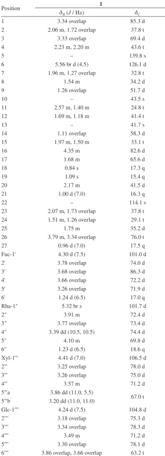
Table 1. 1H and 13C NMR data of compound 1 inCD 3OD
Position 1
dH (J / Hz) dC
1 3.34 overlap 85.3 d
2 2.06 m, 1.72 overlap 37.8 t
3 3.33 overlap 69.4 d
4 2.23 m, 2.20 m 43.6 t
5 – 139.8 s
6 5.56 br d (4.5) 126.1 d
7 1.96 m, 1.27 overlap 32.8 t
8 1.54 m 34.2 d
9 1.26 overlap 51.7 d
10 – 43.5 s
11 2.57 m, 1.40 m 24.8 t
12 1.69 m, 1.18 m 41.4 t
13 – 41.7 s
14 1.11 overlap 58.3 d
15 1.97 m, 1.50 m 33.1 t
16 4.35 m 82.6 d
17 1.68 m 65.6 d
18 0.84 s 17.3 q
19 1.09 s 15.4 q
20 2.17 m 41.5 d
21 1.00 d (7.0) 16.3 q
22 – 114.1 s
23 2.07 m, 1.73 overlap 37.8 t 24 1.51 m, 1.26 overlap 29.1 t
25 1.75 m 35.2 d
26 3.79 m, 3.34 overlap 76.0 t
27 0.96 d (7.0) 17.5 q
Fuc-1' 4.30 d (7.5) 101.0 d
2' 3.78 overlap 74.0 d
3' 3.68 overlap 86.3 d
4' 3.66 overlap 72.2 d
5' 3.26 overlap 71.9 d
6' 1.24 d (6.5) 17.0 q
Rha-1" 5.32 br s 101.7 d
2" 3.91 m 72.4 d
3" 3.77 overlap 73.4 d
4" 3.39 dd (10.5, 10.5) 74.4 d
5" 4.10 m 69.8 d
6" 1.23 d (6.5) 18.6 q
Xyl-1''' 4.41 d (7.0) 106.5 d
2''' 3.25 overlap 78.0 d
3''' 3.26 overlap 75.0 d
4''' 3.57 m 71.2 d
5'''a 3.86 dd (11.0, 5.5)
67.0 t 5'''b 3.20 dd (11.0, 11.0)
Glc-1'''' 4.24 d (7.5) 104.8 d
2'''' 3.18 overlap 75.3 d
3'''' 3.34 overlap 78.3 d
4'''' 3.49 m 71.2 d
5'''' 3.30 overlap 78.1 d
6'''' 3.86 overlap, 3.66 overlap 63.2 t
Cytotoxic effects on cancer cell lines, tube formation assay, and protective effects against anticancer drug-induced nephrotoxicity
The cytotoxicity of samples in HepG2 cells was investigated using the 3-(4,5-dimethylthiazol-2-yl)-2,5-diphenyltetrazolium bromide (MTT) assay.13 In brief, cells were seeded at 1 × 10⁴ cells well-1 in a 96-well plate. The following day, cells were treated with various amounts of the samples and incubated for 24 h at 37 °C in a humidified atmosphere of 5% CO2, 95% air. Subsequently, MTT was added to each well and the plates were incubated for an additional hour at 37 °C. The absorbance of the samples at 450 nm was measured using a microplate reader.
A tube formation assay was conducted following reported methods using HUVECs.14 In brief, cells were seeded (3 × 10² cells well-1) onto a Matrigel-coated plate, medium with or without sample was added, and the plates were then incubated at 37 °C. After 24 h, cells were fixed with 4% paraformaldehyde and stained with Mayer’s hematoxylin. Cell morphology changes and tubular structure formation were observed with a light microscope.
The renoprotective effect against oxidative renal cell damage was evaluated using LLC-PK1 cells as reported previously.15 In brief, LLC-PK1 cells were seeded in 96-well culture plates at 1 × 10⁴ cells per well-1 and allowed to adhere for 2 h. Thereafter, the test sample and/or 25 µmol L-1 cisplatin were added to the culture medium and incubated for 24 h. Cell viability was then measured using a microplate reader.
Results and Discussion
Compound 1 was isolated as a white amorphous powder. The molecular formula was determined to be C50H82O22 from the cationized molecular ion peak at m/z 1057.5192 [M + Na]+ (calcd. for C50H82O22Na: 1057.5195) in the positive-ion HRESIMS, together with its 1H and 13C NMR data. The 1H NMR spectrum of 1 (Table 1) showed two singlet methyl signals at dH 0.84 (s, 3H, CH3-18) and 1.09 (s, 3H, CH3-19), four doublet methyl signals at
dH 0.96 (d, 3H, J 7.0 Hz, CH3-27), 1.00 (d, 3H, J 7.0 Hz, CH3-21), 1.23 (d, 3H, J 6.5 Hz, CH3-Rha), and 1.24 (d, 3H, J 6.5 Hz, CH3-Fuc), four anomeric proton signals at
dH 4.24 (d,1H, J 7.5 Hz, Glc), 4.30 (d,1H, J 7.5 Hz, Fuc), 4.41 (d,1H, J 7.0 Hz, Xyl), and 5.32 (br s, 1H, Rha), and an olefinic proton at dH 5.56 (br d, 1H, J 4.5 Hz, H-6). The 13C NMR spectrum (Table 1) showed a total of 50 carbon signals, in which the characteristic carbon signals at dC 76.0 (C-26), 114.1 (C-22), 126.1 (C-6), and 139.8 (C-5) were assigned using 1H–1H correlation spectroscopy (COSY), heteronuclear single quantum coherence (HSQC), and heteronuclear multiple bond correlation (HMBC) experiments. These characteristic carbon signals, especially an acetalic carbon signal at dC 114.1 (C-22), indicate that compound 1 is a D5 furostanol saponin.11,16,17 Comparison of the 13C NMR data for the aglycone of 1 with those of related furostanol saponins suggests that the aglycone of 1 is a furost-5-ene-1,3,22,26-tetraol,11,16,17 which was confirmed by 2D NMR including COSY, HSQC, and HMBC experiments (Figure 2).
Regarding the identification of four sugar units in compound 1, four anomeric protons at dH 4.24 (Glc),
H H
HO
O O
H
H
OH O
O
O
O O O
OH
O
OH HO
HO OH HO
HO
OH HO
HO HO
H
H C
Figure 2. Selected COSY ( ) and HMBC ( ) correlations of compound 1.
H H
HO
O O
H
H
OH O
O
O
O O O
OH
O
OH HO
HO OH HO
HO
OH HO
HO HO
H
H C
4.30 (Fuc), 4.41 (Xyl), and 5.32 (Rha) showed HSQC correlations with four anomeric carbons at dC 104.8, 101.0, 106.5, and 101.7, respectively. The 1H and 13C NMR data assignable to the sugar units and the evaluation of 2D NMR (COSY, HSQC, and HMBC) suggest that the sugar moieties of 1 consist of glucose, rhamnose, fucose, and xylose.17,18 Acid hydrolysis of 1 with 1 mol L-1 HCl and GC-MS analysis yielded D-xylose, L-rhamnose, D-fucose, and D-glucose.11 The relatively large coupling constants (6.5-7.5 Hz) of each anomeric proton revealed the β configuration of Xyl, Fuc, and Glc, and the broad singlet of the anomeric proton for Rha indicated the α configuration.17,18 The sequence and interglycosidic linkages between the aglycone and the four sugar units were unambiguously defined by HMBC experiments (Figure 2). The sugar sequence of Fuc, Rha, and Xyl and its linkage to C-1 of the aglycone was ascertained by HMBC correlations between dH 4.30 (H-1' of Fuc) and dC 85.3 (C-1), dH 5.32 (H-1" of Rha) and dC 74.0 (C-2' of Fuc), and dH 4.41 (H-1''' of Xyl) and dC 86.3 (C-3' of Fuc). In addition, the HMBC cross-peaks of dH 4.24 (H-1'''' of Glc) with dC 76.0 (C-26) allowed us to identify C-26 as the glucosyl linkage site. The stereochemistry of
1 was confirmed to be the β-orientation of OH-1 and OH-3 by analysis of the nuclear Overhauser effect spectroscopy (NOESY) spectrum showing the correlations of H-1/H-3 and H-1/H-9. Moreover, the β orientation of the hydroxy group of C-22 in the aglycone moiety was determined by the hemiketal carbon signal at dC114.1, approximately 3 to 4 ppm downfield-shifted than that of the α configuration.19,20 The chemical shift difference (Dab = da– db) between the two proton signals of H-26 could be applied to assign the 25R/25S configuration in furostanol saponins.11,21,22 The difference (Dab = 0.45) in the observed chemical shifts of H-26 demonstrated the 25R configuration (Dab < 0.48 for 25R; Dab > 0.57 for 25S).11,21,22 Thus, the structure of 1 was elucidated to be 26-O-β-D-glucopyranosyl-(25R )-furost-5-ene-1β,3β,22β,26-tetraol-1-O-β-D-xylopyranosyl-(1→ 3)-[α-L-rhamnopyranosyl-(1→2)]-β-D-fucopyranoside, here named ophiopogonin T.
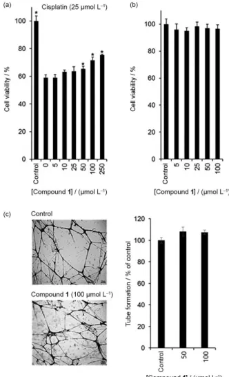
The applicability of medicinal uses for compound 1
was investigated using 3 cell-based assays. In the present study, we investigated the effects of compound 1 on human hepatoma HepG2 cell proliferation, HUVEC tube formation, and cisplatin-induced nephrotoxicity in LLC-PK1 cells. Among the tested 3 assays, compound 1
exhibited potent protective effects against cisplatin-induced kidney cell damage (Figure 3a). The LLC-PK1 cell viability decreased to 60% of the control following treatment with 25 µmol L-1 cisplatin, which was recovered significantly up to 80% of the control in a dose-dependent manner following treatment with compound 1 (Figure 3a).
However, compound 1 (5 to 100 µmol L-1) had no effect on the proliferation of HepG2 cells or tube formation of HUVECs (Figures 3b and 3c). These results reflect the beneficial effects of compound 1 on the side effects of cisplatin without affecting HepG2 cancer cell proliferation or tube formation in HUVECs.
Cisplatin-based treatments have been considered the most effective regimens for advanced/recurrent cervical cancer, however their use is often limited because of severe nephrotoxicity.23,24 In addition, it is recommended that any compounds to be used in combination with cisplatin should have no effect on the anticancer action of cisplatin. Earlier studies have shown that pseudoginsenoside F11 ameliorates cisplatin-induced nephrotoxicity without compromising its antitumor activity.25 It has also been reported that cisplatin-induced LLC-PK1 cell damage is significantly decreased by
Figure 3. Biological activities of compound 1. (a) Effect of compound 1
treatment with ginsenosides Rg3, Rg5, and Rk1.15 Moreover, one of these ginsenosides, Rg3, is known to enhance the inhibitory effects of chemotherapy on esophageal squamous cell carcinoma in mice,26 and the synergistic antitumor effect of ginsenoside Rg3 and cisplatin in a cisplatin-resistant bladder tumor cell line has also been reported.27 Although compound 1 has no effect on HepG2 cancer cell proliferation or tube formation of HUVECs, it can be used in combination with cisplatin, and may potentially exhibit kidney protection effects. These possibilities will be tested in the near future.
Conclusions
A new furostanol saponin, ophiopogonin T (1), was isolated and structurally identified from the tubers of O. japonicus (Liliaceae). Compound 1 showed a significant protective effect against cisplatin-induced cytotoxicity in LLC-PK1 cells without affecting HepG2 cancer cell proliferation or tube formation in HUVECs. To our best knowledge, this is the first report regarding the beneficial effect of a steroidal saponin isolated from O. japonicus on the side effects of one of the most frequently used anticancer drugs, cisplatin.
Supplementary Information
Supplementary data are available free of charge at http://jbcs.sbq.org.br as a PDF file.
Acknowledgments
This research was supported by the Basic Science Research Program through the National Research Foundation of Korea (NRF) funded by the Ministry of Science, ICT & Future Planning (2015R1C1A1A02037383). This study was supported by research grants (PJ010121) provided by the Rural Development Administration (RDA) of Korea. This study was supported by R&D program research grants of Jeollabuk-Do and Imsil-Gun, Republic of Korea.
References
1. Li, N.; Zhang, L.; Zeng, K. W.; Zhou, Y.; Zhang, J. Y.; Che, Y. Y.; Tu, P. F.; Steroids2013, 78, 1.
2. Zhou, C. X.; Zou, L.; Mo, J. X.; Wang, X. Y.; Yang, B.; He, Q. J.; Gan, L. S.; Helv. Chim. Acta2013, 96, 1397.
3. Tian, Q.; Wang, W.; Miao, C.; Peng, H.; Liu, B.; Leng, F.; Dai, L.; Chen, F.; Bao, J.; Plant Sci. 2008, 175, 877.
4. Kou, J.; Tian, Y.; Tang, Y.; Yan, J.; Yu, B.; Biol. Pharm. Bull.
2006, 29, 1267.
5. Li, N.; Zhang, J. Y.; Zeng, K. W.; Zhang, L.; Che, Y. Y.; Tu, P. F.; Fitoterapia 2012, 83, 1042.
6. Chen, X.; Jin, J.; Tang, J.; Wang, Z.; Wang, J.; Jin, L.; Lu, J.;
Carbohydr. Polym. 2011, 83, 749.
7. Duan, C. L.; Li, Y. J.; Li, P.; Jiang, Y.; Liu, J. X.; Tu, P. F.; Helv. Chim. Acta2010, 93, 227.
8. Zhou, Y. F.; Qi, J.; Zhu, D. N.; Yu, B. Y.; Chin. Chem. Lett. 2008,
19, 1086.
9. Wang, J. Z.; Chen, X. B.; Wang, F. P.; Chin. J. Org. Chem. 2008,
28, 1620.
10. Cheng, Z. H.; Wu, T.; Yu, B. Y.; J. Asian Nat. Prod. Res. 2006,
8, 555.
11. Zhang, T.; Kang, L. P.; Yu, H. S.; Liu, Y. X.; Zhao, Y.; Xiong, C. Q.; Zhang, J.; Zou, P.; Song, X. B.; Liu, C.; Ma, B. P.; Steroids 2012, 77, 1298.
12. Tao, J.; Wang, H. Y.; Zhou, H.; Li, S. N.; Life Sci. 2005, 77, 3021.
13. Cheon, G. J.; Kim, K. H.; Park, J. Y.; Lee, D.; Jang, H. J.; Kwak, J. H.; Choi, K. M.; Kim, J. H.; Hwang, G. S.; Ham, J.; Lee, S.; Eom, D. W.; Kang, K. S.; Bull. Korean Chem. Soc. 2015, 36, 431. 14. Prangsaengtong, O.; Park, J. Y.; Inujima, A.; Igarashi, Y.;
Shibahara, N.; Koizumi, K.; J.Evidence-Based Complementary Altern. Med. 2013, 2013, 148297.
15. Park, J .Y.; Choi, P.; Kim, T.; Ko, H.; Kim, H. K.; Kang, K. S.; Ham, J.; J. Agric. Food Chem. 2015, 63, 5964.
16. Agrawal, P. K.; Jain, D. C.; Pathak, A. K.; Magn. Reson. Chem.
1995, 33, 923.
17. Xu, Y. J.; Xu, T. H.; Hao, L. Z.; Zhao, H. F.; Xie, S. X.; Si, Y. S.; Han, D.; Xu, D. M.; Chin. Chem. Lett. 2008, 19, 825. 18. Yu, B. Y.; Qiu, S. X.; Zaw, K.; Xu, G. J.; Hirai, Y.; Shoji, J.;
Fong, H. H.; Kinghorn, A. D.; Phytochemistry1996, 43, 201. 19. Zou, K.; Wang, J. Z.; Du, M.; Li, Q.; Tu, G. Z.; Chem. Pharm.
Bull. 2006, 54, 1440.
20. Dini, I.; Tenore, G. C.; Trimarco, E.; Dini, A.; Food Chem. 2005,
93, 205.
21. Agrawal, P. K.; Steroids2005, 70, 715.
22. Zhang, J.; Ma, B. P.; Kang, L. P.; Yu, H. S.; Yang, Y.; Yan, X. Z.;
Chin. J. Magn. Reson. 2006, 23, 30.
23. Lorusso, D.; Petrelli, F.; Coinu, A.; Raspagliesi, F.; Barni, S.;
Gynecol. Oncol. 2012, 133, 117.
24. Kim, Y. J.; Kim, T. W.; Seo, C. S.; Park, S. R.; Ha, H.; Shin, H. K.; Jung, J. Y.; Nat. Prod. Sci. 2014, 20, 251.
25. Wang, H.; Kong, L.; Zhang, J.; Yu, G.; Lu, G.; Zhang, F.; Chen, X.; Tian, J.; Fu, F.; Sci. Rep. 2014, 4, 4986.
26. Chang, L.; Huo, B.; Lv, Y.; Wang, Y.; Liu, W.; Mol. Clin. Oncol.
2014, 2, 1043.
27. Lee, Y. J.; Lee, S.; Ho, J. N.; Byun, S. S.; Hong, S. K.; Lee, S. E.; Lee, E.; Oncol. Rep. 2014, 32, 1803.
Submitted: September 26, 2015