ABSTRACT
http://dx.doi.org/10.1590/1678-775720150447
Water interaction and bond strength to dentin of
dye-labelled adhesive as a function of the addition
of rhodamine B
Linda WANG1, Odair BIM JÚNIOR1, Adolfo Coelho de Oliveira LOPES2, Luciana Fávaro FRANCISCONI-DOS-RIOS3, Rafael Massunari MAENOSONO1, Paulo Henrique Perlatti D’ALPINO4, Heitor Marques HONÓRIO5, Maria Teresa ATTA1
1- Universidade de São Paulo, Faculdade de Odontologia de Bauru, Departamento de Dentística, Endodontia e Materiais Odontológicos, Bauru, SP, Brasil. 2- Universidade de São Paulo, Hospital de Reabilitação de Anomalias Craniofaciais, Divisão de Prótese, Bauru, SP, Brasil.
3- Universidade de São Paulo, Faculdade de Odontologia, Departamento de Dentística, São Paulo, SP, Brasil. 4- Universidade Anhanguera-Bandeirantes, Grupo de Pesquisa de Biomateriais, São Paulo, SP, Brasil.
5- Universidade de São Paulo, Faculdade de Odontologia de Bauru, Departamento de Odontopediatria, Ortodontia e Saúde Coletiva, Bauru, SP, Brasil.
Corresponding address: Maria Teresa Atta - Alameda Octávio Pinheiro Brisolla, 9-75, Bauru - SP - 17012-901 - Brazil - Phone: +55 14 3235 8358 - Fax: +55 14 3235 8323 - e-mail: [email protected]
6XEPLWWHG6HSWHPEHU0RGL¿FDWLRQ-DQXDU\$FFHSWHG)HEUXDU\
O
for interfacial micromorphology analysis of dental composite restorations on water sorption/solubility (WS/WSL) and microtensile bond strength to dentin (μTBS) of a 3-step total etch and a 2-step self-etch adhesive system. Material and Methods: The adhesivesmg/mL of RB. For the WS/WSL tests, cured resin disks (5.0 mm in diameter x 0.8 mm thick) were prepared and assigned into four groups (n=10): MP, MP-RB, SE, and SE-RB.
μ
prepared and assigned into the same experimental groups (n=10). After the bonding and restoration procedures, specimens were sectioned in rectangular beams, stored in water and tested after seven days or after 12 months. The failure mode of fractured specimens was qualitatively evaluated under optical microscope (x40). Data from WS/WSL and μTBS were assessed by one-way and three-way ANOVA, respectively, and Tukey’s test (D=5%). Results: RB increased the WSL of MP and SE. On the other hand, WS of both MP and SE
μ
for seven days or one year was observed, whereas for SE a decrease in the μTBS means
DBSs with caution, as it can interfere with their physical-mechanical properties, leading to a possible misinterpretation of bonded interface.
Keywords: Fluorescent dyes. Dentin-bonding agents. Water. Absorption. Tensile strength.
INTRODUCTION
For three decades, dental researchers have
to perform in v it r o ultra-morphological assessment of the tooth-adhesive interface via confocal laser scanning microscopy (CLSM)3,18,26. The labeling of dental adhesives refers to a simple mixing process
3,5. These dyes have not
been covalently attached to crosslinking monomers,
adhesive polymerization, the dye molecules get entrapped into the polymer network, labeling it.
under suitable laser excitation19, the path of a labeled adhesive within the bond interface can be easily highlighted in dentin-adhesive specimens prepared for laser scanning microscopy3,24.
absorption wavelength is usually in the green color
into longer, lower energy wavelengths. Besides, RB powder is readily soluble in water and organic solvents, such as ethanol2, which is frequently
adhesives21,27.
Though CLSM is considered a powerful high-resolution and non-destructive method for qualitative investigations on dental bonding, there should be awareness of potential factors limiting the reliability of the bond integrity analysis. A few studies have addressed concerns with the lack of standardization on the concentration of RB and other dyes for adhesive labeling2,3. The amount of RB in the dentin bonding systems (DBSs) must be suitable for the CLSM analysis and, on the other hand, RB must not interfere with the mechanisms of dental bonding or hybridization. Otherwise, it could result in corrupted morphological patterns and misinterpretation of the tooth-adhesive interface3,25. Regarding this matter, the impact of the addition
investigated2. A RB concentration of 0.16 mg/
safe boundary for its association in terms of bond strength and monomer conversion. The same RB concentration was adopted for adhesive labeling in other investigations4,14. However, possible effects
not been addressed in the literature yet. Current DBSs can differ from each other in functional monomers, pH, solvents, and mode of interaction
with the moist dentin substrate17,21. The 3-step etch-and-rinse and the 2-step self-etching systems have been considered the gold standard adhesives, as these materials present improved laboratorial and clinical performances1,13. With regard to the
present higher viscosity and are very hydrophobic
proper dissolution of RB.
The purpose of this study was to evaluate
commercial DBSs on water sorption/solubility and microtensile bond strength to dentin. Drawing upon two hypotheses, this study attempts to investigate the effects of DBS labeling with RB on water sorption, solubility, and bond strength to dentin of two commercial systems (a conventional, 3-step adhesive and a 2-step, self-etching adhesive). The hypotheses tested were as follows: (1) the RB affects the water sorption and the solubility of the
the bond strength to dentin, irrespective of the evaluation time (seven days or 12 months).
MATERIAL AND METHODS
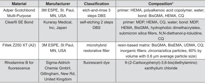
The main materials used in this study are described in Figure 1.
Adhesive labeling with rhodamine B
Rhodamine B (Rhodamine B®, Sigma-Aldrich Chemie GmbH, Gillingham, New Rd, UK) was used according to the manufacturer’s instructions (no
Material Manufacturer &ODVVL¿FDWLRQ Composition*
Adper Scotchbond Multi-Purpose
3M ESPE, St. Paul, MN, USA
etch-and-rinse 3 steps DBS
primer: HEMA, polyalkenoic acid copolymer, water; bond: BisGMA, HEMA, CQ
Kuraray Medical, Inc, Japan
self-etching 2 steps DBS
primer: MDP, HEMA, CQ, water; bond: MDP, HEMA, BisGMA, hydrophobic dimethacrylates,
CQ
Filtek Z250 XT (A2) 3M ESPE, St Paul, MN, USA
microhybrid resin-based matrix: BisGMA, BisEMA, UDMA, CQ;
volume with 0.6 μm average particle size) Rhodamine B for Sigma-Aldrich
Chemie GmbH, Gillingham, New Rd,
United Kingdom
9-(2-Carboxyphenyl)-3,6-bis(diethylamino) xanthylium chloride
*HEMA: 2-Hydroxyethyl methacrylate BisGMA: Bisfenol diglycidyl dimethacrylate CQ: camphorquinone
MDP: 10-methacryloyloxydecyl-dihydrogen phosphate BisEMA:ethoxylated bisphenol-A dimethacrylate UDMA: Urethane dimethacrylate
formula is indicated in Figure 2. Rhodamine B
balance (GR-202, A&D Engineering, Inc., San Jose, CA, USA) inside a small Eppendorf tube. This procedure was performed in duplicate. The two tubes were wrapped in aluminum folium and
was then transferred to the corresponding one. Each tube was carefully adapted to a dental mixer and vigorously mixed for 40 s, in order to dissolve the RB in the resin. After mixing, no RB clusters could be detected in the labeled adhesives with the
experimental DBS was approximately 0.10 mg/mL, just about the same concentration as previously proposed2.
Water sorption (WS) and water solubility (WSL) tests
Ten disk-shaped specimens of each tested adhesive were prepared for the following groups: MP (control adhesive), MP-RB (with 0.10 mg/mL rhodamine), SE (control adhesive), and SE-RB (with 0.10 mg/mL rhodamine). Control and RB-labeled adhesives were directly placed into a stainless steel
with a glass slide8. The experimental groups were light-cured with a light emitting diode curing unit at 1,200 mW/cm2 for 30 s (Radii-cal®, SDI Limited, Bayswater, VIC, Australia). The adhesive disks were subsequently removed from the mold and excess
thickness (h) of each specimen was obtained by measuring three equidistant points on its base with a digital electronic caliper (Mitutoyo Corporation, Tokyo, Japan), and the volume (V) of the specimen was calculated by V=h×(2.5)2×3.14. Water sorption and solubility tests were based on the 4049 ISO standard with the exception of the specimen size.
The adhesive disks were then individually stored in a desiccator (37°C) containing silica gel. Each disk was repeatedly weighted in a calibrated analytical balance (TP-214, Denver Instrument, Denver, CO, USA) in 24-hour intervals, until a constant mass was obtained (m1). Subsequently, the disks were immersed in deionized water in individual vials. During seven days and within 24-hour intervals, the specimens were removed from water, carefully blotted with an absorbent tissue paper, weighted and returned to water until a constant mass was obtained (m2). After this, each specimen was submitted to a new desiccation cycle until a constant mass was obtained (m3). The values of WS and WSL were calculated by equations 1 and 2 respectively:
Also, net water uptake, which represents the sum of water sorption and solubility in percentage, was calculated for each condition.
Microtensile bond strength (μTBS) test and
CLSM of dentin-adhesive interfaces
Extracted sound human third molars, obtained by donation from patients who signed an informed consent beforehand, were included in this study. Ethical protocol was approved by the Ethics Committee for Human Studies (process number 118/2011). The occlusal third of the forty molar crows was cut by a diamond disk (Extec Corp,
machine (Isomet, Buehler Ltd, Lake Bluff, IL, USA),
dentin surfaces were submitted to a water-cooled 600-grit SiC paper abrasion (Buehler Ltd, Lake Bluff, IL, USA) to create standardized surfaces. Then, the specimens were assigned into 4 groups, regarding the bonding protocol as previously described: MP and SE controls (no dye), and RB labeled groups (n=10). WS = (m
2- m3)
V WSL = (m
1- m3)
V
The DBSs were then applied to the dentin surfaces according to the manufacturers’ instructions in Figure 3. Photoactivation was performed for 10 s using the same LED light (Radii-cal®, SDI Limited, Bayswater, VIC, Australia). Composite buildups (3.0 mm in height) were incrementally constructed with a resin composite (FiltekZ250, 3M ESPE, St. Paul, MN, USA). After the bonding procedures, the crowns
parallel to the tooth’s long axis, using the same low-speed saw and diamond disk. At that stage, one slice of each crown from the groups MP-RB and SE-RB was randomly selected to be analyzed via CLSM, using diode laser scanning with a 532 nm laser excitation wavelength (Leica TCS SPE, Leica Microsystems CMS, Mannheim, Germany).
Then, the remaining slices (all groups) were mesiodistally sectioned into rectangular beams with a cross-sectional area of 0.8 mm2 approximately. The dentin-resin specimens were stored in deionized water at 37°C. Half of them were tested after 7-day storage and the other half after 12
to a custom-made testing jig (Bencor Multi T’s like device) with cyanoacrylate glue (Super Bonder Flex Gel Loctite®; Henkel Ltda., São Paulo, SP, Brazil) and subjected to tensile load (50 kgf load cell) at a crosshead speed of 0.5 mm/min until bond failure (Instron, Model 3342, Norwood, MA, USA). In this experiment, bond strength to dentin involved two factors: DBSs (MP or SE) under different conditions (neat adhesives or labelled with RB) and different storage times (7-day or 12 month evaluation), all in two levels.
Failure mode analysis
Fractured dentin-resin interfaces were analyzed
(Dino-Lite Digital Microscope®, AnMo Electronics Corp., New Taipei City, San-Chung District, Taiwan).
(A), mixed failure (M), cohesive failure in resin composite (CC), and cohesive failure in dentin (CD).
Statistical analysis
Data were analyzed with Statistica statistical package 11.0 (Tulsa, OK, USA). The assumptions of equality of variances and normal distribution of errors for all the variables were checked (Kolmogorov-Smirnov). As the assumptions were
WS/WSL and μTBS, and Tukey’s test was carried out for statistical comparisons (D=0.05).
RESULTS
Representative CLSM photomicrographs of dentin-MP and dentin-SE interfaces are shown in Figures 4A and 4B respectively. Adding 0.10 mg/ mL of RB to the adhesive systems tested produced
the laser scanning microscopy. Regarding the interfacial interlocking patterns registered in the photomicrographs, specimens from the group MP-RB presented more and longer resin tags than the ones in the SE-RB group.
The distribution of dye-labeled adhesive throughout demineralized dentin with great resolution and the quality of the hybrid layer imaged was greatly enhanced using the proposed
adhesive layer thicknesses (intense red) are also clearly discernible as well as the characteristics of
produced shorter tags than shown by MP. Also, the
WS and WSL results are shown in Table 1. Rhodamine B caused an increase in WSL for MP and SE. On the other hand, WS of MP or SE was not affected by the addition of the dye. The net water uptake for both adhesives was determined to be similar, irrespective of the presence of the RB or not.
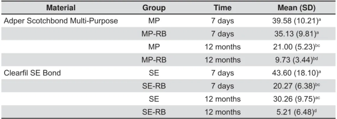
Table 2 presents the results of the μTBS test (in MPa) and comparisons among the experimental groups. By adding RB to Adper Scotchbond
Multi-strength were observed at 7-day analysis, with or without RB associated. After 12 months, no differences between them were found again;
DBS Mode of use
Adper Scotchbond
Multi-Purpose Rinse with water for 15 s;
Remove excess water by blotting with tissue paper;
Apply Adper Scotchbond Multi-Purpose primer to dentin and dry gently for 5 seconds (no waiting); Apply Scotchbond Multi-Purpose adhesive to dentin and light-cure for 10 seconds.
Figure 4- A- Confocal laser scanning microscopy. Dentin-adhesive interface with Adper Scotchbond Multi-Purpose labeled
NA in oil immersion
A B
Water sorption Solubility Net water uptake
(%)** (μg/mm3) (μg/mm3)
MP 90.7 (13.0)a 9.07 14.4 (8.3)b 1.44 10.51
MP-RB 109.8 (5.6)a 10.98 25.9 (9.3)a 2.59 13.57
SE 96.4 (8.5)a 9.64 -7.0(4.0)c -0.70 8.94
SE-RB 96.4 (4.9)a 9.64 19.9 (9.3)a 1.99 11.63
Values are mean (standard deviation), n=10, μg/mm3. Water sorption is given in absolute terms (μg/mm3) and in relative
*90.7 μg/mm3 = 0.0907 mg/mm3 ×100 = 9.07 mg/100 mm3
Table 1- Water sorption (WS) and solubility (WSB) in μg/mm3 of neat and RB-labelled adhesives
Material Group Time Mean (SD)
Adper Scotchbond Multi-Purpose MP 7 days 39.58 (10.21)a
MP-RB 7 days 35.13 (9.81)a
MP 12 months 21.00 (5.23)bc
MP-RB 12 months 9.73 (3.44)bd
SE 7 days 43.60 (18.10)a
SE-RB 7 days 20.27 (6.38)bc
SE 12 months 30.26 (9.75)ac
SE-RB 12 months 5.21 (6.48)d
both groups in comparison with their initial values.
strength to dentin, in both evaluation times. In the DBSs control groups (MP and SE), no difference on bond strength was observed in both testing times. All groups were associated predominantly with adhesive failures, as showed in Figure 5.
DISCUSSION
Dental bonding through hybridization depends primarily on physicochemical interactions between the moist dentin substrate and the resin-based DBSs11,12
polymerization is fundamental for the immediate mechanical performance of the dentin-resin bond interface22,23. Modifying the original composition of
for example, can ultimately interfere with dental bonding and limit the bond integrity analysis by CLSM. According to the present study, the RB concentration of 0.10 mg/mL in the adhesives permitted the detection of the resin distribution in dentin-MP and dentin-SE interfaces by CLSM (Figures 4A and 4B), but it has also negatively
affected some resin properties.
In an attempt to avoid errors in the interpretation of the results, it is relevant to reinforce that rhodamine B did not covalently attach to crosslinking monomers, being inert. During the adhesive polymerization, the dye molecules are entrapped into the polymer network, labeling it. Therefore a previous analysis of degree of conversion (unpublished data) was performed regarding the addition of 0.10 mg/mL of RB, which did not interfere with this property for both the systems.
Dental literature shows that WS/WSL and
μTBS tests are commonly employed to compare characteristics between distinct DBSs, as well as to predict the quality of dental bonding, and even the long-term clinical performance of such resin-based materials6,16. Though RB is not intended for any clinical use under the approach outlined in this paper, the interlocking pattern, which the dye highlights through CLSM observations, is expected to be similar to the bonding patterns normally obtained in the clinical situation – otherwise the morphological assessment could be dubious.
material-dependent.
Control groups of the adhesives MP and SE showed similar WS values. These DBSs pertain to distinct categories (etch-and rinse and
self-systems presenting in separate bottles the same hydrophobic cross-linker resin component, which is known for providing higher polymer stability under wet conditions15. Previous studies have indicated that increasing WS of DBSs may precede degradation processes, which impact the long-term stability of the polymer matrix in wet environments, thus flawing the quality of the dentin-resin interlocking9,11. Based on the present data, RB did not cause any negative effect to WS for any of the tested conditions. However, the addition of RB in the SE adhesive caused an increase in WSL. Based on the analysis of net water uptake, which represents the sum of WS and WSL, it indicates a balanced performance between all tested conditions, which calls for attention to their interpretation. In terms of bond strength, the 3-step etch-and-rinse DBS was less affected from the addition of this dye than the 2-step self-etching one. The elucidation of the
μ
(7-day and 12-month tests) would demand further investigations regarding other polymer properties, and also with the mode of interaction of mild self-etching adhesives with dentin. Their bonding mechanism to dentin relies primarily on the capacity of its self-etching functional monomers to remove minerals of the moist dentin matrix, enabling concomitant resin infiltration and interfacial interlocking10,29. Furthermore, the SE system presents chelating functional monomers in its composition, known for fomenting the occurrence of chemical bonding with residual hydroxyapatite28,30.
if RB can affect the pH of the system SE and impair its self-etching bonding mechanism.
Figures 4A and 4B show a very intense
The concentration of RB seems to be higher than that necessary for a suitable CLSM analysis, and the
micromorphological structures. This can possibly be the reason why the hybrid layer of SE in Figure 4A is not evident. It could be advantageous to investigate some characteristics of RB photophysics, when the dye is dispersed in different cured adhesives.
by a series of factors, such as polarity, viscosity and pH of the microenvironment, and by the 7,20. Therefore,
a preliminary evaluation of the photophysical
based materials could provide valuable information, aiming to determine suitable RB concentrations for
the bond analysis by CLSM.
CONCLUSIONS
for the micromorphologycal analysis, can negatively affect the WSL of both systems and the μTBS of
with their physical-mechanical properties, leading to bias in the bond integrity analysis, especially for overtime bond strength analysis.
ACKNOWLEDGEMENTS
This study was supported by FAPESP - São Paulo Research Foundation (grants 2011/14971-2 and 2012/13160-3). The authors are grateful to Márcia Sirlene Zardin Graeff for her support with
interest regarding the authorship and/or publication of this article.
REFERENCES
1- Breschi L, Mazzoni A, Ruggeri A, Cadenaro M, Di Lenarda R, De Stefano Dorigo E. Dental adhesion review: aging and stability of the bonded interface. Dent Mater. 2008;24:90-101.
2- D'Alpino PH, Pereira JC, Svizero NR, Rueggeberg FA, Pashley
resin-based polymers. J Adhes Dent. 2006;8:285-92.
3- D'Alpino PH, Pereira JC, Svizero NR, Rueggeberg FA, Pashley
based restorations: a literature review. J Dent. 2006;34:623-34. 4- Francisconi LF, Graeff MS, Martins LM, Franco EB, Mondelli RF, Francisconi PA, et al. The effects of occlusal loading on the margins of cervical restorations. J Am Dent Assoc. 2009;140:1275-82.
bonding systems and their handling characteristics on the morphology and micropermeability of the dentine adhesive interface. J Dent. 1999;27:63-71.
6- Ito S, Hoshino T, Iijima M, Tsukamoto N, Pashley DH, Saito T. Water sorption/solubility of self-etching dentin bonding agents. Dent Mater. 2010;26:617-26.
Springer Science+Business Media; 2006. Available from: http:// dx.doi.org/10.1007/978-0-387-46312-4.
8- Malacarne J, Carvalho RM, Goes MF, Svizero N, Pashley DH, Tay FR, et al. Water sorption/solubility of dental adhesive resins. Dent Mater. 2006;22:973-80.
9- Malacarne-Zanon J, Pashley DH, Agee KA, Foulger S, Alves MC, Breschi L, et al. Effects of ethanol addition on the water sorption/ solubility and percent conversion of comonomers in model dental adhesives. Dent Mater. 2009;25:1275-84.
10- Moszner N, Salz U, Zimmermann J. Chemical aspects of self-etching enamel-dentin adhesives: a systematic review. Dent Mater. 2005;21:895-910.
chemical structure on the properties in methacrylate-based dentin adhesives. Dent Mater. 2011;27:1086-93.
13- Peumans M, Kanumilli P, De Munck J, Van Landuyt K, Lambrechts P, Van Meerbeek B. Clinical effectiveness of contemporary adhesives: a systematic review of current clinical trials. Dent Mater. 2005;21:864-81.
14- Sampaio PC, Almeida Júnior AA, Francisconi LF, Casas-Apayco LC, Pereira JC, Wang L, et al. Effect of conventional and
resin-I cavity walls after thermocycling. Oper Dent. 2011;36:403-12. 15- Sauro S, Pashley DH, Mannocci F, Tay FR, Pilecki P, Sherriff M, et al. Micropermeability of current self-etching and etch-and-rinse adhesives bonded to deep dentine: a comparison study using a double-staining/confocal microscopy technique. Eur J Oral Sci. 2008;116:184-93.
16- Scherrer SS, Cesar PF, Swain MV. Direct comparison of the bond strength results of the different test methods: a critical literature review. Dent Mater. 2010;26:e78-93.
17- Spencer P, Ye Q, Park J, Topp EM, Misra A, Marangos O, et al. Adhesive/dentin interface: the weak link in the composite restoration. Ann Biomed Eng. 2010;38:1989-2003.
18- Toledano M, Sauro S, Cabello I, Watson T, Osorio R. A Zn-doped etch-and-rinse adhesive may improve the mechanical properties and the integrity at the bonded-dentin interface. Dent Mater. 2013;29:e142-52.
19- Tsien RY, Ernst L, Waggoner A. Fluorophores for confocal microscopy: photophysics and photochemistry. In: Pawley JB, editor. Handbook of biological confocal microscopy. 3rd ed. New
York: Springer; 2006. p. 338-52.
Weinheim: Wiley-VCH; 2002.
21- Van Landuyt KL, Snauwaert J, De Munck J, Peumans M, Yoshida Y, Poitevin A, et al. Systematic review of the chemical composition of contemporary dental adhesives. Biomaterials. 2007;28:3757-85.
22- Van Meerbeek B, De Munck J, Yoshida Y, Inoue S, Vargas M, Vijay P, et al. Buonocore memorial lecture. Adhesion to enamel and dentin: current status and future challenges. Oper Dent. 2003;28:215-35.
23- Wang Y, Spencer P, Yao X, Brenda B. Effect of solvent content on resin hybridization in wet dentin bonding. J Biomed Mater Res A. 2007;82:975-83.
24- Watson TF. Applications of confocal scanning optical microscopy to dentistry. Br Dent J. 1991;171:287-91.
25- Watson TF. Fact and artefact in confocal microscopy. Adv Dent Res. 1997;11:433-41.
26- Watson TF, Azzopardi A, Etman M, Cheng PC, Sidhu SK. Confocal and multi-photon microscopy of dental hard tissues and biomaterials. Am J Dent. 2000;13(Spec No):19D-24D.
27- Ye Q, Spencer P, Wang Y, Misra A. Relationship of solvent to the photopolymerization process, properties, and structure in model dentin adhesives. J Biomed Mater Res A. 2007;80:342-50. 28- Yoshida Y, Nagakane K, Fukuda R, Nakayama Y, Okazaki M, Shintani H, et al. Comparative study on adhesive performance of functional monomers. J Dent Res. 2004;83:454-8.
29- Yoshida Y, Yoshihara K, Hayakawa S, Nagaoka N, Okihara T, Matsumoto T, et al. HEMA inhibits interfacial nano-layering of the functional monomer MDP. J Dent Res. 2012;91:1060-5.