16 Revista Brasileira de Anestesiologia Vol. 60, No 1, Janeiro-Fevereiro, 2010 LOCKS, ALMEIDA AND PEREIRA
Use of the Ultrasound to Determine the
Level of Lumbar Puncture in Pregnant
Women
Giovani de Figueiredo Locks, TSA, M.D., Maria Cristina Simões de Almeida, TSA, M.D., Amanda Amaro Pereira
INTRODUCTION
Neuroaxis block represents the most common technique in-dicated for cesarean section, since the incidence of maternal morbidity and mortality and neonatal depression is lower than that of general anesthesia1. Among regional anesthetic tech-niques, subarachnoid blocks promote shorter time between induction and delivery and better quality of blockade than epi-dural blockades2. Therefore, subarachnoid blocks have be-come the technique of choice for elective cesarean sections among us.
Traditionally, an imaginary line connecting both iliac crests, called Tuffier’s line, is used to determine the level of the lum-bar puncture. In theory, this line crosses the spine at the level of L4 or L4-L5 space. A puncture below L3-L4 would be below the level of the medullary cone, providing safety to the method. However, it has been demonstrated that anatomical reference has an abnormal distribution and this determination can be inaccurate in a large proportion of patients3. Incorrect determi-nation of the puncture level is a known risk factor for medullary cone injury in spinal blocks. The American Society of Regio-nal Anesthesia recommends that anesthesiologists should be aware of the limitations of the physical exam to determine the puncture level, especially in patients with difficult topographic anatomy. Pregnant and obese patients are included among those4. It has been demonstrated that obesity during preg-nancy increases the incidence of maternal-fetal complications and the prevalence of cesarean sections5.
The use of the ultrasound in anesthesiology has been increa-sing, and its use has been proposed to determine the punctu-re level for spinal blocks6.
The objective of this study was to determine whether identifi-cation of the L3-L4 space by the physical exam differs from that of the ultrasound in obese and non-obese pregnant women.
METHODS
USE OF THE ULTRASOUND TO DETERMINE THE LEVEL OF LUMBAR PUNCTURE IN PREGNANT WOMEN
Revista Brasileira de Anestesiologia 17
Vol. 60, No 1, Janeiro-Fevereiro, 2010
abnormal spinal anatomy, or hypertension), and patients un-dergoing urgent or emergency cesarean sections.
Patients were monitored with pulse oximeter, non-invasive blood pressure, and cardioscope on DII derivation, and ve-nous access was established on an upper limb.
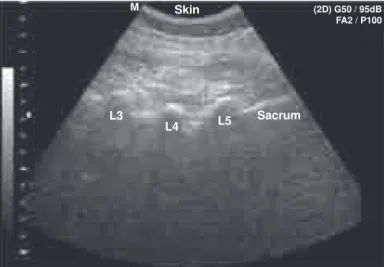
With the patient in the sitting position, an anesthesiologist with more than five years of experience in obstetric anesthesia in-dentified the L3-L4 space using anatomical references, i.e., the level that an imaginary line connecting both upper iliac crests crossed the spine identified L4 or the L4-L5 space. This was followed by lumbar ultrasound with a SonoAce 8000 EX Pri-me (Medison Co., Seoul, South Korea). A convex transducer of 2-5 MHz was used. The transducer was initially placed on the sacral region on a longitudinal paramedian presentation, 2 to 3 cm from the midline, angled to the center of the spinal canal. The sacrum was identified as a continuous hyperechoic line. The transducer was directed cranially, identifying the spi-nous processes of the lumbar vertebrae as a saw-like image in which the teeth of the saw represent the processes and the valleys correspond to the intervertebral spaces (Figure 1)7. Af-ter ultrasonographic identification of the inAf-tervertebral spaces, the level of the clinically estimated L3-L4 space was recorded. Lumbar puncture was performed on the L3-L4 space, identified by ultrasound using the median approach, with a 26G Quin-cke needle; 10 mg of 0.5% hyperbaric bupivacaine associated with 80 µg of morphine were administered, and the anesthesia followed the protocol of the institution.
The degree of obesity was determined according to the clas-sification of the Body Mass Index (BMI) of the World Health Organization, which is adopted by the American College of Obstetrics for pregnant patients.
Patients’ data were analyzed in two groups: Group 1 (G1), non-obese patients with BMI lower than 30 kg.m-2, and Group 2 (G2), obese patients with BMI equal or greater than 30 kg.m-2.
To detect a 27% difference in the accuracy of the clinically-estimated puncture levels between obese and non-obese
patients, with proportion analysis test – accepting a 5% alpha error and 20% beta error – the size of de study population was calculated as 40 patients per group. We considered a prior study8 that estimated the percentage of the correct clini-cal identification of the puncture level around 40%. Data were stored in a data bank of the Microsoft Office Excel v. 7.0 (Se-attle, 2003). Afterwards, the Analysis – Epi Info v 3.3.2 softwa-re (CDC 2005) was used for statistical analysis. The level of statistical significance was 95%.
Data are presented as mean ± standard deviation or absolute frequency (percentage). The Chi-square test was used to de-termine the intergroup association among qualitative parame-ters. The Student t test was used for intragroup differences in quantitative parameters.
RESULTS
From August 2008 to April 2009, 90 patients were included in the study, 43 patients in G1 (non-obese) and 47 in G2 (obe-se). Table I shows the demographic data.
Lumbar intervertebral spaces were identified and counted by ultrasound in all patients. Table II shows the intervertebral spaces identified as L3-L4 on physical exam and the corres-ponding space on ultrasound. A wide variation, without statis-tically significant differences, was observed.
Temporary or permanent neurologic symptoms related with the spinal block were not observed.
Figure 1 – Longitudinal ultrasound of the lumbosacral region identi-fying the upper portion of the sacrum and spinous processes of the lumbar vertebrae.
M Skin
L3
L4 L5 Sacrum
(2D) G50 / 95dB FA2 / P100
Table I – Demographic Data
Group 1 non-obese
Group 2 obese
P
Age (years) 28.16 ± 7.15 29.60 ± 6.24 0.313 BMI (kg.m-2) 26.58 ± 2.08 34.39 ± 3.68 < 0.001
Gestational age (weeks) 39.40 ± 1.31 39.11 ± 1.45 0.325
BMI – Body mass index; Student t test.
Table II – Ultrasound Determination of the Intervertebral Space Clinically Identified as L3-L4
Group 1 non-obese
Group 2 obese
P
L1-L2 3 (7%) 2 (4%) 0.186
L2-L3 14 (33%) 22 (47%)
L3-L4 23 (53%) 23 (49%)
L4-L5 3 (7%) 0 (0%)
Chi-square test.
DISCUSSION
18 Revista Brasileira de Anestesiologia Vol. 60, No 1, Janeiro-Fevereiro, 2010
LOCKS, ALMEIDA AND PEREIRA
only on topographic anatomy the level of accuracy of anesthe-siologists to identify lumbar intervertebral spaces ranges from 29% to 41%3,9,10. Mistaken identifications are usually in the cephalad direction, and the difference can be up to four spa-ces9. Those differences can be explained by two reasons. First, most anthropometric parameters have a normal distri-bution, i.e., a central peak that becomes progressively smal-ler the farther it is from the mean. This is valid for Tuffier’s line and it has been demonstrated radiologically and in cada-vers that this line can vary from the L5-S1 to L3-L411. Second, the position of the patient during the physical exam and X-ray or MRI differs, and most imaging exams are performed with the patient in the supine position. It has been suggested that the L4-L5 distance can increase up to 1 cm when the patient is bent12.
In 2002, Furness demonstrated that the ultrasound could be used to identify the intervertebral spaces13. He used the ul-trasound image in the midline to identify the sacrum and spi-nous processes of the lumbar vertebrae and compared 45 estimates of the intervertebral space. An 84% accuracy in the ultrasound identification against only 37% by palpation was identified.
More recently, two retrospective studies8,14 compared physi-cal exam and ultrasound in obstetric patients. Both evaluated patients in the puerperium, using the epidural or subarach-noid puncture marks for cesarean section or labor analgesia. The authors reported concordance rates of 55.0% and 55% between the vertebral spaces attributed by the anesthesio-logist and that of the ultrasound. Once more, the error of the anesthesiologists varied from one to two cephalad segments. None of the authors observed differences in the correct identi-fication of the intervertebral space when the weight of the pa-tients was analyzed. Those studies can be criticized because post-partum changes in the body of pregnant patients can be seen, therefore explaining possible differences between the blockade and the ultrasound.
This is the difference between the present study and pre-vious studies: performing the ultrasound before the blocka-de and the blocka-delivery and both in the same position. Although concordance rates similar to those reported in the literature were observed – 53% in G1 and 49% in G2 – punctures were performed in the L3-L4 space determined by ultrasound. Our study also analyzed patients in two groups according to the BMI, and the lack of differences between both groups in the concordance rate of the identification of the L3-L4 space were not surprising.
The position of the medullary cone also follows normal distri-bution. In one study that analyzed 635 MRI images, the mean level of the medullary cone was identified at the mid-third of L1, but it varied from the mid-third of T12 to the upper third of L3. This study also observed gender- and age-related diffe-rences in the anatomy of the medullary cone. The level of the medullary cone was lower in females and in the elderly15. An important study by Reynolds16 reported a series of seven cases of neurological damage in spinal or combine block. All patients were females, and six were obstetric cases. Pencil-tip needles were used in all patients and the anesthesiologist
believed he was puncturing the L2-L3 space. Magnetic reso-nance imaging demonstrated spinal cord lesion at the level of T12, in five patients, and L1 in one. It also demonstrated that none of the patients had an abnormally long medullary cone. Patients complained of paresthesia during the blockade, but only one complained of pain during the administration of the anesthetic.
The mechanism proposed for the lesion was direct needle trauma to the medullary cone. It was also suggested that, be-sides the mistaken identification of the intervertebral space, non-traumatic needles were associated with higher probability of neurologic damage, because one millimeter of the needle penetrates in the subarachnoid space before the backflow of CSF. Those data reinforce the idea that the L2-L3 space is not adequate for lumbar puncture.
However, even the correct identification of the L3-L4 space does not exclude the risk of neurologic damage of the medullary cone. The case of a patient who developed “dropped foot” after suba-rachnoid block in L3-L4 for cesarean section17 has been reported. Lumbar MRI showed that the medullary cone of that patient re-ached the level of L4. Symptoms of medullary cone and cauda equina lesions include lumbar pain, pain in the lower limbs, pa-resthesia, and sphincter dysfunction. The incidence of neurologic lesions after subarachnoid block has been estimated in six out of every 10,000 anesthetic procedures, with permanent damage in less than one in 10,00018.
Limitations for the use of the ultrasound include learning the ultrasonographic anatomy, cost, time for execution, and te-chnical limitations, since it is an operator-dependent exam. However, it has been proposed that with increased clinical experience many of these disadvantages can be overcome and it will be possible to increase the reliability of the method7. Note that the ultrasound is not the gold standard for the deter-mination of the level of intervertebral puncture and identifica-tion errors can still be made. For this reason, current care with paresthesia and pain during neuroaxis blocks are still valid. Summarizing, the rate of the correct identification of the L3-L4 space in pregnant women by clinical evaluation is low, both in obese and non-obese pregnant women. Spinal ultrasound before the blockade can reduce the rate of mistaken identi-fication of the L3-L4 space in both groups. Further studies to determine whether the use of the ultrasound to determine the level of the lumbar puncture in pregnant women decreases the risk of medullary cone damage during subarachnoid block are necessary.
REFERÊNCIAS – REFERENCES
1. Tsen LC. Anesthesia for cesarean delivery. ASA Refresher Courses Anesthesiol 2005;33:235-245.
2. American Society of AnesthesiologistsTask Force on Obstetric Anes-thesia. Practice guidelines for obstetric anesthesia: an update report. Anesthesiology 2007;106:843-2863.
3. Van Gessel EF, Forster A, Gamulin Z. Continuous spinal anesthesia: where do spinal catheters go? Anesth Analg 1993;76:1004-1007. 4. Neal JM, Bernards CM, Hadzic A et al- ASRA Practice advisory on
USE OF THE ULTRASOUND TO DETERMINE THE LEVEL OF LUMBAR PUNCTURE IN PREGNANT WOMEN
Revista Brasileira de Anestesiologia 19
Vol. 60, No 1, Janeiro-Fevereiro, 2010
5. Baeten JM, Bukusi EA, Lambe M. Pregnancy complications and ou-tcomes among overweight and obese nulliparous women. Am J Public Health 2001;91:436-440.
6. Watson MJ, Evans S, Thorp JM. Could ultrasonography be used by an anaesthetist to identify a specified lumbar interspace before spinal anaesthesia? Br J Anaesth 2003;90:509-511.
7. Carvalho JCA. Ultrasound-facilitated epidurals and spinals in obste-trics. Anesthesiology Clin 2008;26:145-158.
8. Schlotterbeck H, Schaeffer R, Dow WA et al. Ultrasonographic control of the puncture level for lumbar neuraxial block in obstetric anaesthe-sia. Br J Anaesth 2008;100:230-234.
9. Broadbent CR, Maxwell WB, Ferrie R et al. Ability of anaesthetists to identify a marked lumbar interspace. Anaesthesia 2000;55:1122-1126. 10. Ievins FA. Accuracy of placement of extradural needles in the L3-4 in-terspace: comparison of two methods of identifying L4. Brit J Anaesth 1991;66:381-382.
11. Hogan QN. Tuffier’s line: the normal distribution of anatomic parame-ters. Anesth Analg 1994;78:194-195.
12. Thavasothy M. The reproducibility of the iliac crest as a marker of lumbar spinal level. Anaesthesia 1997;52:811.
13. Furness G, Reilly MP, Kuchi S. An evaluation of ultrasound ima-ging for identification of lumbar intervertebral level. Anaesthesia 2002;57:277-280.
14. Whitty R, Moore M, Macarthur A. Identification of the lumbar in-terspinous spaces: palpation versus ultrasound. Anesth Analg 2008;106:538-540.
15. Soleiman J, Demaerel P, Rocher S et al. Magnetic resonance imaging study of the level of termination of the conus medullaris and the thecal sac: influence of age and gender. Spine 2005;30:1875-1880. 16. Reynolds F. Damage to the conus medullaris following spinal
anaes-thesia. Anaesthesia 2001;56:238-247.
17. Ahmad FU, Pandey P, Sharma BS et al. Foot drop after spinal anes-thesia in a patient with a low-lying cord. Int J Obstet Anesth 2006; 15:233-236.
18. Auroy Y, Narchi P, Messiah A et al. Serious complications related to regional anesthesia: results of a prospective survey in France. Anes-thesiology 1997;87:479-486.
RESUMEN
Locks GF, Almeida MCS, Pereira AA – Uso del Ultrasonido para De-terminación del Nivel de Punción Lumbar en Embarazadas.
JUSTIFICATIVA Y OBJETIVOS: Para determinar el nivel vertebral para la punción lumbar, se utiliza una línea imaginaria que une las crestas ilíacas. Esa línea cruza la columna vertebral en el nivel L4
o en el espacio L4-L5. Esa referencia anatómica puede ser inexacta
en una gran proporción de pacientes. Este estudio quiso determinar si existe alguna diferencia en la determinación del espacio vertebral L3-L4 por el examen físico cuando se le comparó al ultrasonido en
embarazadas obesas y no obesas.
MÉTODO: Se estudiaron pacientes adultas sometidas a la cesárea electiva bajo raquianestesia. Las pacientes se analizaron en dos grupos: obesas y no obesas. Con la paciente en la posición sentada, se deter-minó a través del examen físico, el espacio vertebral L3-L4. Enseguida
se realizó el ultrasonido de la región lumbar. Se identificó el hueso sacro y el transductor fue colocado en dirección craneal para identificar los procesos espinosos de las vértebras lumbares. Quedó registrado el nivel vertebral que había sido estimado clínicamente como L3-L4.
RESULTADOS: Se incluyeron 90 pacientes, siendo 43 no obesas y 47 obesas. En todas las pacientes se pudieron determinar los es-pacios vertebrales lumbares a través del ultrasonido. Los eses-pacios vertebrales identificados como L3-L4 en el examen físico,
correspon-dieron a los estimados como L3-L4 por el ultrasonido en un 53 y un
49% en los grupos de no obesas y obesas, respectivamente. No hubo diferencia significativa entre los grupos.
CONCLUSIONES: El porcentaje de acierto en la identificación del espa-cio vertebral L3-L4 en embarazadas obesas y no obesas es bajo. El