Videofluoroscopy chair for the study of swallowing
and related disorders*
Cadeira especial para o estudo videofluoroscópico da deglutição e suas disfunções
Milton Melciades Barbosa Costa1, Lucia Viviana Canevaro2, Hilton Augusto Koch3, Ricardo DeBonis4
OBJECTIVE: To configure an auxiliary equipment to allow videofluoroscopic study of swallowing and related disorders without the patients’ active collaboration in obtaining the required positions, with reproducibility of views, postures and maneuvers needed for the examination, in spite of limitations imposed by associated diseases. MATERIALS AND METHODS: A Philips BV-22 C-Arm was utilized, with adaptation implemented to allow simultaneous images recording on both digital and analog media. Primary and secondary chairs were specially designed, built and attached to the equipment. RESULTS: Adults and children could be appropriately seated and all necessary radiographic views could be obtained without active positioning or cooperation from the volunteers. Additionally, a calibration system was developed, allowing the input of dimensional parameters for quantification of recorded phenomena. CONCLUSION: The videofluoroscopy utilization spectrum could be extended to agitated patients, and those with behavior similar to patients affected by neuromotor disorders, with greater effectiveness and consequently with lower radiation exposure time.
Keywords: Videofluoroscopy; Swallowing; Dysphagia; Neuromotor disorder; Adapted chair.
OBJETIVO: Configurar equipamento que permita avaliação videofluoroscópica da deglutição e suas desor-dens sem a necessidade de colaboração dos indivíduos na obtenção das posições requeridas, com reprodu-tibilidade das incidências, posturas e manobras necessárias ao exame, independente das limitações impos-tas pelas doenças associadas. MATERIAIS E MÉTODOS: Utilizamos como base um arco em C Philips BV-22. Implementamos adaptações que permitem o registro simultâneo das imagens, em mídia analógica e digital. Cadeiras, principal e secundária, acopladas ao equipamento radiológico foram desenvolvidas. RESULTADOS: Foi possível acomodar adultos e crianças, obtendo-se todas as incidências radiológicas necessárias sem que os voluntários tivessem que se posicionar de modo ativo. Em adição, desenvolvemos sistema de calibração que permite a inserção de parâmetros dimensionais, tornando possível a quantificação dos fenômenos regis-trados. CONCLUSÃO: Pudemos ampliar o espectro de utilização da videofluoroscopia a indivíduos inquietos e de comportamento semelhante a lesionados neuromotores, com maior efetividade e consequente necessi-dade de menor tempo de exposição à radiação.
Unitermos: Videofluoroscopia; Deglutição; Disfagia; Distúrbio neuromotor; Cadeira especial. Abstract
Resumo
* Study developed at Instituto de Ciências Biomédicas and Department of Radiology –Faculdade de Medicina da Universi-dade Federal do Rio de Janeiro (UFRJ), Rio de Janeiro, RJ, Bra-zil. Financial support: Fundação Educacional Charles Darwin.
1. PhD, Titular Professor, Head of the Laboratory of Digestive Motility and Imaging of Instituto de Ciências Biomédicas – Uni-versidade Federal do Rio de Janeiro (UFRJ), Rio de Janeiro, RJ, Brazil.
2. PhD, Physicist at Instituto de Radiodosimetria, Rio de Ja-neiro, RJ, Brazil.
3. PhD, Titular Professor at Department of Radiology – Facul-dade de Medicina da UniversiFacul-dade Federal do Rio de Janeiro (UFRJ) and Santa Casa de Misericórdia do Rio de Janeiro, Rio de Janeiro, RJ, Brazil.
4. Master, Odontologist, Fellow PhD degree at Laboratory of Digestive Motility and Imaging of Instituto de Ciências Biomédi-cas – Universidade Federal do Rio de Janeiro (UFRJ), Rio de Janeiro, RJ, Brazil.
Mailing address: Dr. Milton M.B. Costa. Laboratório de Moti-lidade Digestiva e Imagem, Departamento de Anatomia – ICB-CCS-UFRJ, Ilha do Fundão. Rio de Janeiro, RJ, Brazil, 21941-590. E-mail: [email protected].
Received November 11, 2008. Accepted after revision April 15, 2009.
positioning and freedom of movements for an appropriate study of the swallowing phases both in healthy and dysphagic indi-viduals. Such difficulties are many times aggravated by the frequent association of dysphagia with motor/postural impairment of neurological origin(1–4).
Studies in children with dysphagia, whether of neurological origin or not, are particularly difficult because their natural restlessness associated with the limitations inherent to the available equipment. Inap-propriate equipment configuration for swallowing studies, makes it very difficult to properly position children during the study, and more often than not, they end up being overexposed to radiation at the end Costa MMB, Canevaro LV, Koch HA, DeBonis R. Videofluoroscopy chair for the study of swallowing and related disorders. Radiol Bras. 2009;42(3):179–184.
INTRODUCTION
of a longer than actually necessary exami-nation time. These circumstances can be aggravated by the need for assistance to help in keeping the child properly posi-tioned and to administer the contrast me-dium necessary for the study.
The videofluoroscopic study of swal-lowing must be performed with the patient in the nearest possible position normally adopted to eat(5). Such position should not
be affected by limitations imposed by the equipment. Obtaining a favorable position should always be attempted, particularly to overcome the postural inadequacies gener-ated by the baseline disease.
The limitations that shall be accepted, when all overcoming alternatives have been attempted are those inherent to the patient and his/her disease. Limitations resulting from technical difficulties should not be added to neither confused with the patients’ limitations.
As videofluoroscopy allows noninva-sive, appropriate and clear analysis of the oral, pharyngeal and esophageal phases, it is considered as the method of choice for the evaluation of swallowing disorders(6– 11), and it actually is, on account of its
ef-fectiveness, the best among the currently available methods(12–16).
However, a further reduction in radia-tion exposure and the inclusion of imaging sequences parameters capable of allowing quantitative analyses still unavailable, rep-resent a significant, necessary, and feasible goal in principle, considering the current technological developments. These are steps that would further enhance the con-cept of videofluoroscopy as a tool of excel-lence for the study of the swallowing physi-ology as well as of the physiopathphysi-ology of its related disorders.
The main objective of the present study was the configuration of a radiological equipment to allow the videofluoroscopic evaluation of swallowing and its dysfunc-tions with the patient appropriately posi-tioned and with the possibility of easily and comfortably reproducing incidences, pos-tures and maneuvers, independently of limitations imposed by the disease, making it possible to reduce the examination time with consequent reduction of radiation exposure. A second objective of the present study is the development of possibilities for
the determination of dimensional varia-tions inherent to the generation of radio-logical images, by creating a calibration method to allow the recovery of original dimensions of objects from analyses of the images, making it possible to quantify di-mensions and movements of structures in-volved in swallowing.
MATERIALS AND METHODS
A Philips BV-22 C-Arm model was uti-lized with a 100 kVp x-ray tube and 20 mA maximum intensity, total nominal filtration of 2.5 mmAl equivalent and 2.4 mm focal point with a fixed anode. The available free space between the end of the x-ray tube cone and the image intensifier was 60 cm. The black-and-white video system is based on the NTSC standard, and is com-prised of a Mythos B/W Sony 0.1 lux, f = 3.6 mm minicamera, with 400-line resolu-tion, and a black-and-white 20 inch Philips monitor. Recording resources were added to this video system, allowing simultaneous image capture and analog (Panasonic NV-MV 40 video recorder) or digital (Philips DVDR 3455H DVD recorder) recording. These recorders were connected in series, with control image displayed on a Pana-sonic CT-1383VY color video monitor.
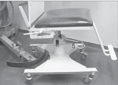
A chair was specially built to be utilized in association with the C-arm for video-fluoroscopic study of the three swallowing phases (oral, pharyngeal and esophageal) in such a manner that the examinations could be performed without active participation of individuals in the movements required for obtaining the radiological incidences (Figure 1).
An accessory chair designed to be fit over the first one was also built to accom-modate children, particularly those requir-ing a caregiver assistance durrequir-ing examina-tion (Figure 2)
The main chair can be fitted in the space between the x-ray tube and the image in-tensifier, allowing the elevation or lower-ing of the seat plane and 360° rotation around its axis, without interfering with the structure of the x-ray equipment. The chair can be removed from the examination po-sition to accommodate a physically dis-abled patient and then be placed back in the original centralized position by means of
Figure 1. On 1, base of the C-arm. On 2, chair with hydraulic elevation, lowering and rotation mechanism to be fitted as an accessory of the ra-diological equipment.
reciprocal converging, concave and convex surface fittings installed on the chair basis and adjusted to the C-arm basis.
In order not to limit the movement of the chair in relation to the equipment to a cen-tralized position, the convex limiter at-tached to the equipment can be removed, allowing the free movement of the chair in the space between the x-ray tube and the image intensifier.
The rolling and lockable supports of the main chair comprise a hydraulic mecha-nism that allows the 360° rotation move-ment as well as a safe 15 cm range of ver-tical movement of the seat plane (Figure 3). A backrest is attached to the wide pad-ded seat, with three segments of decreas-ing widths from the bottom to the top, al-lowing the patient to be rested against it, with freedom of movement in the free space between the x-ray tube and the im-age intensifier.
The radio-transparent backrest of the main chair is articulated to the seat by a double metallic rack system which is posi-tioned outside (below) the exposure field, providing backrest inclination movement from 90° (vertical) to 10° (close to horizon-tal) (Figure 4).
A slot hole at the center of the upper contour of the backrest of the main chair, above the area of radiological exposure, provides attachment means for accessories. The accessory chair for children fits over the main chair. When in place, the acces-sory chair seat forms an angle of 30° in relation to the frontal plane. Such angle provides a safe positioning of the child during the examination, and is comple-mented by an additional safety (abdominal) belt.
The same fitting point used to attach the accessory chair is also used to attach the calibration plate which is assembled in an articulated T-shaped base that enables its movement in frontal and sagittal planes. The 25 × 25 cm plate has a metallic alloy line grid, forming 2 × 2 cm square cells, vertically and horizontally numbered from the center. This plate is adjusted between 0.5 mm thick acrylic plates, to avoid plane distortion. A 10 cm-diameter opening at the center of one of the acrylic plates generates a circular area of lower density for analy-sis of the primary beam centralization.
The x-ray tube cone kept in its original dimensions, was fitted with a receptacle for the insertion of a PTW ionization chamber which is connected to a Diamentor M2 electrometer that measures the kerma-area product (PKA – unit defined as the product
of radiation quantity by the irradiated area)(17,18).
A punctual light source attached to the x-ray tube marks the point of incidence of
the central ray of the primary beam. A re-tractable metric scale laterally attached to the image intensifier allows the measure-ment of the object-intensifier distance, quite useful in the quantification of the recorded images magnifications (Figure 5).
The calibration plate, x-ray tube and the C-arm support base were equipped with spirit levels to allow the correct leveling of the individual parts and also of the whole set up. In order to compensate for eventual floor surface irregularities, the C-arm sup-port base was equipped with leveling feet with threaded inserts.
For the definition of angles generated by oblique incidences, an acrylic protrac-tor was designed and built, which attaches to the slot on the upper part of the main chair backrest, allowing the definition, from the lateral incidences, of the angle between the backrest parallel plane and the median sagittal plane of the patient, with the definition of the number of degrees produced by the several oblique incidences (Figure 6).
The main chair was tested with 20 male adult, healthy volunteers with different bio-types, including wind musicians on whom the possible presence of pharyngeal stress signs is studied. The protocol for the present study was previously approved by the Committee for Ethic in Research of the Universidade Federal do Rio de Janeiro, and all volunteers signed a term of free and informed consent. The accessory chair was tested without x-ray emission, with five children with up to 5 years of age.
Figure 4. Double rack system, in which, with (1) angles between 90° and 60° can be achieved, and with (2) attachment under spring pressure, angles between 60° and 10° can be achieved.
nimble and easy. Furthermore, depending upon particular needs of the child, an as-sistant can help during the study, position-ing himself posteriorly to the segment of the C-arm that holds the x-ray tube.
DISCUSSION
The videofluoroscopic method, with different designations(20–26), is considered
and accepted as the most appropriate method for diagnosis and follow-up of most of dysphagic processes(12–15).
How-ever, and quite often, the method is criti-cized due to the fact of using x-radiation as the image-generating element(9) and, with
the recognition of its qualitative potential it is also desirable that the method provides a quantification of the investigated events. Radiation doses required for the inves-tigation of swallowing dynamics can be considered as being low as compared with those necessary for the performance of the majority of radiological studies accepted and recognized as important, valid and in use in the clinical practice(26). The biased
concept of high doses certainly comes from non-intensified (dark room) fluoroscopy, where the necessary doses to achieve qual-ity images was high indeed. The addition of monitors to the conventional fluoros-copy process, with the use of a camera and RESULTS
Adult individuals seated on the adapted chair could be mobilized for the required incidences — anteroposterior, postero-an-terior, right- and left-lateral, antero-oblique and posterior-oblique, derived from each profile — without having to cooperate in an active manner to obtain the necessary positioning.
The distance between a plane of inter-est of an exposed individual and the image intensifier may be defined both for frontal and lateral incidences, by means of the metric scale attached to the image intensi-fier. Once the distance between the desired plane and the intensifier is known, the cali-bration plate is placed on its T-shaped fenestered base, so its grid pattern (with known measures) is registered in an analo-gous plane to the known one to be occupied by the study object. The dimensional varia-tions related to the plate and its image al-lows the quantification of dimensional variations occurring in the object. The di-mensional variations of the plate are simi-lar to those of the exposed object. The im-ages of the plate with the hypotransparent central area also serve the purpose of de-tecting possible distortions and irregulari-ties, even the subtle ones, in the peripheral zone of the obtained images. In the system
adopted in the present study, the measured distortion was in the order of 3% (maxi-mum tolerance: 10%, referenced on the Order (Portaria) 453)(19).
The reciprocal fitting (chair/equipment) allowed its removal and repositioning in the previous centralized position. The free horizontal movement of the chair after re-moval of the reciprocal fitting has a range of 30 cm. Its median sagittal plane can be moved 15 cm closer to the surface of the image intensifier, generating images with smaller distortions and dimensions closer to the actual size of the objects.
The free vertical displacement required for the observation of structures involved in the three phases of swallowing, is achieved by the hydraulic mechanism of the chair. Such displacement range can be increased by associating the hydraulic movement of the chair with the electrically provided vertical movement of the x-ray tube-intensifier structure.
The punctual light source attached to the equipment allowed the correct localiza-tion of the areas to be irradiated, thus avoid-ing the need for additional radiation expo-sure.
The accommodation of children on the accessory chair proved to be effective, and the mobility of the system, simulating sev-eral incidences, proved to be extremely
Figure 5. On 1, PTW ionization chamber placed in a housing that reconstitu-tes the x-ray tube cone. An arrow on 1 indicareconstitu-tes punctual luminous beam ori-ginating from a light source attached to the x-ray tube. On 2, x-ray tube. On 3, the black and white monitor. On 4, slots on upper part of the chair backrest that provides attachment points for accessories. On 5, image intensifier. On 6, calibration plate, with arrow indicating the incidence of the punctual luminous beam on the calibration plate. On 7, backrest of the main chair.
an image intensifier, resulted in a signifi-cant reduction in the radiation exposure, both for the patient and the radiologist. The image analysis that previously required high radiation doses and slow adaptation to a totally dark room, has been replaced by the analysis on a monitor screen in a nor-mally lit room, with much lower doses than those previously required(26–31).
However, videofluoroscopy can be per-formed with even lower doses. Usually, unnecessary exposure ends up happening due to technical difficulties in comply with an evaluation protocol utilizing inappropri-ate, improvised radiological equipment to perform the study. Additionally, there are those difficulties found in the evaluation of children and also those specific to patients with motor deficits associated with swal-lowing disorders.
Technical difficulties in the examination increase the exposure times, limit the infor-mation that can be obtained and cause rep-etitions that produce new and undesirable exposures. Furthermore, an inappropriate positioning of the patient may negatively interfere, generating additional difficulties in swallowing or enhancing the existing ones, causing difficulties imposed by the precarious structural condition to be erro-neously perceived as diseases. This fact is particularly relevant for children and pa-tients with motor deficits.
An appropriate evaluation of the swal-lowing dynamics and related disorders comprise the study of oral, pharyngeal and esophageal phases requiring a protocol in-volving the evaluation of several regions with different densities and incidences that cannot be neglected with the sole intent of reducing radiation exposure time. It is pos-sible to perform a swift exam, thus reduc-ing exposure times, with patients capable of properly positioning themselves, under-standing and acting according to requests necessary for compliance with the protocol of an appropriate investigation of the swal-lowing phases. However, children, patients with motor deficits and those with a low cooperation capacity end up requiring ad-ditional exposure times, which should al-ways be kept at the minimum necessary levels.
The development of adaptations with the objective of providing the radiological
equipment with specificity for the video-fluoroscopic study of swallowing is cer-tainly the correct way to reduce or even eliminate technical restrictions, with con-sequent lower radiation exposure for both the patient and the investigator.
This solution was first proposed by Cox & Petty(32), with the development of a
spe-cial chair to be utilized with patients with severe neuromotor diseases. The chair pro-posed by the present study is more com-plete, incorporating an accessory chair for evaluation of children, and other resources such as elements that allow the inclusion, in the images, of parameters for the desir-able real time quantification of the phe-nomena and linear measurement of dimen-sional variations of the recorded structures. Based on the results of the present study, the developed chair in association with the C-arm, will effectively meet the position-ing needs, particularly for those patients incapable of active participation and coop-eration, whether adults or children, allow-ing them to be positioned and evaluated with freedom in the different necessary radiological incidences that so far were inaccurate and difficult to obtain. Besides benefiting patients that could not undergo the procedure due to the severity of their motor deficits, the implementation of the present project will facilitate the study pro-tocols accomplishment, reducing the re-quired time for their performance and the final radiation dose employed.
The quantification of events in the pha-ryngeal and esophageal phases has been a focus of radiological methods(33,34). The
utilization of this chair and the current stage of videofluoroscopy will allow the selec-tion and quantificaselec-tion of the phenomena whose measurement adds information to the qualitative data allowed by videofluor-oscopic evaluation(35,36).
Videofluoroscopy, in principle defined as a method capable of recording the swal-lowing dynamic on magnetic tapes (VHS), has progressed to a method that can be defined as capable of real-time recording on magnetic media of the swallowing dy-namics images. The standardization of the number of images in a given unit of time, the advent of digitalization and the possi-bility of insertion in the image of an ele-ment of known measures to be compared
with the structure to be measured, make the quantification of dimension and time varia-tions, an easy task.
With the chair adapted to the C-arm, it is possible, with the use of the calibration plate, to establish the produced magnifica-tion at every point, between the x-ray tube and the image intensifier, and compare the dimensional variations of this pattern with the variations occurring in the object under study, positioned in the same localization between the x-ray tube and the intensifier. It is also easy to measure the duration of an event recorded in its dynamics. The time interval recorded on analog or digital me-dia has a known number of frames per unit of time (second). In the NTSC-M system, the most frequently utilized in videofluor-oscopy, the number of framesper second is 29.97, with each framelasting 33 thou-sandths of a second. Thus, by counting the number of frames in an event and multiply-ing this number by 0.033, the duration of the event in seconds is determined.
The permanent control of the distance between the object under study and the im-age intensifier, and the knowledge of the magnification values of the calibration plate provided by the chair at any point of the space between the x-ray tube and the image intensifier, at any of the radiologi-cal incidences, allows the radiologi-calculation, by analogy, of the dimensional magnifica-tions. By knowing the number of frames recorded in a unit of time, the duration in seconds of a dynamic event can be easily determined provided it is possible to iden-tify in the sequence of images, the fames depicting the beginning and the end of the phenomenon.
CONCLUSIONS
The main and accessory chairs coupled to the radiological equipment were capable of accommodating and allowing all re-quired radiological incidences without the active participation of individuals, whether adults or children, as it would happen with neuromotor impaired patients with exten-sive limitations. This fact not only increases the range of application of videofluoro-scopy, but also makes it more effective, with shorter examination times and, con-sequently, lower radiation exposure.
The inclusion of a calibration plate, with the possibility of quantification, in-creases the excellence of the method which is considered as the one with greatest quali-tative effectiveness for allowing the noninvasive morphofunctional analysis of the three phases of swallowing.
REFERENCES
1. Lawrence ES, Coshall C, Dundas R, et al. Esti-mates of the prevalence of acute stroke impair-ments and disability in a multiethnic population. Stroke. 2001;32:1279–84.
2. Aviv JE, Sacco RL, Thomson J, et al. Silent laryn-gopharyngeal sensory deficits after stroke. Ann Otol Rhinol Laryngol. 1997;106:87–93.
3. Daniels SK, Brailey K, Priestly DH, et al. Aspi-ration in patients with acute stroke. Arch Phys Med Rehabil. 1998;79:14–9.
4. Ramsey DJC, Smithard DG, Kalra L. Early assess-ments of dysphagia and aspiration risk in acute stroke patients. Stroke. 2003;34:1252–7.
5. Costa MMB. Uso de bolo contrastado sólido, lí-quido e pastoso no estudo videofluoroscópico da dinâmica da deglutição. Radiol Bras. 1996;29: 35–9.
6. Beck TJ, Gayler BW. Image quality and radiation levels in videofluoroscopy for swallowing stud-ies: a review. Dysphagia. 1990;5:118–28. 7. Jones B, Kramer SS, Donner MW. Dynamic
im-aging of the pharynx. Gastrointest Radiol. 1985; 10:213–24.
8. Linden P. Videofluoroscopy in the rehabilitation of swallowing dysfunction. Dysphagia. 1989;3: 189–91.
9. Linden P, Siebens AA. Dysphagia: predicting la-ryngeal penetration. Arch Phys Med Rehabil. 1983;64:281–4.
10. Robbins JA, Logemann JA, Kirshner HS. Swal-lowing and speech production in Parkinson’s dis-ease. Ann Neurol. 1986;19:283–7.
11. Wilson PS, Bruce-Lockhart FJ, Johnson AP. Videofluoroscopy in motor neurone disease prior to cricopharyngeal myotomy. Ann R Coll Surg Engl. 1990;72:375–7.
12. Bisch EM, Logemann JA, Rademaker AW, et al. Pharyngeal effects of bolus volume, viscosity, and temperature in patients with dysphagia resulting from neurologic impairment and in normal sub-jects. J Speech Hear Res. 1994;37:1041–9.
13. Gottlieb D, Kipnis M, Sister E, et al. Validation of the 50 ml drinking test for evaluation of post-stroke dysphagia. Disabil Rehabil. 1996;18:529– 32.
14. Macedo-Filho ED. Avaliação endoscópica da deglutição no diagnóstico da disfagia orofaríngea. In: Costa MMB, Leme EMO, Koch HA. Temas em deglutição e disfagia: abordagem multidisci-plinar. Rio de Janeiro: PAEDD; 1998. p. 77–82.
15. Noll SF, Bender CE, Nelson MC. Rehabilitation of patients with swallowing disorders. In: Brad-dom RL, editor. Physical medicine and rehabili-tation. Philadelphia: Saunders; 2000. p. 535–57. 16. Costa MMB, Monteiro JS. Exame videofluoros-cópico das fases oral e faríngea da deglutição. In: Costa M, Castro LP. Tópicos em deglutição e disfagia. Rio de Janeiro: Medsi; 2003. p. 273–84.
17. International Commission on Radiological Pro-tection. ICRP Publication 73: Radiological pro-tection and safety in medicine, 73. Annals of the ICRP. 1997;26(2).
18. International Atomic Energy Agency. Dosimetry in diagnostic radiology. An international code of practice. Technical Reports Series No. 457. Vienna: IAEA; 2007.
19. Cowen AR, Clarke OF, Coleman NJ, et al. Leeds x-ray test objects instruction manual. Leeds: University of Leeds; 1992.
20. Lefebvre MM, Sauvegrain J, Fortier-Beaulieu M. Indications et résultats du radiocinema et de la radiotélévision en pathologie digestive infantile. Arch Mal Appar Dig. 1962;51:1044–6. 21. Skolnick ML. Videofluoroscopic examination of
the velopharyngeal portal during phonation in lateral and base projections – a new technique for studying the mechanics of closure. Cleft Palate J. 1970;7:803–16.
22. Curtis DJ, Sepulveda GU. Epiglottic motion: video recording of muscular dysfunction. Radi-ology. 1983;148:473–7.
23. Palmer JB, Tanaka E, Siebens AA. Motions of the posterior pharyngeal wall in swallowing. Laryn-goscope. 1988;98:414–7.
24. Winnberg A, Pancherz H, Westesson PL. Head posture and hyo-mandibular function in man. A
synchronized electromyographic and videoflu-orographic study of the open-close-clench cycle. Am J Orthod Dentofacial Orthop. 1988;94:393– 404.
25. Yotsuya H. An x-ray TV cinematographical study on relation of the movements of the hyoid bone, the tongue radix, the epiglottis and the soft pal-ate during deglutition (author’s transl). Shikwa Gakuho. 1981;81:1–46.
26. Costa MMB, Nova JLL, Carlos MT, et al. Video-fluoroscopia: um novo método. Radiol Bras. 1992;25:11–8.
27. Airth GR. Image requirements. In: Hospital Physicists’ Association (conference reports se-ries-26) Quality control in diagnostic radiology. London: Hospital Physicists’ Association; 1976. p. 26–7.
28. Costa MMB, Canevaro LV, Azevedo ACP, et al. Valores típicos do “produto dose área” (DAP) obtidos durante o estudo videofluoroscópico da deglutição. Radiol Bras. 2003;36:17–20.
29. Costa MMB, Canevaro LV, Azevedo ACP. Avalia-ção dosimétrica do método videofluoroscópico aplicado ao estudo da dinâmica da deglutição. Radiol Bras. 2000;33:353–7.
30. Costa MMB, Canevaro LV, Azevedo ACP. Dosi-metric assessment of swallowing examinations with videofluoroscopy. International Conference on the Radiological Protection of Patients in Di-agnostic and Interventional Radiology, Nuclear Medicine and Radiation Therapy; 2001 Mar; Malaga, Spain.
31. Costa MMB. Dose referência: quantificação das doses de raios-x necessárias ao estudo videofluo-roscópico da deglutição. SIICSalud, Expertos del Iberoamérica; 2004. [Acessado em 20 de maio de 2007]. Disponível em: http://www.siicsalud.com/ dato/dat042/05414002.htm
32. Cox MS, Petty J. A videofluoroscopy chair for the evaluation of dysphagia in patients with severe neuromotor disease. Arch Phys Med Rehabil. 1991;72:157–9.
33. Lynch CS, Chammas MC, Mansur LL, et al. Bio-mecânica ultra-sonográfica da deglutição: estudo preliminar. Radiol Bras. 2008;41:241–4. 34. Sakate M, Teixeira AS, Yamashita S, et al. Um
novo método de avaliação do “tempo esofágico” com ultra-sonografia por abordagem externa. Radiol Bras. 2008;41:309–12.
35. Costa MMB, Moreno MPR. Videomed. Software sem registro de patente desenvolvido pelo Núcleo de Computação Eletrônica da Universidade Fe-deral do Rio de Janeiro. Rio de Janeiro: NCE/ UFRJ; 2000.