ORIGINAL ARTICLE
Association between gentamicin resistance and stress tolerance in water
isolates of
Ralstonia pickettii and R. mannitolilytica
Pompeyo Ferro1&Ivone Vaz-Moreira1&Célia M. Manaia1
Received: 16 February 2018 / Accepted: 9 July 2018 / Published online: 21 July 2018 # Institute of Microbiology, Academy of Sciences of the Czech Republic, v.v.i. 2018 Abstract
Members of the species Ralstonia pickettii and R. mannitolilytica, although ubiquitous and lacking major virulence factors, have been associated with nosocomial outbreaks. Tolerance to metals, antibiotics, and disinfectants may represent an advantage for their ubiquity and opportunistic pathogenic potential. In this study, we compared five strains that differed on the origin (hospital effluent, tap water, mineral water) and in the susceptibility to aminoglycosides, regarding their tolerance to metals and disinfec-tion. The growth kinetics and biofilm formation capacity were tested in four R. pickettii strains and one R. mannitolilytica at sub-inhibitory concentrations of aminoglycosides or arsenite. The survival to UV radiation, chlorine, or hydrogen peroxide was also compared in aminoglycoside resistant and susceptible strains. Aminoglycoside-resistant strains presented a higher tolerance to arsenite than the susceptible ones and either aminoglycosides or arsenite was observed to stimulate the biofilm formation. Sub-inhibitory concentrations of the aminoglycoside gentamicin or arsenite significantly decreased the growth rate and yield, but only arsenite caused a significant increase of the lag phase. Hydrogen peroxide presented higher disinfection effectiveness against aminoglycoside susceptible than against resistant strains, an effect that was not observed for UV or chlorine. Although this conclusion needs validation based on a larger number of isolates, including clinical, the results suggest that aminoglycoside resistance may be associated with traits that influence Ralstonia spp. fitness in the environment.
Introduction
Members of the phylum Proteobacteria, mainly some fami-lies and genera of the classes Alpha-, Beta-, and Gammaproteobacteria, are among the most prevalent bacteria in water habitats (Vaz-Moreira et al.2014; Vaz-Moreira et al.
2017). Some species of the genus Ralstonia within the class Betaproteobacteria, in particular, the species Ralstonia pickettii and R. mannitolilytica, are frequently observed in aquatic habitats, specifically in wastewater, potable water, sur-face water, and mineral water (Becerra-Castro et al.2015; Falcone-Dias et al.2012; Ryan et al.2011; Vaz-Moreira et al.2017). These Ralstonia species are comprised of ubiqui-tous bacteria that have been found in a wide variety of envi-ronments, such as in plastic (Poly Vinyl Chloride) pipes
forming biofilms structures, aerospace samples, purified wa-ter, saline solutions, skin disinfectants, biological samples, caps of blood culture bottles, or even in human patients in the respiratory tract or the oral cavity (Adley et al. 2005; Anderson et al. 1990; Boutros et al. 2002; Coenye et al.
2002; Coman et al.2017; Daxboeck et al.2005; Koenig and Pierson 1997; Kulakov et al. 2002; Labarca et al., 1999; Maroye et al. 2000; McNeil et al.,1985; Mijnendonckx et al. 2013; Riley and Weaver 1975; Ryan et al. 2006; Stelzmueller et al.,2006; Verschraegen et al.1985). The ubiq-uitous character of these bacteria is related to the requirement for minimal nutrient resources and explains the transmission from various sources to humans (Daxboeck et al.2005). The capacity to grow in moist environments and to form biofilm has also been proposed as a reason for the Ralstonia spp. persistence in some environments (Adley et al. 2005). Motility, a characteristic of Ralstonia spp. is also sometimes associated with the increased capacity to form biofilm (Guttenplan and Kearns 2013; O'Toole and Kolter1998). Although the decrease of motility after prolonged preservation and sub-culture has been reported in Ralstonia spp. (Ryan
2009; Vaneechoutte et al.2001), the association between mo-tility and biofilm formation has been proposed in this genus. * Célia M. Manaia
cmanaia@porto.ucp.pt
1
CBQF - Centro de Biotecnologia e Química Fina– Laboratório Associado, Escola Superior de Biotecnologia, Universidade Católica Portuguesa, Rua Arquiteto Lobão Vital, 172,
In particular, in some Ralstonia spp., aerotaxia was observed to regulate the biofilm formation (Yao and Allen2007).
R. pickettii and R. mannitolilytica are not considered pri-mary pathogens and, hence, are not screened in routine mon-itoring analyses in hospitals (Coenye et al.2002; Orme et al.
2015; Waugh et al.2010). Nevertheless, it has been argued that the low frequency of infection episodes attributed to Ralstonia spp. may be a consequence of misidentifications of these bacteria, being suggested that members of this group may be more widespread, invasive and severe than previously thought (Coman et al.2017; Daxboeck et al.2005; Ryan et al.
2006). A recent study reported an association between the presence of intestinal Ralstonia pickettii and an augmented glucose intolerance in obesity (Udayappan et al., 2017). Although members of R. pickettii and R. mannitolilytica lack major virulence factors, and rarely are reported as causing infection, members of these species have been considered the most pathogenic species of the genus (Vaneechoutte et al. 2004). Indeed, nosocomial outbreaks attributed to R. pickettii or R. mannitolilytica have been reported regularly over the last 30 years (Coman et al.2017; De Baere et al.,
2001; Fernandez et al.,1996; Khajuria et al.2014; Labarca et al., 1999; Riley and Weaver 1975; Ryan et al. 2006; Vaneechoutte et al.2001; Verschraegen et al.1985).
In part, the ubiquitous character and potential to infect humans may be associated with the capability to stand envi-ronmental stresses observed in R. pickettii and R. mannitolilytica. For instance, the capacity to survive in hospi-tal disinfectants, as chlorhexidine and ethacridine lactate (acrinol) (Ryan et al.2006) or to participate in bioremediation processes through the breakdown of xenobiotic compounds was described in R. pickettii (Ryan et al. 2007). Also metal tolerance seems to be a relevant property of these bacteria illustrated by the fact that the microbiota enriched from a hospital effluent in copper led to the isolation of monospecies cultures of metal resistant Ralstonia spp., in spite of the com-plex microbial community of such an effluent (Becerra-Castro et al.2015). The array of antibiotic resistance phenotypes to gentamicin, chloramphenicol, colistin, tobramycin, polymyx-in B, and many others observed polymyx-in members of these species are probably also related with their ubiquity (Daxboeck et al.
2005; Pan et al.2011; Ryan et al.2009; Stelzmueller et al.,
2006).
Given this background information, the occurrence of R. pickettii and R. mannitolilytica throughout the urban water cycle (wastewater and tap water) and in pristine water sources (mineral water) (Becerra-Castro et al.2015; Falcone-Dias et al.2012; Kulakov et al.2002; Ryan et al.2006; Vaz-Moreira et al.2017) raises interest on their ecology. Able to withstand the drinking water treatment, bacteria of these species can reach humans through the water consumption. This work was based on the hypothesis that traits, such as tolerance to metals and antibiotics, to disinfection or the capacity to
produce biofilm in the presence of antibiotics, may differ among Ralstonia spp. strains and hence affect their response to stress and environmental fitness. As part of the experimen-tal design, a set of five Ralstonia spp. strains, isolated from hospital wastewater, mineral water, and tap water, were tested for their response under the abovementioned stress types.
Materials and methods
Bacterial strains
Five Ralstonia spp. isolates were selected for this study, four Ralstonia pickettii, two from hospital wastewater, one from mineral water and one from tap water, and one Ralstonia mannitolilytica from tap water (Table1). Cultures were main-tained and preserved in nutritive (Luria-Bertani) broth supple-mented with 15% (v/v) glycerol.
The identification of the strains was made based on the 16S rRNA gene sequence analysis using the primers 27F and 1492R as previously described (Ferreira da Silva et al.
2007). The sequences were compared with the public da-tabase EzBioCloud (Yoon et al. 2017). The five strains were characterized based on selected biochemical tests using the commercial kits API 20E, API 20NE, and API ZYM (bioMérieux) following the manufacturer’s instruc-tions. Capsule presence was tested by negative staining (McKinney 1953) in cultures grown in the absence and presence of sub-inhibitory concentrations of gentamicin. These additional characterizations were done in an attempt to find some traits that could be associated with the anti-biotic and metals resistance phenotypes.
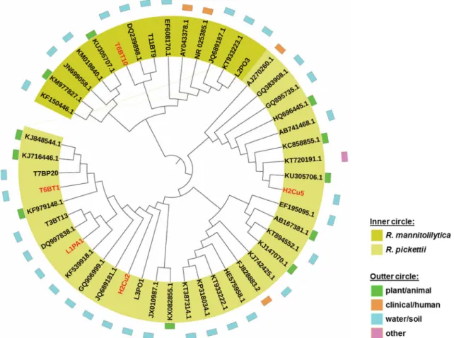
The 16S rRNA gene sequences of the studied strains were compared with other good quality (> 1000 bp) sequences of R. pickettii and R. mannitolilytica strains of different origins available in the GenBank (http://www.ncbi.nlm.nih.gov/). The nucleotide sequence analysis was performed using the MEGA6 software (Tamura et al.2013), based on the model of Jukes and Cantor (Jukes and Cantor1969), and a dendro-gram was created using the neighbor-joining method. The iTol software v3.2.4 (Letunic and Bork2016) was used to repre-sent the isolates source in the dendrogram.
Determination of antibiotic and metal resistance
phenotypes
The antibiotic resistance phenotypes were determined by disk diffusion method as recommended by the Clinical Laboratory Standards Institute (CLSI2015), for 12 antibiotics: nalidixic acid (NA, 30μg); ciprofloxacin (CIP, 5 μg); streptomycin (STR, 10μg); gentamicin (GEN, 10 μg); tetracycline (TET, 30μg); cephalothin (CP, 30 μg); meropenem (MER, 10 μg); ceftazidime (CEF, 30 μg); ticarcillin (TIC, 75 μg); colistin
sulfate (CT, 50μg); sulfamethoxazole (SUL, 25 μg), and sulfamethoxazole/trimethoprim (SXT, 23.75/1.25μg). The in-terpretation criteria (R, resistance; S, susceptible) based on inhibition zone diameters were as follows (mm): NA30: R≤ 13, S≥ 19; CIP5: R ≤ 15, S ≥ 21; STR10: R ≤ 11, S ≥ 15; GEN10: R≤ 12, S ≥ 15; TET30: R ≤ 11, S ≥ 15; CP30: R ≤ 14, S≥ 18; MER10: R ≤ 15, S ≥ 19; CEF30: R ≤ 14, S ≥ 18; TIC75: R≤ 15, S ≥ 24; CT50: R ≤ 10, S ≥ 11; SUL25: R ≤ 12, S≥ 17; SXT25: R ≤ 10, S ≥ 16. In each assay, the reference strain Pseudomonas aeruginosa DSM 1117 was used for quality control.
Determination of minimum inhibitory concentrations
(MICs)
The minimum inhibitory concentrations (MICs) were deter-mined using the Etest or the microdilution method at 30 °C. The Etest (BioMérieux, France) or MICE (OXOID, United Kingdom) were used for the antibiotics gentamicin (CN 256–0.015 μg/mL, OXOID, MA0116F), streptomycin (SM 0.064–1024 μg/mL, BioMérieux, 526,800), ceftazidime (TZ 0.016–256 μg/mL, BioMérieux, 412293), meropenem (MEM 32–0.002 μg/mL, OXOID, MA0121F), and sulfamethoxazole (SX 0.064–1024 μg/mL, BioMérieux, 412458). The microdilution method was used for tetracycline and metals (Andrews 2001), using bacterial suspensions of absorbance 0.08–0.1 at 625 nm in Mueller-Hinton broth supplemented with 0.1–32 mg/L of tetracycline, 0.001–2 mmol/L of NaAsO2, 0.01–10 mmol/L of NiCl2·6H2O or 1–14 mmol/L of CuSO4·5H2O. For concentrations of CuSO4·5H2O above 5 mmol/L, the MICs were tested in Tris-buffered Mueller-Hinton broth. The MICs were determined as the minimum concentration that inhibited visible bacterial growth after 24 h of incubation.
Based on preliminary distinctive results between the tested strains, the aminoglycoside gentamicin, and the metal arsenite were selected to assess their effects as stressors and will be from this point forward designated as stressors. Each of the
five strains was assayed in stressor-free (SF) culture medium and in the presence of gentamicin or arsenite at concentrations close to the MIC value.
Stressors and growth kinetics
Cultures were assayed in Mueller-Hinton broth or in this cul-ture medium supplemented with adequate concentrations and volume of stressor solution. Therefore, strains H2Cu2, T6BT1 and L1PA1 were assayed in 125 mg/L gentamicin or 1.1 mmol/L As3+; strain H2Cu5 was assayed in 6 mg/L gen-tamicin or 0.01 mmol/L As3+; and strain T6BT10 was assayed in 0.4 mg/L gentamicin or 0.01 mmol/L As3+. Bacterial sus-pensions with an initial absorbance of 0.05 at 610 nm (A610) were incubated at 30 °C with orbital shaking (~ 70 rpm) and were monitored every hour until reached the stationary phase (~ 24 h). Growth curves and kinetic parameters (growth rate, lag phase, and yield) were determined in triplicate in indepen-dent assays. Growth curves were represented as log values of A610 in function of time. The lag phase was the period of time necessary to start the exponential phase. The growth rate (μ) was determined based on the slope of the curve during the exponential growth phase, according to the equation Ln Nt− Ln N0=μ(t − t0), where N is the number of cells at time t. The growth yield corresponded to the maximum A610 reached.
Stressors and biofilm formation
The capacity of each strain to form biofilm was tested in modified Luria-Bertani broth (mLB) (tryptone 5 g/L, yeast extract 2.5 g/L and sodium chloride 1 g/L) over a range of different stressor concentrations: 0.01, 0.05, 0.5 and 1.1 mmol/L As3+; 25, 75 and 125 mg/L of GEN; and 125, 250, 500 and 750 mg/L of STR, concentrations bellow the MICs for the strains H2Cu2, L1PA1 and T6BT1; and of 0.01 mmol/L of As3+; 0.4 and 6 mg/L of GEN; and 50 mg/L of STR for the strains H2Cu5 and T6BT10. The assays were Table 1 Group of isolates tested in this study
Strain Species Source of isolation Isolation medium and conditions Abundance in the source (order of magnitude, CFU’s/mL)
Reference
H2Cu2 R. pickettii Hospital wastewater Culture enrichment in modified Luria-Bertani broth with Cu2+(2.5 mmol/L)
103 Becerra-Castro et al.2015
H2Cu5 R. pickettii Hospital wastewater Culture enrichment in modified Luria-Bertani broth with Cu2+(2.5 mmol/L)
103 Becerra-Castro et al.2015
L1PA1 R. pickettii Mineral water Pseudomonas isolation agar with 32 mg/L amoxicillin
101 Falcone-Dias et al.2012
T6BT1 R. pickettii Tap water Tergitol 7-agar 10−1 Vaz-Moreira et al.2017
performed in clear flat bottom 96-well polystyrene microtiter plates (Orange Scientific, Belgium) as described by Simões et al. (2007). Briefly, the microtiter wells were filled with 200μL of bacterial suspension (A610 = 0.1; prepared from overnight cultures in mLB at 30 °C) in mLB or in mLB supplemented with one of the stressors, incubated for 48 h at 30 °C and measured the absorbance at 620 nm (A620) in a microplate reader (FLUOstar optima, BMG Labtech, Germany). After that, the plates were washed with phosphate buffer and air-dried for 30 min. To assess and compare the biofilm forma-tion, the biomass was fixed with methanol, left to dry, stained with crystal violet, washed again and the dye resuspended with glacial acetic acid prior to measuring the absorbance at 570 nm (A570). A negative control consisting of non-inoculated culture medium and a reference culture (Pseudomonas aeruginosa DSM 1117) were included in each assay. Each experiment was performed at least six times for each strain. The quantification of the biofilm formation was performed as described by Rode et al. (2007), through the calculation of a ratio A570/A620, referring to absorbance at 570 nm (to measure the biofilm formation) and absorbance at 620 nm (to measure the bacterial growth). The absorbance values were corrected by the subtraction of the respective absorbance measured in the negative control (non-inoculated culture medium). With the procedure used, the possible con-tribution of the growth yield for the capacity to form biofilm was normalized by the use of the ratio A570/A620, referring to absorbance at 570 nm (measure of the biofilm formation) and absorbance at 620 nm (measure of the bacterial growth).
Disinfectants and inactivation
The effectiveness of the germicide UV radiation, chlorine or hydrogen peroxide was tested in saline solution (0.85% (w/v) NaCl) bacterial suspensions of A610 = 0.1. Suspensions were prepared from 24 h Plate Count Agar (PCA) cultures. Samples collected at the beginning and over the assay were cultivated for enumeration on PCA and incubated at 30 °C for 24–48 h. For UV disinfection was used a germicide UV lamp with a wavelength of 254 nm, under which were exposed PCA plates onto which were spread 100μL of a bacterial suspension with about 10 to 300 CFU/mL. Exposure times were of periods of 0, 15, 30, 45, 60, 90 and 150 s.
To test the effect of chlorine was used a solution of 10 mg/L sodium hypochlorite prepared from commercial bleach with a concentration of sodium hypochlorite equivalent to 5% (50 g/ L). Bacterial suspensions (A610 = 0.1) prepared in saline so-lution were exposed to sodium hypochlorite at a final concen-tration of 5 mg/L. A solution of 1.5% (w/v) sodium thiosulfate was used to neutralize the effect of chlorine at different expo-sure times of 0, 2, 7, 12, 17, 25 and 60 min. Cultures were plated immediately after the addition of the neutralizing agent.
The effect of hydrogen peroxide was tested using a 0.1% solution prepared from a 30% stock (Carlo Erba Reagents, Italy). Bacterial suspensions (A610 = 0.1) were exposed to hy-drogen peroxide at a final concentration of 0.05% (v/v). A fresh-ly prepared solution of bovine liver catalase (0.1 g/L) was used in a ratio 0.1/5 (v/v) to eliminate residual hydrogen peroxide (Fiorentino et al.2015) after exposure times of 0, 2, 7, 12, 17, 25 and 60 min. Cultures were plated after catalase addition.
Statistical analyses
The effect of different stressors and the behavior of different strains were compared based on the parametric test one-way ANOVA or the non-parametric tests Kruskal-Wallis and Mann-Whitney, depending if the results followed or not a normal distribution. The capacity to form biofilm in the pres-ence and abspres-ence of stressors was compared based on the non-parametric test Mann-Whitney. The effect of disinfectants on cells inactivation was compared based on parametric one way ANOVA test with post hoc test Tuckey. All the statistical analyses were performed with the SPSS software package, version 23.0 (IBM SPSS software, Chicago, IL).
Results
Ralstonia spp. tolerance to antibiotics and heavy
metals
Based on the 16S rRNA gene sequence analyses, the R. pickettii and R. mannitolilytica strains studied clustered to-gether with others from sources such as plant/animal, clini-cal/human, water and soil, or other environments (e.g., air) (Fig. 1). Although not related to the isolation origin, three phylogenetic sub-groups could be distinguished, one that in-cluded strains H2Cu2, L1PA1 and T6BT1, sharing a 16S rRNA gene sequence identity of 99.7–99.9%, other including strain H2Cu5, with a 16S rRNA identity with first group of 99.0–99.2% and another one of R. mannitolilytica, which, non-surprisingly included the strain T6BT10 with a 16S rRNA gene sequence identity of 97.8–98.2% with the R. pickettii isolates tested. These differences were not confirmed at the biochemical phenotype for which the five strains displayed a similar profile (data not shown).
All strains were observed to be resistant to colistin and ticarcillin and susceptible to the quinolones (nalidixic acid, cip-rofloxacin), sulfonamides (sulfamethoxazole and sulfamethox-azole/trimethoprim), beta-lactams (cephalothin and ceftazi-dime) and tetracycline. Variable phenotypes were observed for meropenem and aminoglycosides susceptibility. Strains R. pickettii H2Cu5 and R. mannitolilytica T6BT10, both suscepti-ble to gentamicin, differed on the susceptibility to streptomycin observed only in R. mannitolilytica T6BT10. Hence,
gentamicin resistance was observed in the group of phylogenet-ically closely related strains H2Cu2, L1PA1 and T6BT1, with MIC-gentamicin values > 256 mg/L, while strains H2Cu5 and T6BT10, in distinct phylogenetic sub-groups, presented lower MIC values for both gentamicin and streptomycin (Table2). MIC values for As3+were about 30 times higher in gentamicin resistant than in gentamicin susceptible isolates, while no dif-ferences among strains were observed for Ni2+or Cu2+. This finding suggested that aminoglycoside and arsenite resistance mechanisms might be associated.
Stressors and growth kinetic
Based on the hypothesis that a common mechanism of resis-tance could be used by these strains for gentamicin and arse-nite, growth kinetic parameters were determined in the ab-sence and in the preab-sence of each of those stressors (Table
3). In the absence of any stressor, the growth rates for the five strains were similar (~ 0.4 h−1) (Table3). Either gentamicin or arsenite led to significant (p < 0.05) reductions in the growth rate, being the highest reductions observed in the Fig. 1 Environmental distribution of Ralstonia pickettii and Ralstonia mannitolilytica, including the strains studied (in red)
Table 2 Minimum inhibitory concentrations (MICs) for antibiotics and metals determined for the Ralstonia spp. strains under study
Strain MICs
GEN STR TET MER CEF SUL As3+ Ni2+ Cu2+
(mg/L) (mg/L) (mg/L) (mg/L) (mg/L) (mg/L) (mmol/L) (mmol/L) (mmol/L)
H2Cu2 > 256 > 1024 1 > 32 6 24 1.4 4 12
H2Cu5 6 56 0.25 6 6 4 0.05 4 12
L1PA1 > 256 > 1024 1 > 32 6 24 1.4 4 12
T6BT1 > 256 > 1024 1 16 8 24 1.4 4 12
T6BT10 0.5 4 8 > 32 4 4 0.05 4 12
GEN, gentamicin; STR, streptomycin; TET, tetracycline; MER, meropenem; CEF, ceftazidime; SUL, sulfamethoxazole and metal salts of As3+, Ni2+, and Cu2+
presence of sub-inhibitory concentrations of gentamicin (Table3). The lag phases in the absence of stressor ranged 0.7–0.9 h. In the presence of arsenite, but not in the presence of gentamicin, these values significantly (p < 0.05) increased (to 2.4–3.4 h) in the strains with highest MIC-As3+
values (Table3). In absence of stressors, growth yield ranged 2.4– 2.9. These values that were significantly (p < 0.05) reduced in the presence of arsenite for strain H2Cu2 (to 2.2) or in the presence of gentamicin for strains H2Cu2, H2Cu5, L1PA1, and T6BT1 (to 0.5–0.9). In general, the reduction of growth yield was more pronounced in the presence of gentamicin than of arsenite. These differences in the growth parameters in the presence of gentamicin or arsenite suggest that even if a com-mon resistance mechanism is used to grow in the presence of each of those stressors, probably distinct functions are targeted in the cell by the antibiotic or the metal.
Effect of stressors in the capacity of biofilm formation
The capacity to form biofilm may be an advantage in Ralstonia spp. to face adverse conditions (Adley et al.2005; Anderson et al.1990; Di Domenico et al.,2016; Ryan et al.
2011). Hence, it was hypothesized that the stressors amino-glycosides and arsenite could stimulate the capacity to pro-duce biofilm (Fig.2). The low concentrations of stressor tol-erated by the aminoglycoside susceptible strains H2Cu5 and T6BT10 were not observed to induce in those strains an in-creased capacity to form biofilm. In contrast, the strains resis-tant to the aminoglycosides gentamicin and streptomycin (H2Cu2, T6BT1 and L1PA1) presented significant increases in the capacity to form biofilm, with increases of 2–4 times for the lower concentrations and 5–11 times for the highest con-centrations of aminoglycosides tested, in comparison with the non-stressor assays (Fig. 2). In the same way, 1.1 mmol/L arsenite a significant, although lower (1.5–1.9 times), increase of biofilm formation. The capacity to form biofilm can be associated with the production of polysaccharide capsules that facilitate the adherence to surfaces and the formation of biofilms (Moxon and Kroll1990). It was thus hypothesized that the increased capacity to form biofilm could be due to an observable overproduction of capsule polysaccharides in the presence of sub-inhibitory concentration of gentamicin. However, this hypothesis was not proved, eventually because the method used to observe capsules was not sufficiently sensitive.
Disinfectants and inactivation
The hypothesis beyond these assays was that aminoglyco-side and arsenite resistant strains would present a higher resilience against the different types of disinfectant—UV radiation, chlorine, and peroxide disinfection (Fig. 3). However, it was observed that only peroxide disinfection
Table 3 V ariations on the bacterial growth paramete rs gro w th rate, p h ase lag, and y ield, under sub-inhibitory concentrat ions of arsenite (As 3+ ) or g entamicin (GEN) or control co nditions (stress or free, SF) Strain (stres sor co n centration) Growth rate (per hour) P hase lag (hours) Y ield (A610) SF As 3+ GEN S F As 3+ GEN S F As 3+ GEN H2 Cu2 1 25 mg/ L GE N o r 1 .1 mm ol/ L As 3+ 0.4 ± 0.03 1;a,b 0.3 ± 0.02 2;a 0.1 ± 0.01 3;a 0.9 ± 0.2 1;a 3.4 ± 0 .5 2;a 2 .0 ± 0 .7 1, 2;a 2 .9 ± 0 .1 1;a 2 .2 ± 0 .2 2 ;a 0 .5 ± 0 .1 3; a H2 Cu5 6 mg/ L GE N o r 0 .0 1 m m o l/ L A s 3+ 0.3 ± 0.01 1;a 0.3 ± 0.04 1;a 0.1 ± 0.01 2;b 0.7 ± 0.4 1;a 1.0 ± 0 .3 1;b 2 .2 ± 2 .5 1; a 2 .5 ± 0 .3 1;a 2 .0 ± 0 .3 1 ;a 0 .9 ± 0 .2 2; a L 1 P A 1 125 mg/L GEN o r 1 .1 mmol/L As 3+ 0.4 ± 0.04 1;a,b 0.3 ± 0.04 1;a,b 0.2 ± 0.01 2;c 0.7 ± 0.4 1;a 3.0 ± 0 .9 2;a 0 .8 ± 0 .1 1; a 2 .9 ± 0 .4 1;a 2 .5 ± 0 .5 1 ;a 0 .8 ± 0 .1 2; a T6BT1 125 mg/L GEN or 1.1 mmol/L As 3+ 0.4 ± 0.02 1;a,b 0.3 ± 0.01 2;a 0.2 ± 0.01 3;b 0.9 ± 0.3 1;a 2.4 ± 0 .3 2;a 1 .4 ± 0 .4 1; a 2 .7 ± 0 .1 1;a 2 .4 ± 0 .3 1 ;a 0 .7 ± 0 .1 2; a T6BT10 0.4 m g/L G EN or 0.01 mmol/L As 3+ 0.4 ± 0.01 1;b 0.4 ± 0.01 1,2;b 0.4 ± 0.01 2;d 0.8 ± 0.1 1;a 0.8 ± 0 .1 1;b 0 .7 ± 0 .1 1; a 2 .4 ± 0 .5 1;a 2 .1 ± 0 .4 1 ;a 1 .9 ± 0 .6 1; b A610 , bacterial suspension absorbance at 610 nm; Statistically significant dif feren ces b etween stres s conditions (SF , As 3+ , and GEN) are indicated b y the numbers: 1 , 2 , 3 ; and sig n ifican t d if fe re nce s bet w ee n str ains are indicated by the letters: a, b, c, d
supported that hypothesis. Neither UV radiation nor chlo-rine were observed to produce a distinct effect on the gen-tamicin resistant or susceptible strains (Fig.3). The UV radiation promoted a reduction of 1–2 log at each 15 min of exposure till the maximum period tested of 45 min (Fig.3a). In the presence of 5 mg/L chlorine, it was ob-served a sharp culture inactivation (2 min), to reach after 7 min of exposure, counts < 10 CFU/mL (Log 1) (Fig.3b). In contrast to the other two disinfectants, hydrogen peroxide revealed higher antibacterial effectiveness against the gen-tamicin and arsenite susceptible strains than against the re-sistant. Susceptible strains decreased to counts below the quantification limit (one log-unit) after 7 min of exposure, in contrast to the resistant strains that required 12 min to reach < one log-unit (Fig.3c).
Discussion
Ralstonia spp. are ubiquitous, mainly in aquatic environ-ments, including drinking water (Vaz-Moreira et al.2017), and have been reported as contaminants of clinical sterile so-lutions or materials (Boutros et al.2002; Labarca et al.,1999; McNeil et al.,1985) or as the prevalent species in hemodial-ysis water samples (Vincenti et al.,2014). Ralstonia pickettii strains from different clinical and environmental origins were reported as being resistant to gentamicin, ticarcillin and meropenem, although highly susceptible to ciprofloxacin, tet-racycline and sulfamethoxazole/trimethoprim (Ryan and Adley2013), confirming the phenotypes of the strains exam-ined in this study. The observation that phylogenetically and phenotypically close Ralstonia strains, isolated from distinct Fig. 2 Influence of stressors on
the ability of biofilm formation, for the strains H2Cu2 (a), T6BT1 (b), and L1PA1 (c). The quantification of the biofilm formation was performed through the calculation of a ratio A570/
A620, referring to absorbance at
570 nm (measure of the biofilm formation) and absorbance at 620 nm (measure of the bacterial growth). The non-inoculated control presented a ratio A570/
A620of 1.0 ± 0.1; and the P.
aeruginosa presented ratios A570/
A620of 3.1 ± 0.7 for stressor-free
(SF); 2.2 ± 0.9 for gentamicin (GEN) 6 mg/L; 2.8 ± 0.7 for streptomycin (STR) 50 mg/L; 0.2 ± 0.09 for meropenem 4 mg/L; 1.9 ± 0.9 for arsenite (As) 0.01 mmol/L; 0.3 ± 0.05 for copper 6.0 mmol/L; and 0.7 ± 0.2 for nickel 2.5 mmol/L
aquatic environments, differed on the susceptibility to genta-micin (Tables1and2), suggested that it might be due to gene acquisition. Indeed, in a parallel study, whole genome se-quencing from strains H2Cu2 and H2Cu5 showed that only the aminoglycoside resistant isolate contains genes associated with resistance to arsenic, and toxic compounds, encoding lysozyme inhibitors, or phages/prophages receptors (Vaz-Moreira et al.2016).
The correlation observed between the resistance to arsenite and gentamicin may also be an indication of a possible mech-anism of co-resistance (genetic linkage between two or more resistance genes) or cross-resistance (same genetic determi-nant confers resistance to both antibiotics and metals), fre-quently reported for heavy metals and antibiotics
(Baker-Austin et al. 2006; Dib et al.2008; Seiler and Berendonk
2012; Zhou et al.2015) specially in contaminated environ-ments (Ahemad and Malik2013).
Supposedly the physiological response to arsenite and ami-noglycosides involves diverse mechanisms, as the response for biofilm formation and growth kinetic did not respond in the same mode for both antimicrobials (Table 2). However, the stress imposed by sub-inhibitory concentrations of amino-glycosides or of metals increased the capacity to form biofilms of the aminoglycoside resistant isolates (Fig.2). This can be due to an unspecific stress response, not related with the re-sistance mechanism, in particular enhanced production of ex-tracellular polymeric substances (EPS) which lead to cell ad-hesion (Baker-Austin et al.2006; Donlan2002; Donlan and Costerton 2002; Lindsay and von Holy 2006), and biofilm formation (Balaban 2008; Donlan and Costerton 2002; Lindsay and von Holy2006). Similar results were observed for Pseudomonas aeruginosa and Escherichia coli isolates, increasing their capacity to form biofilm in the presence of aminoglycosides (Aka and Haji2015; Hoffman et al.2005). However, Paul et al. (2014) observed a negative influence of 0.01 mmol/L arsenite on the capacity to form biofilm of Pseudomonas spp. or Rhizobium spp. strains. Some authors refer to the importance of the bacteria motility on the capacity to form biofilm (Guttenplan and Kearns2013; O'Toole and Kolter1998; Yao and Allen2007). Indeed, Ralstonia spp. are motile and this property might influence the variable capacity to form biofilm observed in the present study. However, it was not possible to assess differences in motility capabilities in the tested strains or to investigate if stressors interfere with flagel-la and therefor with biofilm formation. Considering the histo-ry of Ralstonia spp. as important colonizers of highly oligo-trophic environments, other conditions to explore could be the effect of nutritional stress.
In contrast to what was observed for hydrogen peroxide, the survival to UV radiation or chlorine disinfection did not differ in aminoglycoside resistant or susceptible strains (Fig.3). This observation is probably related with the inacti-vation mechanisms involved, DNA/RNA damage for UV, cell metabolism for chlorine and oxidative stress for hydrogen peroxide (Estrela et al.2002; Hijnen et al.2006; McDonnell and Russell1999). These results suggest that the mechanisms involved in the aminoglycoside resistance are probably not directly associated with the mechanisms of survival to UV radiation or chlorination. Studies performed with a higher number of isolates, from different origins, may give more consistency to these results. The capability of Ralstonia spp. to survive in some environments subjected to disinfection processes, as for example, the wastewater or drinking water systems, is probably determinant for their capacity to spread or be transmitted to humans. It is curious to note that amino-glycoside resistant strains have increased capacity to form biofilm in the presence of some environmental stressors, since Fig. 3 Bacterial inactivation with (a) UV radiation, (b) chlorine (5 mg/L),
this may be a relevant factor to facilitate the survival and spread of aminoglycoside resistant Ralstonia spp. strains in the environments subjected to stress conditions, as antimicro-bial challenges. These characteristics combined with the oli-gotrophic character may contribute for the Ralstonia ubiquity in aquatic habitats.
Conclusions
The aminoglycosides resistance was associated with the highest tolerance to arsenite.
Sub-inhibitory concentrations of gentamicin or arsenite significantly decreased the growth rate and yield, while arse-nite but not gentamicin caused a significant increase of the lag phase. The biofilm formation was stimulated in the presence of aminoglycosides or arsenite, in the aminoglycoside resis-tant but not in the susceptible strains.
Disinfection with UV or chlorine presented identical effec-tiveness in aminoglycoside resistant or susceptible strains. In contrast, hydrogen peroxide presented higher effectiveness against aminoglycoside susceptible than resistant strains.
The results support the hypothesis that antibiotic resistance is associated with improved tolerance to stress.
Acknowledgements This work was financially supported by National Funds from FCT - Fundação para a Ciência e a Tecnologia through pro-jects UID/Multi/50016/2013 and NanoDiaBac ENMed/0001/2014, and IVM grant (SFRH/BPD/87360/2012); by FCT through the project WaterJPI/0001/2013 - STARE (Stopping Antibiotic Resistance Evolution); and by Erasmus Mundus Project SUD-UE (EMA2-STRAND 1, LOT 14) through PF grant (2013-2589).
References
Adley C, Ryan M, Pembroke J, Saieb F (2005) Ralstonia pickettii: bio-film formation in high-purity water. Biobio-films: Persistence and Ubiquity, pp 261–271
Ahemad M, Malik A (2013) Prevalence of heavy metal and antibiotic resistance in bacterial isolates from metal polluted soils. Microbiol J.
https://doi.org/10.3923/mj.2013
Aka ST, Haji SH (2015) Sub-MIC of antibiotics induced biofilm forma-tion of Pseudomonas aeruginosa in the presence of chlorhexidine. Braz J Microbiol 46:149–154
Anderson RL, Holland BW, Carr JK, Bond WW, Favero MS (1990) Effect of disinfectants on pseudomonads colonized on the interior surface of PVC pipes. Am J Public Health 80:17–21
Andrews JM (2001) Determination of minimum inhibitory concentra-tions. J Antimicrob Chemother 48(Suppl 1):5–16
Baker-Austin C, Wright MS, Stepanauskas R, McArthur JV (2006) Co-selection of antibiotic and metal resistance. Trends Microbiol 14: 176–182
Balaban N (2008) Control of biofilm infections by signal manipulation vol 2. Springer
Becerra-Castro C, Machado RA, Vaz-Moreira I, Manaia CM (2015) Assessment of copper and zinc salts as selectors of antibiotic
resistance in Gram-negative bacteria. Sci Total Environ 530-531: 367–372
Boutros N, Gonullu N, Casetta A, Guibert M, Ingrand D, Lebrun L (2002) Ralstonia pickettii traced in blood culture bottles. J Clin Microbiol 40:2666–2667
CLSI (2015) Performance standards for antimicrobial susceptibility test-ing; twenty-fifth informational supplement M100-S25. vol 35. Clinical and Laboratory Standards Institute, USA
Coenye T, Vandamme P, LiPuma JJ (2002) Infection by Ralstonia species in cystic fibrosis patients: identification of R. pickettii and R. mannitolilytica by polymerase chain reaction. Emerg Infect Dis 8: 692–696
Coman I, Bilodeau L, Lavoie A, Carricart M, Tremblay F, Zlosnik JE, Berthiaume Y (2017) Ralstonia mannitolilytica in cystic fibrosis: a new predictor of worse outcomes. Respir Med Case Rep 20:48–50 Daxboeck F, Stadler M, Assadian O, Marko E, Hirschl AM, Koller W (2005) Characterization of clinically isolated Ralstonia mannitolilytica strains using random amplification of polymorphic DNA (RAPD) typing and antimicrobial sensitivity, and comparison of the classification efficacy of phenotypic and genotypic assays. J Med Microbiol 54:55–61
De Baere T et al (2001) Classification of Ralstonia pickettii biovar 3/ 'thomasii'strains (Pickett 1994) and of new isolates related to noso-comial recurrent meningitis as Ralstonia mannitolytica sp. nov. Int J Syst Evol Microbiol 51:547–558
Di Domenico EG et al (2016) Development of an in vitro assay, based on the biofilm ring test®, for rapid profiling of biofilm-growing bacte-ria. Front Microbiol 7:1429
Dib J, Motok J, Zenoff VF, Ordonez O, Farias ME (2008) Occurrence of resistance to antibiotics, UV-B, and arsenic in bacteria isolated from extreme environments in high-altitude (above 4400 m) Andean wet-lands. Curr Microbiol 56:510–517
Donlan RM (2002) Biofilms: microbial life on surfaces. Emerg Infect Dis 8:881–890
Donlan RM, Costerton JW (2002) Biofilms: survival mechanisms of clinically relevant microorganisms. Clin Microbiol Rev 15:167–193 Estrela C, Estrela CR, Barbin EL, Spano JC, Marchesan MA, Pecora JD (2002) Mechanism of action of sodium hypochlorite. Braz Dent J 13:113–117
Falcone-Dias M, Vaz-Moreira I, Manaia CM (2012) Bottled mineral wa-ter as a potential source of antibiotic resistant bacwa-teria. Wawa-ter Res 46: 3612–3622
Fernandez C et al (1996) Nosocomial outbreak of Burkholderia pickettii infection due to a manufactured intravenous product used in three hospitals. Clin Infect Dis 22:1092–1095
Ferreira da Silva M, Vaz-Moreira I, Gonzalez-Pajuelo M, Nunes OC, Manaia CM (2007) Antimicrobial resistance patterns in Enterobacteriaceae isolated from an urban wastewater treatment plant. FEMS Microbiol Ecol 60:166–176
Fiorentino A, Ferro G, Alferez MC, Polo-Lopez MI, Fernandez-Ibanez P, Rizzo L (2015) Inactivation and regrowth of multidrug resistant bacteria in urban wastewater after disinfection by solar-driven and chlorination processes. J Photochem Photobiol B 148:43–50 Guttenplan SB, Kearns DB (2013) Regulation of flagellar motility during
biofilm formation. FEMS Microbiol Rev 37:849–871
Hijnen WA, Beerendonk EF, Medema GJ (2006) Inactivation credit of UV radiation for viruses, bacteria and protozoan (oo)cysts in water: a review. Water Res 40:3–22
Hoffman LR, D'Argenio DA, MacCoss MJ, Zhang Z, Jones RA, Miller SI (2005) Aminoglycoside antibiotics induce bacterial biofilm for-mation. Nature 436:1171–1175
Jukes TH, Cantor CR (1969) Evolution of protein molecules. In: Munro (ed) Mammalian protein metabolism, vol 3. Academic Press, New York
Khajuria A, Praharaj AK, Grover N, Kumar M (2014) Emergence of VIM-2 metallo-beta-lactamase producing Ralstonia pickettii clinical isolate in India. Indian J Med Microbiol 32:191–193
Koenig DW, Pierson DL (1997) Microbiology of the space shuttle water system. Water Sci Technol 35:59–64
Kulakov LA, McAlister MB, Ogden KL, Larkin MJ, O'Hanlon JF (2002) Analysis of bacteria contaminating ultrapure water in industrial sys-tems. Appl Environ Microbiol 68:1548–1555
Labarca JA et al (1999) A multistate nosocomial outbreak of Ralstonia pickettii colonization associated with an intrinsically contaminated respiratory care solution. Clin Infect Dis 29:1281–1286
Letunic I, Bork P (2016) Interactive tree of life (iTOL) v3: an online tool for the display and annotation of phylogenetic and other trees. Nucleic Acids Res 44:W242–W245
Lindsay D, von Holy A (2006) Bacterial biofilms within the clinical setting: what healthcare professionals should know. J Hosp Infect 64:313–325
Maroye P, Doermann HP, Rogues AM, Gachie JP, Megraud F (2000) Investigation of an outbreak of Ralstonia pickettii in a paediatric hospital by RAPD. J Hosp Infect 44:267–272
McDonnell G, Russell AD (1999) Antiseptics and disinfectants: activity, action, and resistance. Clin Microbiol Rev 12:147–179
McKinney RE (1953) Staining bacterial polysaccharides. J Bacteriol 66: 453
McNeil MM et al (1985) Nosocomial Pseudomonas pickettii colonization associated with a contaminated respiratory therapy solution in a special care nursery. J Clin Microbiol 22:903–907
Mijnendonckx K, Provoost A, Ott C, Venkateswaran K, Mahillon J, Leys N, Van Houdt R (2013) Characterization of the survival ability of Cupriavidus metallidurans and Ralstonia pickettii from space-related environments. Microb Ecol 65:347–360
Moxon ER, Kroll JS (1990) The role of bacterial polysaccharide capsules as virulence factors. In: Bacterial capsules. Springer, Berlin Heidelberg, pp 65–85
Orme J, Rivera-Bonilla T, Loli A, Blattman NN (2015) Native valve endocarditis due to Ralstonia pickettii: a case report and literature review. Case Rep Infect Dis 2015:324675
O'Toole GA, Kolter R (1998) Flagellar and twitching motility are neces-sary for Pseudomonas aeruginosa biofilm development. Mol Microbiol 30:295–304
Pan W, Zhao Z, Dong M (2011) Lobar pneumonia caused by Ralstonia pickettii in a sixty-five-year-old Han Chinese man: a case report. J Med Case Rep 5:377
Paul D, Poddar S, Sar P (2014) Characterization of arsenite-oxidizing bacteria isolated from arsenic-contaminated groundwater of West Bengal. J Environ Sci Health A Tox Hazard Subst Environ Eng 49:1481–1492
Riley PS, Weaver RE (1975) Recognition of Pseudomonas pickettii in the clinical laboratory: biochemical characterization of 62 strains. J Clin Microbiol 1:61–64
Rode TM, Langsrud S, Holck A, Moretro T (2007) Different patterns of biofilm formation in Staphylococcus aureus under food-related stress conditions. Int J Food Microbiol 116:372–383
Ryan MP (2009) Genotypic and phenotypic analysis of Ralstonia pickettii high purity water isolates. University of Limerick
Ryan MP, Adley CC (2013) The antibiotic susceptibility of water-based bacteria Ralstonia pickettii and Ralstonia insidiosa. J Med Microbiol 62:1025–1031
Ryan MP, Pembroke JT, Adley CC (2006) Ralstonia pickettii: a persistent Gram-negative nosocomial infectious organism. J Hosp Infect 62: 278–284
Ryan MP, Pembroke JT, Adley CC (2007) Ralstonia pickettii in environ-mental biotechnology: potential and applications. J Appl Microbiol 103:754–764
Ryan MP, Pembroke JT, Adley CC (2009) Novel Tn4371-ICE like ele-ment in Ralstonia pickettii and genome mining for comparative elements. BMC Microbiol 9:242
Ryan MP, Pembroke JT, Adley CC (2011) Genotypic and phenotypic diversity of Ralstonia pickettii and Ralstonia insidiosa isolates from clinical and environmental sources including high-purity water. Diversity in Ralstonia pickettii. BMC Microbiol 11:194
Seiler C, Berendonk TU (2012) Heavy metal driven co-selection of anti-biotic resistance in soil and water bodies impacted by agriculture and aquaculture. Front Microbiol 3:399
Simões LC, Simões M, Vieira MJ (2007) Biofilm interactions between distinct bacterial genera isolated from drinking water. Appl Environ Microbiol 73:6192–6200
Stelzmueller I et al (2006) Ralstonia pickettii-innocent bystander or a potential threat? Clin Microbiol Infect 12:99–101
Tamura K, Stecher G, Peterson D, Filipski A, Kumar S (2013) MEGA6: molecular evolutionary genetics analysis version 6.0. Mol Biol Evol 30:2725–2729
Udayappan SD et al (2017) Intestinal Ralstonia pickettii augments glu-cose intolerance in obesity. PLoS One 12:e0181693
Vaneechoutte M, De Baere T, Wauters G, Steyaert S, Claeys G, Vogelaers D, Verschraegen G (2001) One case each of recurrent meningitis and hemoperitoneum infection with Ralstonia mannitolilytica. J Clin Microbiol 39:4588–4590
Vaneechoutte M, Kampfer P, De Baere T, Falsen E, Verschraegen G (2004) Wautersia gen. nov., a novel genus accommodating the phy-logenetic lineage including Ralstonia eutropha and related species, and proposal of Ralstonia [Pseudomonas] syzygii (Roberts et al. 1990) comb. nov. Int J Syst Evol Microbiol 54:317–327
Vaz-Moreira I, Nunes OC, Manaia CM (2014) Bacterial diversity and antibiotic resistance in water habitats: searching the links with the human microbiome. FEMS Microbiol Rev 38:761–778
Vaz-Moreira I, Tamames J, Martinez JL, Manaia CM (2016) Draft ge-nome sequences of two Ralstonia pickettii strains with different aminoglycoside resistance phenotypes. Genome Announc 4: e01257–e01216
Vaz-Moreira I, Nunes OC, Manaia CM (2017) Ubiquitous and persistent Proteobacteria and other Gram-negative bacteria in drinking water. Sci Total Environ 586:1141–1149
Verschraegen G, Claeys G, Meeus G, Delanghe M (1985) Pseudomonas pickettii as a cause of pseudobacteremia. J Clin Microbiol 21:278– 279
Vincenti S et al (2014) Non-fermentative gram-negative bacteria in hos-pital tap water and water used for haemodialysis and bronchoscope flushing: prevalence and distribution of antibiotic resistant strains. Sci Total Environ 499:47–54
Waugh JB, Granger WM, Gaggar A (2010) Incidence, relevance and response for Ralstonia respiratory infections. Clin Lab Sci 23:99 Yao J, Allen C (2007) The plant pathogen Ralstonia solanacearum needs
aerotaxis for normal biofilm formation and interactions with its to-mato host. J Bacteriol 189:6415–6424
Yoon SH, Ha SM, Kwon S, Lim J, Kim Y, Seo H, Chun J (2017) Introducing EzBioCloud: a taxonomically united database of 16S rRNA gene sequences and whole-genome assemblies. Int J Syst Evol Microbiol 67:1613–1617
Zhou Y, Xu YB, Xu JX, Zhang XH, Xu SH, Du QP (2015) Combined toxic effects of heavy metals and antibiotics on a Pseudomonas fluorescens strain ZY2 isolated from swine wastewater. Int J Mol Sci 16:2839–2850