Polyoxometalates as potential next‐generation metallodrugs in the combat against cancer
Texto
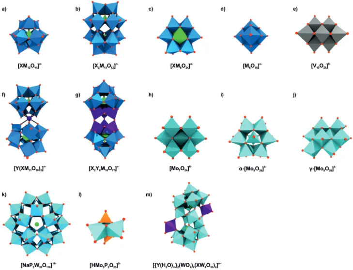
Imagem
![Figure 3. Structures of POM–quinolone hybrids. a) [Cu 2 (Enro) 3 H 2 O][](https://thumb-eu.123doks.com/thumbv2/123dok_br/18848572.929331/6.892.75.434.89.384/figure-structures-of-pom-quinolone-hybrids-cu-enro.webp)


Documentos relacionados
The iterative methods: Jacobi, Gauss-Seidel and SOR methods were incorporated into the acceleration scheme (Chebyshev extrapolation, Residual smoothing, Accelerated
Ousasse apontar algumas hipóteses para a solução desse problema público a partir do exposto dos autores usados como base para fundamentação teórica, da análise dos dados
The probability of attending school four our group of interest in this region increased by 6.5 percentage points after the expansion of the Bolsa Família program in 2007 and
didático e resolva as listas de exercícios (disponíveis no Classroom) referentes às obras de Carlos Drummond de Andrade, João Guimarães Rosa, Machado de Assis,
destaca que a participação ou representação do usuário na formação do padrão técnico é quase inexistente em função das dificuldades associadas ao conhecimento e aos
The irregular pisoids from Perlova cave have rough outer surface, no nuclei, subtle and irregular lamination and no corrosional surfaces in their internal structure (Figure
i) A condutividade da matriz vítrea diminui com o aumento do tempo de tratamento térmico (Fig.. 241 pequena quantidade de cristais existentes na amostra já provoca um efeito
Alguns ensaios desse tipo de modelos têm sido tentados, tendo conduzido lentamente à compreensão das alterações mentais (ou psicológicas) experienciadas pelos doentes
