BJRS
07-02A (2019) 01-07ISSN: 2319-0612
Accept Submission: 2019-01-16
X-ray diffraction pattern and relative crystallinity of
irradiated arrowroot starch
A. G. Barroso; R. H. L. Garcia; N. L. Del Mastro
Instituto de Pesquisas Energéticas e Nucleares (IPEN/CNEN-SP)/ Centro de Tecnologia das Radiações/ Av. Professor
Lineu Prestes 2242/05508-000 São Paulo, SP
Corresponding author: [email protected]
ABSTRACT
After cereals, tubers and roots are the major source of starch for food and industrial uses. Arrowroot refers to any plant of the genus Marantha, but the term is most commonly used to describe the easily digested starch ob-tained from the rhizomes of Marantha arundinacae. The rhizomes of this herbaceous plant contain about 20% of starch. As few studies exist on arrowroot starch, the objective of this preliminary work was to study the X-ray diffraction (XRD) patterns of arrowroot starch when treated by γ-radiation with doses up to 15 kGy in a 60Co source. The XRD patterns of the arrowroot starch exhibited A-type crystalline arrangements with strong peaks at approximately 15º, 17º, 18º and 23º (2θ). A slight increase of diffractogram peaks intensity was noticed after the irradiation process. The cristallinity index was calculated using Bruker DIFFRAC.EVA version 4.2 software. Relative crystallinity seems to increase with irradiation at low doses that could be attributed to different radia-tion sensitivity among the amorphous and crystalline regions of the arrowroot starch molecule. Present results will contribute to elucidate the behavior under radiation treatment of this starchy component increasingly em-ployed by the food industry.
1. INTRODUCTION
Starch is constituted of two biopolymers, amylose, essentially a linear macromolecule, and amylo-pectin, a highly branched macromolecule. These two biopolymers form a semicrystalline structure in the starch granule, which consists of crystalline and amorphous lamellas [1]. Starches from dif-ferent botanical origin vary in total starch and amylose content. Original from tropical regions of South America, arrowroot (Marantha arundinacae) also called araruta, is the flour made from the tuber of the same name. Brazilian Indians believed that araruta neutralized the poison used by the enemies [2]. In cookery it is used for thickening sauces and in cakes, cookies and desserts. Today, we know that the arrowroot tuber extracts have immune stimulatory effects in vivo and in vitro [3] and it is effective in disease prevention and a useful biomedical source material [4-6]. Also, arrow-root starch is starting to be used as polymer matrix in the production of biodegradable films [7]. According to Gordillo et al. [8], arrowroot starch exhibited high purity (starch content >99%) with an amylose content >40%.
X-ray diffraction (XRD) is a nondestructive technique used to identify and quantify crystalline and amorphous phases in solids. As few studies exist on arrowroot starch, the objective of this prelimi-nary work was to study the XRD patterns of arrowroot starch when treated by ionizing radiation.
2. MATERIALS AND METHODS
Arrowroot or araruta starch was obtained in bulk from local food market as a fine powder. The starch were gamma irradiated at doses of 0 - 15kGy, dose rate about 1 kGy.h-1, using a 60Co
Gam-macell 220, Atomic Energy of Canada Ltd (AECL), in polyethylene bags at room temperature, dose uniformity factor of 1.13. Data of X-ray diffraction were acquired from a Bruker D8 diffractometer, using monochromatic Cu-Kα radiation (wavelength = 1.541 A˚). The starch samples were packed tightly in appropriate round aluminum specimen holders (0,61 cm3) and exposed (density about 1.49 g/cm³) to the X-ray beam with the X-ray generator running at 40 kV and 30 mA. The scanning regions of the diffraction angle 2 theta were from 5° to 37°, which covers all the significant
diffrac-tion peaks of starch crystallites. Other operadiffrac-tion condidiffrac-tions were as follows: Stepsize of 0.04 and 10 s per step, Soller and divergence slit 1.0 mm, receiving slit 0.4 mm. The ‘d’ spacings were comput-ed according to Bragg’s law. The degree of crystallinity of samples was estimatcomput-ed following the method of Nara & Komiya (1983). The crystallinity index (CI) was calculated dividing the value of the crystalline area (obtained from the difference of total and amorphous area) by the total area of the diffractogram. Amorphous area was determined using the background function and a Bruker DIFFRAC.EVA version 4.2 software, with curvature and threshold parameters equal to unity. To improve accuracy, each sample was analyzed 5 times using different quotas of sample.
The first step for CI determination was to subtract the background, which has contributions due to incoherent and air scattering, electronic noise and other unwanted signals. To accomplish this step an XRD of aluminum plate (that is virtually free of amorphous material) was carried out, and its background signal was subtracted from all diffractograms of this work (light blue line at Figure 1).
The second step was the determination of the amorphous fraction of the diffractograms, using the EVA software, using the parameters curvature and threshold equal to unity. This step created the green dashed line at Figure 1. The last step was to integrate the total area of the diffractograms (blue line at Figure 1), subtract the amorphous area and divide by the diffractogram total area.
3. RESULTS AND DISCUSSION
XRD data for non-irradiated arrowroot is displayed in Figure 1. As can be seen, the X-ray diffrac-tion patterns of the arrowroot starch exhibited A-type crystalline arrangements with strong peaks at approximately 15º, 17º, 18ºand 23º (2θ) and weaker peaks at approximately 10º, 11.5º, 20º, 26.5º, 30.5º and 34º (2θ). This pattern is quite similar to the corn starch from the reference #39-1911 from the PDF catalog, with its peaks shown in red at Figure 1. The 20 diffractograms obtained in this work had very subtle visual differences (Figure 2).
Figure 1: X-Ray Diffractogram of non-irradiated arrowroot starch.
Figure 2: Detail of X-Ray Diffractograms of arrowroot starch.
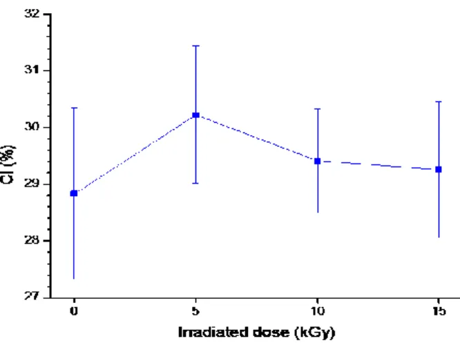
Data of relative crystallinity are displayed in Figure 3. Calculated CI from arrowroot starch samples irradiated with 0, 5, 10 e 15 kGy were respectively 29.1±1.6; 30.6±1.0; 29.5±1.0 and 29.4±1.2. As the standard deviations of present results are rather high, it is not possible to assure that the per-ceived increase of the CI for samples irradiated with 5 kGy was significant. This could be attributed
to differences in radiation sensitivity among the amorphous and crystalline regions of the arrowroot starch molecule. However, this effect is negligible at the higher radiation doses used in this work.
Figure 3: Average Crystallinity Index (CI) calculated from irradiated arrowroot starch.
As Pepe et al. [9] pointed out, some treatments can cause the rearrangement of amylose and amylo-pectin chains in the starch, and therefore may modify its X-ray pattern, crystallinity, swelling pow-er, amylose leaching, pasting, and gelatinization properties, as well as its susceptibility to enzymatic or acidic hydrolysis. Also, different radiation sensitivity of the amorphous and crystalline regions of the arrowroot starch molecule can be expected.
4. CONCLUSION
XRD was applied to study the structure of arrowroot starch. XRD pattern exhibited A-type crystal-line arrangements with strong peaks at approximately 15º, 17º, 18º and 23º (2θ). The crystalcrystal-line content of arrowroot starch was within the expected range of common starches of 20-45%. In the present work, relative crystallinity do not increased significantly with radiation treatment, although samples irradiated at 5 kGy seem to present a slight increase. Present results will contribute to
elu-cidate the behavior under radiation treatment of this starchy component increasingly employed by the food industry.
ACKNOWLEDGMENT
The authors would like to thank Eng. Elizabeth Somessari for her assistance with the irradiation procedure.
REFERENCES
1. JANE, J; KASEMSUWAN, T.; LEAS, S. et al. Anthology of starch granule morphology by scanning electron microscopy. Starch/Starke, v 46, p 121–129, 1994.
2. KIJAC, M.B. The south america table: the flavor and soul of authentic home cooking
from patagonia to Rio de Janeiro. The Harvard Common Press, Boston, MA, 2003.
3. KUMALASARI, I.D.; HARMAYAN, E.; LESTARI, L.; ARSANTI, R. et al. Evaluation of immunostimulatory effect of the arrowroot (Marantaarundinacea. L) in vitro and in vivo.
Cytotech-nology, v. 64, p. 131-137, 2012.
4. KALAPPURA, U.G.; AUGUSTINE, R.; MATHEW, K.T. Microwave properties of arrowroot and its medical applications. Microw Opt Technol Lett, v. 51, p. 1267–1270, 2009.
5. LEE, M.Y.; K.H.CHAN, K.H. Effects of cheonggukjang containing arrowroot isoflavones on bone metabolism in ovariectomized rats. Food Sci Biotechnol. v. 20, p. 335–34, 2011.
6. NISHAA, S.; VISHNUPRIYA, M.; SASIKUMAR, J.M.; CHRISTABEL, H.P.; GOPALA-KRISHNAN, V.K. Antioxidant activity of ethanolic extract of Marantaarundinacea L. tuberous rhizomes. Asian J Pharm Clin Res, v. 5, p 85–88 , 2012.
7. SANTOS, W.J.; MIRANDA, A.P.; GONÇALVES, B.; et al. Propriedades mecânicas de fil-mes obtidos a partir de fécula de araruta (marantaarundinaceae), nanoargila e glycerol, In:
CON-GRESSO BRASILEIRO DE ENGENHARIA E CIÊNCIA DOS MATERIAIS, 21º
8. GORDILLO, C. A. S.; VALENCIA, G. A.; ZAPATA, R. A. V.; HENAO, A. C. A. Physico-chemical Characterization of Arrowroot Starch (Marantaarundinacea Linn) and Glycerol/Arrowroot Starch Membranes. Int J Food Eng, v. 10 (4), p. 727-735, 2014.
9. PEPE, L. S.; J. MORAES, J.; ALBANO, K. M.; TELIS, V. R. N.; FRANCO, C. M. L. Effect of heat-moisture treatment on the structural, physicochemical, and rheological characteristics of arrowroot starch. Food Sc Technol Internat, v. 22(3), p. 256-265, 2015.