ISSN 1 5 1 7 - 7 0 7 6
Revista Matéria, v. 12, n. 1, pp. 128 – 133, 2007 http://www.materia.coppe.ufrj.br/sarra/artigos/artigo10845
In vitro Bioactivity of PMMA-Based Composite in
Simulated Plasma with Albumin
Poliana P. Lopes1, Bárbara J. M. Leite Ferreira2, Nuno A. F. Almeida2, Tatiane E. Silva1, Márcio C. Fredel1, Maria Helena F. V. Fernandes2, Rui N. Correia2.
1
Departamento de Engenharia Mecânica, Universidade Federal de Santa Catarina - UFSC, Trindade / Caixa Postal: 476 - Florianópolis/SC - 88040-900, Brasil;
e-mail: [email protected], [email protected], [email protected].
2
CICECO e Departamento de Engenharia Cerâmica e do Vidro, Universidade de Aveiro, Campus Universitário de Santiago, 3810-193 Aveiro, Portugal.
e-mail: [email protected], [email protected], [email protected], [email protected].
ABSTRACT
A new composite of poly(methyl methacrylate) – co -(ethyl-hexyl-acrylate) filled with glass of composition (wt.%) 61,33% 3CaO.P2O5 - 23,03% SiO2 – 15,64% MgO was developed. The formation of
CaP (calcium phosphate) layers on implants is crucial for their integration in bone. Simulated biological fluids to study in vitro formation of those layers do not usually contain important organic substances, such as proteins. In the present work in vitro bioactivity was studied in a simulated body fluid in presence (SBFA) and in absence of albumin (SBF). The effect of protein on calcium phosphate formation was followed by ion concentration analyses (ICP), X-ray diffraction (XRD) and scanning electron microscopy coupled with X-ray energy dispersive spectroscopy (SEM-EDS). The results showed the precipitation of a CaP layer on the composite in SBFA. Further crystallization of this layer, initially amorphous, seems to be hindered by the presence of albumin.
Keywords: In vitro bioactivity, albumin, PMMA-based composite, calcium phosphate.
1 INTRODUCTION
Poly(methyl methacrylate) bone cement is extensively used as cement in orthopaedic surgery [1-2]. However, it tends to thicken the intervening fibrous tissue layer, which leads to aseptic loosening of the implant in some cases. In order to avoid this problem, bioactive PMMA bone cement has been formulated by incorporation of hydroxyapatite or bioactive glass [3-5]. These composites are known to spontaneously nucleate a CaP layer when in contact with Kokubo’s Simulated Body Fluid (SBF) [6].
The fluids usually chosen to simulate plasma do not contain proteins [7]. Addition of proteins like bovine serum albumin (BSA) to the fluid in contact with implant materials may affect mineralisation through adsorption on materials and/or formation of complexes with dissolved ions, namely calcium, in physiological conditions [8]. The biomaterial surface can be quickly coated with protein before other interactions occur [9], thus modifying the reactions with the environment. Albumin was selected for the present investigation because it is the protein with the highest concentration in plasma [10]. Moreover, it performs many functions including transport, namely of calcium and drugs [8].
2 MATERIALS AND METHODS
2.1 Preparation of Composite
The bioactive composite was formulated using a solid:liquid ratio of 1:1. The liquid components consisted of methyl methacrylate (MMA) and ethy-hexyl-acrylate (EHA) monomer, and were purchased from Aldrich Chemical Company. A bioactive glass of composition (wt.) 61,33% 3CaO.P2O5 - 23,03% SiO2
– 15,64% MgO was prepared by the classic melt quenching method. It was melted in a platinum crucible in air at 1550 ºC for 1 hour, quenched in water in order to produce a glass frit and milled to less than 10 μm particle size. Benzoyl peroxide (PBO), used as polymerization initiator, was supplied by Merck. Only the MMA was purified to extract the hydroquinone stabilizer; all the other reagents were used as received.
The preparation of the composite begins with the addition of the monomers, then PBO is added, and finally the glass is incorporated into the mixture. For polymerization, the mixture is cast into a parallel plate glass mold and heated for 24 h at 70 ºC.
2.2 In Vitro Bioactivity
The assessment of in vitro bioactivity was carried out by soaking pieces of 5x5x3 mm3, mounted vertically, in 15 ml of tris-buffered “SBF”, proposed by Kokubo et al., maintained at 37 ºC in sterile polyethylene containers. SBF is an acellular, aqueous solution with an ionic composition that closely resembles that of human plasma, shown in Table 1, and buffered to physiological pH 7,25 – 7,4. Microorganism contamination of samples is avoided by filtering the SBF with a 0,22 μm Milipore® system [11].
BSA was obtained from SERVA and used without filtering. The proteic solution was prepared dissolving 4 mg/mL of bovine serum albumin into SBF to yield “SBFA” and used immediately.
Soaking periods were 3, 7 and 14 days. After immersion the specimens were removed from the fluid and the ionic concentration of phosphorous (P), silicon (Si), calcium (Ca) and magnesium (Mg) of solutions were determined for each period.
Table 1: Ionic Concentrations (mM) in SBF and Human Plasma.
Na+ K+ Mg+2 Ca+2 Cl- HCO3- HPO4-2 SO4-2
Plasma 142.0 5.0 1.5 2.5 103.0 27.0 1.0 0.5
SBF 142.0 5.0 1.5 2.5 147.8 4.2 1.0 0.5
3 RESULTS AND DISCUSSION
3.1 Modification in SBF Composition
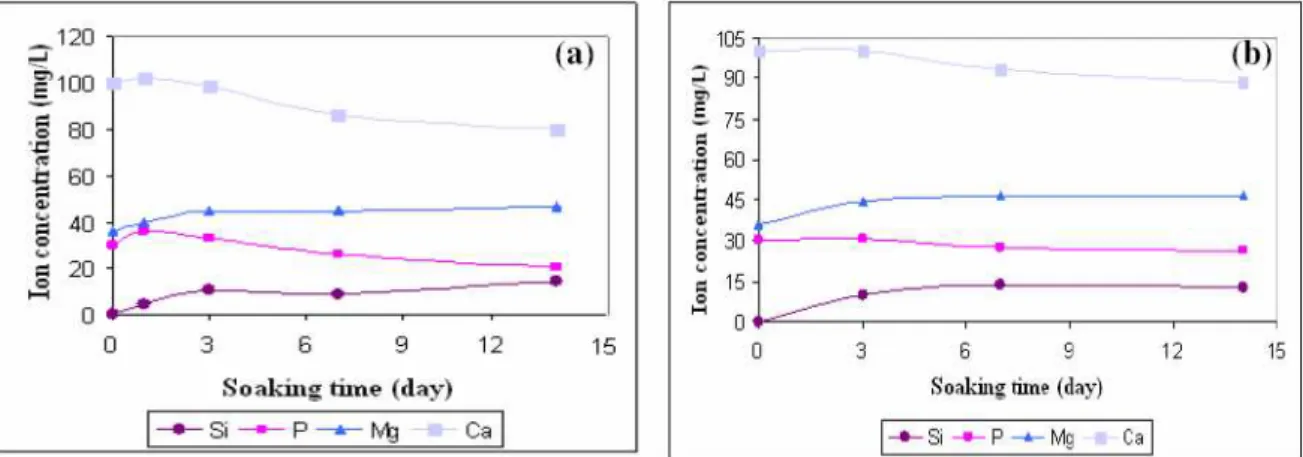
Changes in the concentrations of calcium, silicon, phosphorous and magnesium ions in SBF and SBFA due to immersion of the composites are shown in Fig. 1. The calcium and phosphate ions are required for the calcium phosphate layer generation. The reduction of their concentrations in SBF indicates the formation of CaP on the surface of the composite [12-14]. Initial increases in Si and Mg take place due to filler dissolution.
When BSA was present in the solution, the consumption of Ca and P was greatly decreased when compared to the samples immersed in pure SBF. However, a slight decrease in both (Ca and P) concentrations indicates that the CaP nucleation was not totally prevented in the presence of BSA.
Figure 1: Variation of ionic concentrations (a) in SBF and (b) in SBFA.
3.2 Formation of Apatite Layer
Further information on the structure of the CaP layer was obtained by the XRD and SEM-EDS measurements. Figure 2 shows the X-ray diffraction patterns of composites before and after soaking in SBF and SBFA for several times. The peaks started to appear after 3 days of imersion and sharpened for the longest period. The XRD spectra of composite exhibited the characteristic peaks at 2θ= 26° and 32°, attributable to hydroxyatite [3, 5, 17]. These results indicate that apatite crystals are deposited on the surface of material. However, the diffraction peaks are less defined for tests in the presence of albumin; therefore, albumin induces the formation of deposits with low cristallinity. On the other hand, the low intensity of the peaks may be due to a lower thickness of the calcium phosphate layer.
Figure 2: XRD patterns of the composite soaking (a) in SBF and (b) in SBFA.
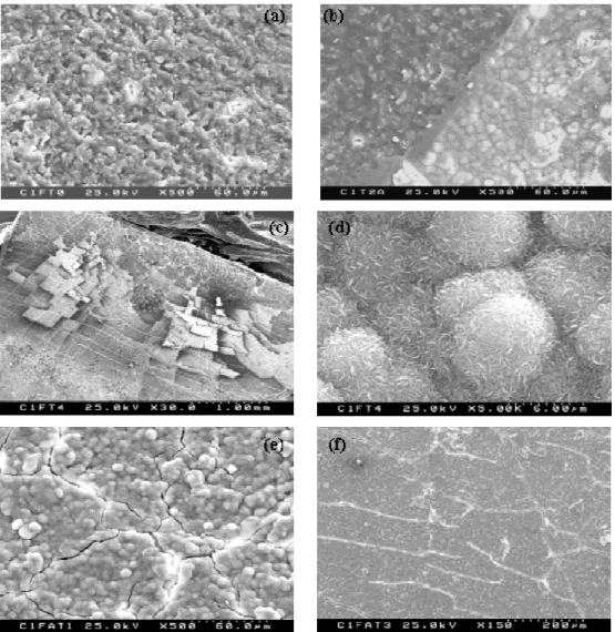
Figure 3: SEM microghaphs (a) before immersion, (b) after 3 days in SBF, (c) 14 days in SBF, (d) is a magnification of (c), (e) after 3days in SBFA (f) 14 days in SBFA.
The relative concentration in silicion, magnesium, calcium and phosphorus in the deposits are determined by EDS analyses. We observed for both cases (SBF and SBFA) a decrease in the silicion and magnesium content and an increase in calcium and phosphorus contents with the soaking periods. After 14 days the calculated Ca/P ratio was approximately 1,45 and 1,53 for SBF and SBFA respectively.
4 CONCLUSIONS
Composites of PMMA-co-EHA matrix filled with 61,33% 3CaO.P2O5 - 23,03% SiO2 - 15,64%
MgO suitable to be used as biomaterials have been studied. The combined application of ICP, XRD and SEM-EDS techniques allowed monitoring the formation of the layer, after immersion in SBF and also SBFA, as being constituted by calcium and phosphorus. The results indicate that addition of albumin in SBF did not prevent the calcium phosphate nucleation. Moreover, when the protein is present in solution, the effect on crystallization of the layer may be inhibited.
5 ACKNOWLEDGMENT
The author acknowledges the collaboration of Mr. Eugenio Soares and LCA, University of Aveiro, by ion concentration analyses (ICP). Conceição B. P. Costa (X-ray Diffraction) and Marta Carmona (Electron Microscopy) for valuable technical and professional assistance.
6 REFERENCES
[1] DEB, S., AIYATHURAI, L., ROETHER, J. A., et al., “Development of High-Viscosity, Two-Paste Bioactive Bone Cements”, Biomaterials, v. 26, pp. 3713-3718, 2005.
[2] LEITE FERREIRA, B. J. M., DUARTE, M. G. M., GIL, M. H., et al., “In Vitro Bioactivy in Glass-Ceramic/PMMA-co-EHA Composites”, Key Engineering Materials, v. 254-256, pp. 581-584, 2004. [3] LADRÓN DE GUEVARA-FERNÁNDEZ, S., RAGEL, C.V., VALLET-REGÍ, M., “Bioactive Glass-Polymer Materials for Controlled Release of Ibuprofen”. Biomaterials, v. 24, pp. 4037-4043, 2003. [4] MÉNDEZ, J. A., FERNÁNDEZ, M., GONZÁLEZ-CORCHON, A., et al., “Injectable Self-Curing
Bioactive Acrylic-Glass Composites Charged Specific Anti-Inflammatory / Analgesic Agent”.
Biomaterials, v. 25, pp. 2381-2392, 2004.
[5] OHTSUKI, C., MIYAZAKI, T., KYOMOTO, M., et al., “Development of Bioactive PMMA-Based Cement by Modification with Alkoxysilane and Calcium Salt”. Journal Material Science: Material
Medicine, v. 12, pp. 895-899, 2001.
[6] ARCOS, D., RAGEL, C. V., VALLET-REGÍ, M., “Bioactivity in Glass/PMMA Composites Used as Drug Delivery System”, Biomaterials, v. 22, pp. 701-708, 2001.
[7] AREVA, S., PELTOLA, T., SAILYNOIJA, E., et al., “Effect of Albumin and Fibrinogen on Calcium Phosphate Formation on Sol-Gel-Derived Titânia Coatings In Vitro”, Chemical and Materials, v. 14, pp. 1614-1621, 2002.
[8] MARQUES, P.A.A.P., SERRO, A.P., SARAMAGO, B.J., et al., “Mineralisation of Two Phosphate Ceramics in HBSS: Role of Albumin”, Biomaterials, v. 24, pp. 451-460, 2003.
[9] ROACH, P., FARRAR, D., PERRY, C.C., “Interpretation of Protein Adsorption: Surface-Induced Conformation Changes”. Journal of the American Chemical Society, v. 127, pp. 8168-8173, 2005. [10] MANSUR, H.S., ORÉFICE, R.L., LOBATO, Z.P., et al., “Adsorption/Desorption Behavior of Bovine
Serum Albumin and Porcine Insulin on Chemically Patterned Porous Gel Networks”, Kluwer
Academic Publishers, Manufactured in The Netherlands, pp. 105-116, 2001.
[11] VALLET-REGÍ, M., RAGEL, C. V., SALINAS, A. J., “Glasses with Medical Applications”, Journal of
Inorganic Chemistry, pp. 1029-1042, 2003.
[13] SILVA JR, P.E., OREFICE, R.L., “Compósitos Bioativos Obtidos a Partir da Inserção de Vidro Bioativo em Matriz de Poli(Metacrilato de Metila)”, Polímeros: Ciência e Tecnologia, v. 11, pp. 109-115, 2001.
[14] VALLET-REGÍ, M., ROMERO, A. M., RANGEL, C.V., et al., “XRD, SEM-EDS, and FTIR Studies of In Vitro Growth of an Apatite-Like Layer on Sol-Gel Glasses”, Journal of Biomedical Materials
Research, v. 44, pp. 416-421, 1999.
[15] KOKUBO, T., KIM, HM; KAWASHITA, M., “Novel Bioactive Materials with Different Mechanical Properties”, Biomaterials, v. 24, pp. 2161-2175, 2003.
[16] TAKADAMA, H., KIM, HM., KOKUBO, T., “X-ray Photoelectron Spectroscopy Study on the Process of Apatite Formation on a Sodium Silicate Glass in Simulated Body Fluid”. Journal of the American
Ceramic Society, v. 8, pp. 1933-1936, 2002.