Original article
RBAFSSOCIEDADE BRASILEIRA DE ATIVIDADE FÍSICA E SAÚDE
Brazilian Journal of Physical Activity and Health
RBAFS
Revista Brasileira de Atividade Física & Saúde
SOCIEDADE BRASILEIRA DE ATIVIDADE FÍSICA E SAÚDE
Brazilian Journal of Physical Activity and Health
Effect of aerobic physical training on cardiac
vagal reactivation in young sedentary
Efeito do treinamento físico aeróbio sobre
a reativação vagal cardíaca em jovens
sedentárias
Mário Augusto Paschoal1Tuanny Teixeira Pinheiro2,3
Gabriela Mariani Brigliador4
Thaís Maria Alvarenga Caruso5
Layse Nakazato Guedes de Lima2 Abstract
The objective was to compare the cardiac parasympathetic behavior during the recovery phase of an incremental exercise (IE), before and after an aerobic training (AT) program. For this, fifteen healthy sedentary young people, aged between 18 and 25 years, underwent the IE in treadmill with initial velocity of 4.0 km/h and increments of 1.0 km/h/min until exhaustion. After the MIE the heart beats were recorded during 10min. The register was sent to a computer to be processed the heart rate variability (HRV) analysis using the index pNN50, RMSSD and HF (u.n.) of the times 0-5min and 5-10min post-effort. After, underwent an AT with 12 sessions of 40min at intensity equivalent to 65% of HR peak. Subsequently, the IE was repeated until they reached the same speed of IE of the first phase before AT. The data were compared using Krus-kal-Wallis test with significant level of p<0.05. The HRV analysis 0-5min and 5-10min showed no differences between the data, with: a) 0-5min: pNN50 (0.3±0.7 % pre AT and 0.4±1.1 % post AT), RMSSD (8.4±5.5ms pre AT and 9.6±7.5ms post AT), HF(u.n.) 27.6±17.0% pre AT and 28.2± 13.8% post AT); and b) 5-10min: pNN50 (0.1±0.4% pre AT and 0.4±0.8% post AT), RMSSD (8.0±4.6ms pre AT and 10.6±7.9ms post AT), HF(u.n.) 27.6±18.3% pre AT and 29.8± 17.5% post AT). The application of a short duration AT was not effective to increase the parasympathetic nervous system interference on the heart during the recovery phase after IE.
Keywords
Parasympathetic nervous system; Exercise; Heart rate. Resumo
O objetivo foi comparar o comportamento parassimpático cardíaco durante a fase de recuperação de um exercício incremental (EI), realizado antes e após programa de treinamento aeróbio (TA). Para isso, 15 jovens sedentárias saudáveis, com idades entre 18 e 25 anos, se submeteram a um EI em esteira, com ve-locidade inicial de 4,0Km/h e acréscimos de 1,0Km/h/min, até atingirem a exaustão. Após, seus batimentos cardíacos foram registrados durante 10min e enviados a um computador para processamento da análise da variabilidade da frequência cardíaca (VFC), pelos índices pNN50, RMSSD e AF (u.n.) dos tempos 0-5min e 5-10min pós-esforço. Depois, submeteram-se a um TA de 12 sessões de 40min com intensidade equivalente a 65% da FC pico. Após, o EI foi repetido até que atingissem a mesma velocidade do EI da fase pré TA. Os dados pré e pós TA foram comparados por meio do teste de Kruskal-Wallis. As análises da VFC 0-5min e VFC 5-10min, não mostraram diferença entre os dados, com: a) 0-5min: pNN50 (0,3±0,7 % pré TA e 0,4±1,1 % pós TA), RMSSD (8,4±5,5ms pré TA e 9,6±7,5ms pós TA), AF(u.n.) 27,6±17,0% pré TA e 28,2± 13,8% pós TA); e b) 5-10min: pNN50 (0,1±0,4 % pré TA e 0,4±0,8 % pós TA), RMSSD (8,0±4,6ms pré TA e 10,6±7,9ms pós TA), AF(u.n.) 27,6±18,3% pré TA e 29,8± 17,5% pós TA). A apli-cação do programa de TA de curta duração não se mostrou efetiva na ampliação da interferência do sistema nervoso parassimpático sobre o coração durante a fase de recuperação após EI.
Palavras-chave
Sistema nervoso parassimpático; Exercício; Frequência cardíaca.
Rev Bras Ativ Fis Saúde p. 403-413 DOI: http://dx.doi.org/10.12820/1413-3482.2012v17n5p403
1 Faculdade de Fisioterapia, PUC-Campinas, Campinas, SP, Brasil
2 Faculdade de Fisioterapia, PUC-Campinas, Campinas, SP, Brasil
3 Residente da UNIFESP, São Paulo, SP, Brasil 4 Aluna de Iniciação Científica – bolsista PIBIC/CNPq
5 Aluna de Iniciação Científica – bolsista FAPIC/PUC
IntRoductIon
Over recent decades, the interest in studying heart rate variability (HRV) as an index for evaluating autonomic cardiac function and the prognosis for diseases of the cardiovascular system has increased enormously1. These studies have
una-nimously found that lower HRV is related to a poor prognosis and contributes towards predicting mortality2-5.
Within this context, studies seeking to evaluate HRV and heart rate (HR) have started to appear, with analysis not only at rest but also during stable dyna-mic exercise and during recovery periods following intense incremental effort1,6-8.
It has been observed that HR elevation during effort and its reduction during the post-effort phase are modulated by the autonomous nervous system9,10. Equally, it
has been seen that lower HR recovery is also a strong predictor of mortality1 and
sudden death11.
Specifically in relation to the autonomic cardiac adjustments that occur during the period immediately after an intense effort, it is known that in order to promote a reduction in HR values to a condition close to what was observed before the effort, there is a progressive elevation of parasympathetic cardiac activity relating to the time that has elapsed between the end of the effort and the recovery period9,10,12-17.
However, although studies have sought to establish relationships between trai-ning volume and the magnitude of the reduction in the HR value after effort1,15,
and between the latter and autonomic modulation over this period, few studies have be concerned with investigating whether short-duration aerobic training programs would be efficient for promoting changes to the magnitude of post-ef-fort vagal reactivation in healthy individuals.
Thus, the present study sought, through HRV analysis, to investigate the phe-nomenon of post-effort cardiac vagal reactivation, before and after a short pro-gram of aerobic training, in order to ascertain whether it might interfere with the HR responses documented.
Secondarily, this investigation sought to provide new support regarding the question of the duration of aerobic training needed for possible changes to vagal modulation to become established in the heart.
In this regard, according to the responses obtained, new proposals relating to the number of aerobic training sessions needed for improvement of autonomic cardiac modulation might be established. This might be needed in situations such as preparation of patients for cardiac surgery or recovery processes subsequent to acute myocardial infarction.
Because of these issues and their clinical-functional relevance, it can be un-derstood that even though the results would be obtained from healthy individuals, they might serve as parameters for scheduling treatment, physical training and correlated studies.
Methods
This longitudinal study was conducted between August 2010 and June 2011 at the physiotherapy outpatient clinic of the Pontifical Catholic University of Campinas, state of São Paulo, and was approved by the Ethics Committee for Research In-volving Human Beings of the university’s Life Sciences Center, under protocol no. 757/09. The investigation was conducted within the ethical standards required by
the Declaration of Helsinki of 1964, and in accordance with Resolution 196/96 of the Brazilian Ministry of Health.
The following stages were followed:
Selection of volunteers
This was done in accordance with preestablished inclusion criteria. From among the individuals who came for anthropometric and clinical examinations, 15 volun-teers aged 18 to 25 years who were students at our university were chosen.
The inclusion criteria were that all the subjects should be sedentary, as assesed through a brief interview; should not be smokers; should not be using medications that might interfere with cardiorespiratory responses; should not have any cardio-respiratory diseases, as confirmed through the clinical examinations performed; should not be obese, as shown by their body mass index (BMI); and should not be pregnant.
Anthropometric evaluation
This was conducted to make height and weight measurements so as to calculate BMI. The BMI values selected as the inclusion criterion were between 20 and 30 kg/m2, in order to avoid the presence of obesity, which is considered to be a
fac-tor that alters cardiac autonomic modulation18. For this evaluation, the volunteers
were positioned without footwear on a Filizola® scale (São Paulo, Brazil) that was
precalibrated in 100-gram steps. Height was also measured while the subject was standing on this apparatus, by means of a metal rod marked out in centimeters.
Clinical assessment
HR and arterial blood pressure (ABP) measurements were made with the volun-teer in dorsal decubitus, after a three-minute resting period. Lung and heart sou-nds were also investigated by means of a stethoscope. HR was measured by means of palpation of the radial wrist, over a one-minute period, and ABP was measured in accordance with the norms of the Sixth Brazilian Hypertension Guidelines19,
differing only in relation to the volunteer’s body position that was used for the measurements. A standard mercury-column sphygmomanometer (Wan Med®, São Paulo, Brazil) and a stethoscope (Littman Classic II®, Sumaré, Brazil) were used.
After the clinical assessment, each volunteer was given an information docu-ment explaining the restrictions that they should adhere to, starting 24 hours be-fore undergoing the incremental exercise (IE) protocol. These requirements were as follows: no consumption of chocolate, coffee, foods containing caffeine or soft drinks containing cola; no exercising beyond the ordinary day-to-day activities; to try to have at least eight hours of sleep on the night preceding the IE; and to avoid using any medication.
Moreover, the IE protocol could not be done over the period from three days before menstruation (according to each volunteer’s menstrual cycle) to three days afterwards.
Effort protocol performed before and after the aerobic training (AT)
program (incremental exercise, IE)
The protocol followed before the AT program was developed on a treadmill (Super ATL, Inbrasport®, Porto Alegre, Brazil), with an initial velocity of 4.0 km/h, main-tained for two minutes, followed by increases of 1.0 km/h every minute thereafter until the volunteer was exhausted20.
The same protocol was repeated after the AT, and was halted at the time when the volunteer reached the same velocity or absolute load that had been attained before the AT program.
During the IE, all heartbeats were recorded using a HR meter (Polar S180i®, Kempele, Finland) with the aim of calculating the value of 65% of the peak HR obtained through this protocol. These records were then transmitted to a compu-ter through an incompu-terface that enabled graphical presentation of the normal R-R intervals (iRR) relating to the effort protocol, by means of the Polar Precision Per-formance® software (Kempele, Finland).
Data-gathering in relation to analysis of cardiac parasympathetic activity
At the end of the effort protocols, the volunteers left the treadmill and walked around for one minute to recover. They then lay down on a mattress and remained there for another nine minutes. Throughout this period, their heartbeats were re-corded for data analysis.These data were gathered during both post-effort phases (before and after AT), so that the data on cardiac parasympathetic modulation could be compared. The HRV analyses for these periods were performed both in the time domain (TD) and in the frequency domain (FD), and the calculations were done by applying the fast Fourier transform algorithm. In this, the values for the high-frequency (HF) variable were calculated in normalized units (nu).
It was decided to calculate the vagal modulation in normalized units because this format makes it possible to express the percentage contribution of the pa-rasympathetic nervous system in relation to the total potential. This excludes the very low frequency (VLF) band, since this band has dubious value in short-dura-tion records21.
The Polar Precision Performance® software was used for the HRV analysis. The
calculation used for obtaining values on normalized units was as follows: HF/ (total potential – VLF) x 100. This followed the Task Force recommendations21.
The values of the HRV parameters relating to the records covering the 10-mi-nute period after completing the effort protocols before and after the AT were analyzed comparatively as two periods: 0 to 5 min and 5 to 10 min.
In the time domain analysis, parameters that reflected the vagal modulation on the heart were evaluated, including the following: the standard deviation values for the normal RR intervals (SD of the iRR, in ms); the square root of the mean of the sum of the squares of the differences between adjacent normal iRR values (rMSSD, in ms); and the percentage of adjacent iRR values with a difference in duration greater than 50 ms (pNN50).
In the frequency domain, the high-frequency (HF) spectral components, i.e. the band between 0.15 Hz and 0.4 Hz, which also expresses cardiac vagal activity, and the ratio between the low and high-frequency components (LF/HF) of the HRV were evaluated21.
Data-gathering in relation to HR values
The mean HR relating to the time intervals of 0-5 min and 5-10 min of the re-covery phase subsequent to incremental effort and the total HR (total number of heartbeats relating to the periods) were extracted from the reports generated by the software and are presented in Table 2.
in-vestigation was that the mean HR consisted of mean values for the variable over the time periods of 0-5 min and 5-10 min after the incremental effort, and did not show an absolute value over these periods. However, total HR showed the real value for the number of heartbeats over the entire period, thereby providing further data for illustrating the behavior of the variable in question.
To document the rapid reduction in HR immediately after cessation of effort, this was analyzed at the times of 15 s, 30 s, 60 s and 120 s (Figure 1). Likewise, the median values for delta HR were calculated from the ratios between peak HR and the HR after one and two minutes of the post-effort recovery phase (Figure 2).
Aerobic training (AT)
This consisted of 12 sessions of 40 minutes in duration, of which 20 minutes were on a treadmill and 20 minutes were on a cycle ergometer (Johnson JPB 5100®, Shanghai, China), without intervals and performed three times a week on alterna-te days. It should be emphasized that all the volunalterna-teers complealterna-ted the 12 sessions of AT.
Although the IE was developed on a treadmill and part of the training was de-veloped on a cycle ergometer, the intensity prescribed for this ergometer was also controlled by means of cardiac monitoring (FT1 Polar®, Kempele, Finland). In
this, target zones for the training were established, limited by HRs of 5 bpm above and below the individually calculated value. If the HR were to leave this zone, the apparatus would produce a signal warning the volunteer to make adjustments to adhere to the expected effort level.
Statistical analysis
The anthropometric and clinical data were presented in tables with means and standard deviations, solely to describe the characteristics of the sample used in this study.
The HR values of the recovery phase after the maximum effort, before and after the AT, were analyzed regarding their distributions. Through logarithmic transformation, these values were shown to have non-normal characteristics and were then analyzed by means of the Kruskal-Wallis test, with a significance level established at p < 0.05. For this, the Graph Pad Prism 4.0® software was used (San
Diego, USA).
The same test was used to analyze the data relating to cardiac parasympathetic activity, by means of the HRV indices.
Results
Table 1 (below) presents the ages, anthropometric data and clinical data on the study participants. It can be seen that these parameters were appropriate for heal-thy individuals, since they are within the limits of normality.
Table 2 shows the means and standard deviations relating to the time and frequency domains of the HRV, corresponding to the heartbeat records of the first and last five-minute periods of the recovery phase subsequent to IE, with compa-risons between the times before and after AT.
The data were compared within and between the times before and after AT, involving the times from 0 to 5 min after the incremental test (named the initial
five minutes) and the times from 5 to 10 min after the incremental test (named the final five minutes. In comparing the times before and after AT, no differences were observed in any of the variables, while in comparing 0-5 min and 5-10 min before AT and comparing 0-5 min and 5-10 min after AT, the differences (p < 0.05) were limited to the variables of iRR, mean HR and total HR.
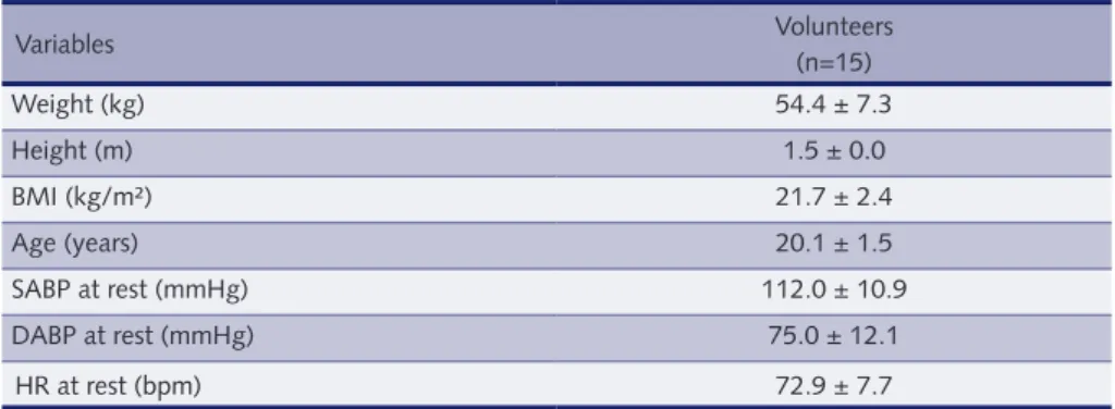
table 1 – Means and standard deviations of the volunteers’ ages and anthropometric and clinical data.
Variables Volunteers (n=15) Weight (kg) 54.4 ± 7.3 Height (m) 1.5 ± 0.0 BMI (kg/m²) 21.7 ± 2.4 Age (years) 20.1 ± 1.5 SABP at rest (mmHg) 112.0 ± 10.9 DABP at rest (mmHg) 75.0 ± 12.1 HR at rest (bpm) 72.9 ± 7.7
BMI = body mass index; SABP = systolic arterial blood pressure; DABP = diastolic arterial blood pressure; HR = heart rate
table 2 – Means and standard deviations of the variables studied, in the time domain and frequency domain, corresponding to the initial five minutes (0-5 min) and final five minutes (5-10 min) after the incremental tests, before and after aerobic training.
Variables
Initial 5 min (0-5 min) after test,
before AT (n = 15) Initial 5 min (0-5 min) after test, after AT (n = 15) Final 5 min (5-10 min) after test, before AT (n = 15) Final 5 min (5-10 min) after test, after AT (n = 15) iRR (ms) 479.0 ± 50.7 498.1 ± 52.7 593.6 ± 58.2* 618.7 ± 71.7** pNN50 (%) 0.3 ± 0.7 0.4 ± 1.1 0.1 ± 0.4 0.4 ± 0.8 rMSSD (ms) 8.4 ± 5.5 9.6 ± 7.5 8.0± 4.6 10.6 ± 7.9 Mean HR (bpm) 126.7 ± 13.4 121.6 ± 11.5 102.1 ± 10.3* 98.1 ± 10.2** Total HR (bpm) 635.3 ± 66.8 610.5 ± 58.4 507.7 ± 54.9* 491.7 ± 50.9** HF (nu) % 27.6 ± 17.0 28.2 ± 13.8 27.6 ± 18.3 29.8 ± 17.5 LF/HF ratio 4.5 ± 3.6 3.6 ± 2.8 4.8 ± 4.7 5.3 ± 8.0 iRR = R-R intervals; ms = milliseconds; pNN50 = percentage of adjacent iRR values with difference in duration greater than 50 ms; rMSSD = square root of the mean of the sum of the squares of the differences between adjacent normal iRR values; HR = heart rate; bpm = beats per minute; LF = low frequency; HF = high frequency, in normalized units (nu); AT = aerobic training. * p < 0.05 in comparing the values for the initial and final five-minute periods before aerobic training (AT). ** p < 0.05 in comparing the values for the initial and final five-minute periods after aerobic training (AT).
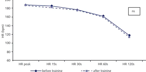
The median HR values obtained at the peak effort and at 15 s, 30 s, 60 s and 120 s after the effort before and after AT are shown in Figure 1. It can be seen that the curve for HR behavior had practically the same shape before and after the 12 training sessions that the volunteers attended. For this reason, no diffe-rence was observed over the first two minutes of the recovery phase after the incremental effort.
Figure 2 shows the delta HR values relating to the decreases in HR one and two minutes after the effort. They were extracted by subtracting the peak HR value from the HR values observed at these times. It should be noted that there was no difference between the data.
Figure 1 – Median HR values obtained at the peak effort and 15 s, 30 s, 60 s and 120 s afterwards, before and after the 12 sessions of aerobic training among healthy sedentary volunteers.
-90 -80 -70 -60 -50 -40 -30 -20 -10 0 HR ( bpm) before AT after AT
afterwards: peak to 1 min afterwards: peak to 2 min
Figure 2 – Comparison of median values for delta HR reductions. In white, the amount of the reduction in HR from the end of the incremental test (time zero) to the end of the first minute after the effort, both before and after the period of short-duration aerobic training (AT) (12 sessions). In grey, the amount of the reduction in HR from the end of the incremental test (time zero) to the end of the second minute after the effort, both before and after the period of short-duration aerobic training (12 sessions).
dIscussIon
The main finding from the present study is that the AT that was proposed, con-sisting of only 12 sessions and done at a relative intensity of 65% of peak HR, did not give rise to modification of cardiac parasympathetic modulation during the recovery phase following IE.
The total time and the training schedule and its intensity can be highlighted as the main factors that may have interfered with the results22. These factors are
widely known to alter the organic responses to physical training, but they cannot be tested or controlled for in a single study.
In this regard, Uusitalo et al23 had already reported that undertaking an
exer-cise program of mild intensity would be enough for healthy adults to present some improvement in autonomic cardiac function. However, they stated that there might be large variations in these modifications, which would make it difficult to document them accurately.
Yamamoto et al10 monitored the HR responses among seven healthy
analysis, whether the autonomous nervous system might be the agent responsible for diminishing the post-effort HR as a result of adaptation to training. The in-tensity applied was greater than that of the present study (80% of VO2 max), and
changes to autonomic modulation of HR were detected after one week of training. Therefore, as laid out above, exercise intensity is without doubt one factor to be considered in all studies along this line, since the biological responses are different when the stimuli are also different.
Corroborating the above assertion, other studies23-30 have also shown a
signi-ficant relationship between modifications in vagal-sympathetic modulation and physical training done by healthy individuals. In these studies, increased cardiac parasympathetic activity was revealed, with or without diminished sympathetic activity. However, they were all conducted for longer times than that of the present study and, it must be noted, the change in parasympathetic tonus was not docu-mented during the recovery period after intense effort but, rather, in standardized autonomic functional tests.
One issue that probably has limited the development of studies that might confirm the existence of increased vagal modulation on the heart under post-effort conditions is that there is a transient reduction in HR during the first instants af-ter the activity, which thus impairs HRV analysis using the fast Fourier transform. For this algorithm to be effective, a condition of stationarity of the tracing is nee-ded21. In fact, some studies31 have proposed using a short-time Fourier transform,
with a method resembling the fast Fourier transform, in order to analyze dynamic modifications to the HRV during non-stationary periods. In this manner, possib-le imprecisions resulting from analyses performed during non-stationary periods would be controlled for.
Nevertheless, despite this possible interference that tends to occur during the initial moments after the effort when the analysis is done using the traditional me-thod31, we believe that this did not have any significant influence on the data, since
these were monitored until 10 minutes after the end of the incremental protocol. In addition to these methodological issues involving the initial moments of the post-effort phase, some authors have taken a more pragmatic stance in this regard, and have suggested that the HRV would remain small for 30 minutes after intense effort32. This would make it difficult to detect subtle differences in cardiac
parasympathetic tonus with this toll during this period of investigation. Hence, further studies to assess these issues better are justified.
Another point analyzed in the present study, relating to the characteristics of the physical training undertaken (and which therefore might interfere with the HRV analyses), is the velocity of the exponential decline in HR. This reduction in HR may result from various factors, such as: an effect from the intrinsic properties of the circulation33; adjustments that depend on the magnitude of the venous
re-turn and the depth and frequency of respiration20; the integrated sum of the
reac-tivation of vagal tonus and withdrawal of sympathetic tonus16; and the cessation of
inputs coming from the central command and from the afferent signals that origi-nate in the mechanical receptors of the skeletal musculature involved in the work34.
All these factors together are activated during the recovery phase following physical effort, and the type, intensity and frequency of the physical training used modify these responses to a greater or lesser extent.
The results from the HRV analysis and the HR responses presented in Table 2 can be partially compared with those from studies like that of Uusitalo et al35.
The-se authors evaluated female athletes who performed exhaustive endurance training for six to nine weeks and also did not show modifications in cardiac autonomic control or in intrinsic HR. The fact that these individuals were athletes is a factor that needs to be taken into consideration, since the possible cardiac autonomic modifications would already be present and would be unlikely to change. In this respect, they different from sedentary individuals like those of the present study, whose initial response to physical training always tends to be more effective29.
It needs to be emphasized that in the present study there were no differences in analyzing the HRV after effort, comparatively between the time periods of 0-5 min and 5-10 min, either before or after AT. On the other hand, the mean HR and total HR were found to be different between the periods of 0-5 min and 5-10 min, both before and after AT. In other words, comparing the periods of 0-5 min and 5-10 min only before AT or only after AT, they differed.
However, when these data were compared over the same period, such as 0-5 min before AT compared with 0-5 min after AT, it was seen that there was no significant difference. This suggests that the differences in HR and total HR only related to the duration of time over which the variables were recorded, and not a possible effect resulting from the training undertaken.
In relation to the median HR values observed between 0 and 120 s after the incremental effort (Figure 1) and between the delta peak HR values at the ends of the first and second minutes after the effort (periods that are considered to be important with regard to predictions of cardiovascular diseases)2,3, there was no
difference in the comparison between before and after AT.
In this regard, the volunteers were shown to be healthy, taking into considera-tion the affirmaconsidera-tions of Cole et al2 and Nishime et al4. These authors reported that
in cases in which the documented reductions in heartbeat were not more than 12 beats, during the period of the first minute after the effort, there was a significant relationship with increased mortality rate.
In the present investigation, the delta peak HR values up to the first minute after the effort (Figure 2) did not differ from before to after the AT, such that they were -23.6 bpm before AT and -29.7 bpm after AT. Thus, these were greater than what Cole et al2 put forward as a cardiovascular risk factor.
The delta HR values are concordant with what was presented by Sugawara et al36. Although these authors documented higher delta HR values at the initial
moments of the recovery, in comparing from before to after physical training, the duration of training used in their investigation was twice what was used in the present study.
Likewise, the delta peak HR values up to the second minute after effort (Figu-re 2) confirmed that the volunteers’ cardiovascular function was normal, such that both before and after AT, there was a continuous decline in HR over this period.
Regarding limitations of the present study, the absence of a control group can be highlighted. A control group might have provided greater support for discus-sing the results. Furthermore, the data from the present study do not apply to populations of elderly people37 or people who have already undergone training38,
since it is known that the vagal reactivation after effort in young women with little aerobic capacity is different from what is found in those populations. Likewise, because the present study focused only on possible modifications to cardiac auto-nomic function resulting from the length of exposure to training, other hypotheses such as different intensities and/or volumes of training were not tested.
Other points to be considered are the possibility that the volunteers might be-come familiarized with the effort protocol and the short period of HRV analysis after the effort.
conclusIon
Application of a short AT program consisting of 12 sessions, done three times a week on alternate days, at an intensity equivalent to 65% of peak HR, was not shown to be sufficiently effective for amplifying the interference magnitude of the parasympathetic response on the heart, as measured using HRV analysis during the immediate recovery period subsequent to incremental physical effort, among healthy sedentary young adults.
In addition to this, new studies with different proposed intensities, times and durations of aerobic physical training should be conducted and compared. For these reasons, the present study may be important for serving as a parameter for future investigations along this line.
RefeRences
1. Goldberger JJ, Le FK, Lahiri M et al. Assessment of parasympathetic reactivation after exer-cise. Am J Physiol Heart Circ Physiol 2006;290:H2446–H2452.
2. Cole CR, Blackstone EH, Pashkow FJ, Snader CE, Lauer MS. Heart rate recovery immedi-ately after exercise as a predictor of mortality. N Engl J Med 1999;341:1351-1357.
3. Cole CR, Foody JM, Blackstone EH, Lauer MS. Heart rate recovery after submaximal testing as a predictor of mortality in a cardiovascular healthy cohort. Ann Intern Med 2000;132:552-555. 4. Nishime EO, Cole CR, Blackstone EH, Pashkow FJ, Lauer MS. Heart rate recovery and
treadmill exercise score as predictors of mortality in patients referred for exercise ECG. JAMA 2000;284:1392-8.
5. Hull SS Jr, Vanoli E, Adamson PB et al. Do increases in markers of vagal activity imply protec-tion from sudden death?: The case of scopolamine. Circulaprotec-tion 1995;91:2516–2519.
6. Gregoire J, Tuck S, Yamamoto Y, Hughson R. Heart rate variability at rest and exercise: influ-ence of age, gender and physical training. Can J Appl Physiol 1996;21(6):455-70.
7. Paschoal MA, Gonçalves NVO, Petrelluzzi KFS, Machado RV. Controle autonômico cardíaco durante a execução de atividade física dinâmica de baixa intensidade. Rev Soc Cardiol Estado de São Paulo 2003;5(supl A):1-11.
8. Paschoal MA, Siqueira JP, Machado RV, Petrelluzzi KFS, Gonçalves NVO. Efeitos agudos do exercício dinâmico de baixa intensidade sobre a variabilidade da frequência cardíaca e pressão arterial de indivíduos normotensos e hipertensos leves. Rev Ciênc Méd 2004;13(3):223-234. 9. Borrensen J, Lambert MI. Autonomic control of heart rate during and after exercise:
measure-ments and implications for monitoring training status. Sports Med 2008;38(8):633-46. 10. Yamamoto K, Miyachi M, Saito T, Yoshioka A, Onodera S. Effects of endurance training on
resting and post-exercise cardiac autonomic control. Med Sci Sports Exerc 2001;33(9):1496-502. 11. Jouven X, Empana J, Schwartz P et al. Heart-rate profile during exercise as a predictor of
sud-den death. N Engl J Med 2005;352:1951-1958.
12. Ng J, Sundaram S, Kadish AH, Goldberger JJ. Autonomic effects on the spectral analysis of heart rate variability. Am J Physiol Heart Circ Physiol 2009;297:H1421-H1428.
13. Ricardo DR, Almeida MB, Franklin BA, Araújo CGS. Initial and final exercise heart rate transients: influence of gender, aerobic fitness and clinical status. Chest 2005;127(1):318-27. 14. Shelter K, Marcus R, Frolicker VF et al. Heart rate recovery: validation and methodology
issues. J Am Coll Cardiol 2001;38:1980-7.
15. Imai K, Sato H, Hori M et al. Vaguely mediated heart rate recovery after exercise is accelerated in athletes but blunted in patients with chronic heart failure. J Am Coll Cardiol 1994;24:1529-35. 16. Perini R, Orizio C, Comandè A et al. Plasma norepinephrine and heart rate dynamics during
17. Buchheit M, Gindre C. Cardiac parasympathetic regulation: respective associations with car-diorespiratory fitness and training load. Am J Physiol Heart Circ Physiol 2006;291: H451–H458. 18. Chethan HA, Murthy N, Basavaraju K. Comparative study of heart rate variability in normal
and obese young adult males. Int J Biol Med Res 2012; 3(2):1621-1623. 19. VI Diretrizes Brasileiras de Hipertensão. Rev Bras Hipertens 2010;17(1)11-17.
20. Wasserman K, Hansen JE, Sue DY, Whipp BJ, Casaburi R. Principles of exercise testing and interpretation. Lea & Febiger, 2a. ed, Philadelphia, USA, 1994.
21. Task Force of European Society of Cardiology and North American Society of Pacing and Electrophysiology. Heart rate variability – Standards of measurement, physiological interpre-tation, and clinical use. Circulation 1996;93:1043-1065.
22. Talanlan JL, Galloway SDR, Heigenhauser GJF, Bonen A, Spriet LL. Two weeks of high-in-tensity aerobic interval training increases the capacity for fat oxidation during exercise in wom-en. J Appl Physiol 2007;102:1439-1447.
23. Uusitalo ALT, Laitinen T, Väisänen SB, Länsimies E, Rauramaa R. Effects of endurance train-ing on heart rate and blood pressure variability. Cli Physiol & Func Im 2002;22:173-9. 24. Paschoal MA, Polessi EA, Simioni FC. Evaluation of heart rate variability in trained and
sed-entary climacteric women. Arq Bras Cardiol 2008;90(2):74-79.
25. Melanson EL, Freedson OS. The effect of endurance training on resting heart rate variability in sedentary adult males. Eur J Appl Physiol 2001;85:442-9.
26. Stein PK, Ehsani AA, Domitrovich PP et al. Effect of exercise training on heart rate variability in healthy older adults. Am Heart J 1999;138:567-76.
27. Levy WC, Cerquera MD, Harp GD et al. Effect of endurance exercise training on heart rate variability at rest in healthy young and older men. Am J Cardiol 1998;82:1236-41.
28. Al-Ani M, Munir SM, White M et al. Changes in R-R variability before and after endurance training measured by power spectral analysis and by the effect of isometric muscle contraction. Eur J Appl Physiol 1996;74:397-403.
29. De Meersman RE. Heart rate variability and aerobic fitness. Am Heart J 1993;125:726-31. 30. Gallo Jr L, Maciel BC, Marin-Neto JA et al. Sympathetic and parasympathetic changes in
heart rate control during dynamic exercise induced by endurance training in man. Brazilian J Med Biol Res 1989;22:631-43.
31. Mainardi LT, Bianchi AM, Cerutti S. Time-frequency and time-varying analysis for assessing the dynamic responses of cardiovascular control. Crit Rev Biomed Eng 2002; 30(1-3):175-217. 32. Javorka M, Zilla I, Balharek T, Javorka K. Heart rate recovery after exercise: relations to heart
rate variability and complexity. Braz J Med Bio Res 2002;35(8)991-1000.
33. Savin WM, Davidson DM, Haskell WL. Autonomic contribution to heart rate recovery from exercise in humans. J Appl Physiol 1982;53:1572-1575.
34. Rowell LB, O’Leary DS. Reflex control of the circulation during exercise: chemorreflexes and mechanoreflexes. J Appl Physiol 1990;69(2):407-18.
35. Uusitalo ALT, Uusitalo AJ, Ruscko HK. Exhaustive endurance training for 6-9 weeks did not change in intrinsic heart rate and cardiac autonomic modulation in female athletes. Int J Sports Med 1998;19:532-40.
36. Sugawara J, Murakami H, Maeda S, Kuno S, Matsuda M. Change in pos-exercise vagal reac-tivation with exercise training and detraining in young men. Eur J Appl Physiol 2001;85(3-4):259-63.
37. DeLorey DS, Kowalchuk JM, Paterson DH. Effect of prior heavy-intensity exercise on pulmo-nary O2 uptake and muscle deoxygenation kinetics in young and older adult humans. J Appl Physiol 2004;97(3):998-1005. Epub 2004 May 7.
38. Melo RC, Santos MDB, Silva E, Quitério RJ, Moreno MA et al. Effects of age and phys-ical activity on the autonomic control of the heart in healthy men. Braz J Med Biol Res 2005;38(9):1331-8.
Correspondence Address Prof. Dr. Mário Augusto Paschoal Faculdade de Fisioterapia da Pontifícia
Universidade Católica de Campinas Avenida John Boyd Dunlop s/n Campus II – Jardim Ipaussurama CEP: 13059-900 – Campinas, SP. e-mail: fisioni@puc-campinas.edu.br fone: (19) 3343-6820 Received 08/15/2012 Revised 10/17/2012 11/16/2012 12/21/2012 Accepted 12/21/2012