LETTER TO THE EDITOR
CD44 glycoprotein in cancer: a molecular
conundrum hampering clinical applications
Rita Azevedo
1,2, Cristiana Gaiteiro
1,2,3, Andreia Peixoto
1,2,4, Marta Relvas‑Santos
1, Luís Lima
1,4,
Lúcio Lara Santos
1,2,5,6and José Alexandre Ferreira
1,2,4,5,7,8*Abstract
CD44 is a heavily glycosylated membrane receptor playing a key role in cell adhesion, signal transduction and cytoskeleton remodelling. It is also one of the most studied glycoproteins in cancer, frequently explored for stem cell identification, and associated with chemoresistance and metastasis. However, CD44 is a general designation for a large family of splicing variants exhibiting different degrees of glycosylation and, potentially, functionally distinct roles. Moreover, structural diversity associated with ambiguous nomenclature has delayed clinical developments. Herein, we attempt to comprehensively address these aspects and systematize CD44 nomenclature, setting milestones for biomarker discovery. In addition, we support that CD44 may be an important source of cancer neoantigens, most likely resulting from altered splicing and/or glycosylation. The discovery of potentially targetable CD44 (glyco)isoforms will require the combination of glycomics with proteogenomics approaches, exploring customized protein sequence databases generated using genomics and transcriptomics. Nevertheless, the necessary high‑throughput analytical and bioinformatics tools are now available to address CD44 role in health and disease.
Keywords: CD44, Glycosylation, Cancer biomarkers, Nomenclature, CD44 isoforms
© The Author(s) 2018. This article is distributed under the terms of the Creative Commons Attribution 4.0 International License (http://creativecommons.org/licenses/by/4.0/), which permits unrestricted use, distribution, and reproduction in any medium, provided you give appropriate credit to the original author(s) and the source, provide a link to the Creative Commons license, and indicate if changes were made. The Creative Commons Public Domain Dedication waiver (http://creativecommons.org/ publicdomain/zero/1.0/) applies to the data made available in this article, unless otherwise stated.
The transmembrane glycoprotein receptor CD44 plays a key role in cell adhesion to the extracellular matrix and interacts with growth factors and several extracellular ligands, including hyaluronic acid, collagen, osteopontin and many metalloproteinases to drive signal transduc-tion and cytoskeleton rearrangements [1]. By interact-ing with co-factors and adaptor proteins, CD44 has been further implicated in lymphocyte homing, haematopoie-sis, cell migration and adhesion, tumour invasion and metastasis [1]. Several isoforms of CD44 can be gener-ated through the insertion of alternative exons at the variable region in a process regulated at both tissue and cellular levels. While ubiquitously expressed in healthy adult and foetal tissues, the molecular plasticity of alter-natively spliced CD44 accounts for diversified functional roles. However, the intricate correlation between CD44
isoforms and underlying biological functions is yet to be fully disclosed. It has been long described that malignant transformation and progression are accompanied by a deregulation of CD44 splicing mechanisms, comprehen-sively addressed in recent reviews [2, 3]. The events lead-ing to CD44 isoform molecular remodelllead-ing have direct implications in several cancer hallmarks and appear to vary according to the type of lesion, supporting the existence of disease-specific molecular fingerprints and potentially targetable biomarkers [4]. Not surprisingly, CD44 has been a hot topic in cancer research, frequently associated with more aggressive phenotypes and widely explored for cancer stem-cell identification [5]. Particular focus has been set on narrowing CD44 screening to its cancer-associated isoforms envisaging the necessary sen-sitivity and specificity for clinical applications. However, the lack of protocols for its full isoform discrimination at the protein level, and the existence of many function-ally distinct isoforms poses a major drawback. Currently, these hurdles can only be partially circumvented by tar-geting CD44 transcripts using variant-specific probes or
Open Access
*Correspondence: [email protected]‑saude.pt
1 Experimental Pathology and Therapeutics Group, Portuguese Institute of Oncology, Rua Dr. António Bernardino de Almeida, 4200‑072 Porto, Portugal
by emerging RNAseq approaches. Analytical difficulties are aggravated by vast post-translational modifications, with emphasis on the very high glycosylation density of variable regions. Moreover, glycans often present a non-templated and context-dependent nature, with several glycoforms coexisting for the same protein on a given biological milieu, further increasing CD44 molecular and functional diversity (Table 1). This poses a major chal-lenge for identification by conventional immunoassays as well as high-throughput proteomics, which has delayed the definition of CD44 isoforms in health and disease.
The absence of nomenclature standardization also emerges as a key issue, making inter-study comparisons and clinical translation almost impossible. Showcasing some examples, CD44H and CD44E terminologies arise from the first observations in hematopoietic and epi-thelial cells; gp116 and gp85 distinguish glycoforms by molecular weights (116 and 85 kDa, respectively); CD44 hematopoietic cell E-/L-selectin ligand (HCELL) refers to CD44 isoforms expressed in hematopoietic and cancer cells showing elevated sialofucosylated glycans content and high affinity for E-/L-selectin ligands [6, 7]. All these designations fail to provide clear insights on the molecu-lar nature of the isoforms. Notwithstanding, some studies adopt a nomenclature based on commercial names of the used monoclonal antibodies, highlighting the targeted variable exon, being CD44v3, CD44v6, CD44v9 amongst the most associated with cancer, including chemoresist-ance and prognosis [8, 9]. Moreover, these studies often disregard that the analysis of a specific variable exon can result in the detection of all isoforms containing it instead of one particular protein [10]. In addition, most variable regions may be significantly hindered by dense glycosylation, biasing detection. On the other hand, reports exploring the transcriptome have emerged as a powerful tool for determining CD44 isoforms diversity;
however, few have provided the necessary validation at the protein level due to above mentioned analytical diffi-culties. Another important source of nomenclature ambi-guity results from the direct translation of results from
Mus musculus studies to Homo sapiens disregarding that
mice CD44 gene presents an extra variable exon (v1) not present in humans. Further contributing to ambivalence, UniProt and NCBI protein databases adopt different des-ignations for the same isoform (illustrated in Table 1). Altogether these aspects impact negatively on our under-standing of the biological and clinical relevance of CD44 isoforms in cancer and other diseases, urging nomencla-ture standardization.
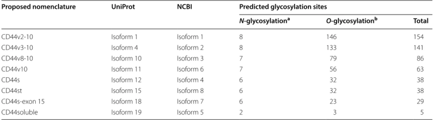
Facing these challenges, we have conducted a com-prehensive in silico analysis of CD44 isoforms through NCBI and UniProt databases, using BLAST and TMHMM Server v2.0 tools. The human CD44 gene is located on the short arm of chromosome 11 [GRCh38. p7, NC_000011.10 (35138870-35232402)] and its pre-cursor mRNA consists of 19 exons. Namely, exons 1–16 encode the extracellular domain, exon 17 encodes the highly conserved transmembrane domain, and exons 18 and 19 encode the cytoplasmic domain. In silico analysis in NCBI has predicted over twenty-one possible mRNA transcripts derived from the alternative splicing of exons 6–14 and 18, eleven of which were also found in UniProt. However, only eight have been experimentally confirmed (detailed in Table 1 and Fig. 1), specifically six isoforms with variable extracellular domain extensions, one iso-form with a truncated cytoplasmic tail, and one isoiso-form truncated at the extracellular domain. Here we attempt to standardize the nomenclature for the above described isoforms through a logical terminology. Following pre-existing designations, we propose that human CD44 iso-forms originated by alternative splicing of the variable region should highlight the included exons. Conversely, Table 1 Proposed CD44 nomenclature for experimentally observed isoforms, its correspondence with UniProt and NCBI databases and predicted N- and O-glycosylation sites
a N-glycosylation sites predicted using NetNGlyc server 1.0 (http://www.cbs.dtu.dk/servi ces/NetNG lyc/) b O-glycosylation sites predicted using NetOGlyc server 4.0 (http://www.cbs.dtu.dk/servi ces/NetOG lyc/)
Proposed nomenclature UniProt NCBI Predicted glycosylation sites
N-glycosylationa O-glycosylationb Total
CD44v2‑10 Isoform 1 Isoform 1 8 146 154 CD44v3‑10 Isoform 4 Isoform 2 8 133 141 CD44v8‑10 Isoform 10 Isoform 3 7 79 86 CD44v10 Isoform 11 Isoform 6 7 56 63 CD44s Isoform 12 Isoform 4 6 32 38 CD44st Isoform 15 Isoform 8 6 32 38
CD44s‑exon 15 Isoform 18 Isoform 7 6 23 29
isoforms lacking the variable region should present a nominal designation. Figure 1 constitutes a schematic representation of the experimentally determined human CD44 isoforms, and the proposed nomenclature is described below:
• CD44v2-10 The canonical CD44 isoform includes a peptide sequence encoded by exons 6–14, while splicing out exon 18. This isoform has a predicted molecular weight of 82 kDa but presents
exten-sive glycosylation, thereby arising to approximately 250 kDa or higher.
• CD44v3-10 Also known as epican, this isoform results from the retention of exons 7–14 (v3–v10) and splicing out of exon 18. Its unmodified form has 77 kDa, resulting in an up to 200 kDa glycoprotein after post-translational modifications.
• CD44v8-10 Also known as CD44E or CD44R1, this isoform is originated through retention of exons 12–14 (v8–v10) and splicing out of exon 18. It has
Fig. 1 Schematic representation of experimentally confirmed human CD44 pre‑mRNA and respective isoforms. Blue filled boxes represent constant region exons, while white filled boxes represent exons of the variable region present in the designated CD44 isoform. Dark blue filled boxes with reduced box size represent truncated exons from the constant region. The blue line represents missing exon(s). Exon 18, filled black, contains an early 3’UTR and only makes part of CD44st isoform
53 kDa in its unaltered form, while reaching 130 kDa in its glycosylated form.
• CD44v10 Also known as gp116 or CD44R2, this iso-form retains the variable exon 14 (v10), while splicing out exon 18. The unglycosylated form has approxi-mately 47 kDa whereas the glycosylated forms have been observed at 120 kDa.
• CD44s Also known as CD44H or gp85, the standard form of CD44 splices out all variable exons and exon 18. Originally with 39 kDa, the subsequent post-translational addition of N-linked and O-linked oli-gosaccharides gives rise to a 85–90 kDa glycoprotein. • CD44st Also known as short-tail or tail-less, is a
32 kDa isoform splicing out the variable region and exon 19, while retaining exon 18. Importantly, exon 18 contains a stop codon that originates a truncated cytoplasmic tail, consequently leading to the loss of intracellular protein domains and signalling motifs necessary for interaction with cytoskeletal compo-nents.
• CD44s-exon15 A CD44s homolog of 37 kDa lacking the peptide sequence encoded by exon 15.
• CD44sol This CD44 soluble isoform only retains exons 1–4, while presenting truncated forms of the exons 3 and 4. The modification of the two later exons leads to a smaller extracellular domain as well as to the loss of transmembrane and cytoplasmic domains. This 16 kDa isoform is often shed to bodily fluids through matrix metalloprotease activity.
In summary, the wide array of structurally similar CD44 isoforms, associated with dense glycosylation and inadvertent lack of nomenclature consensus has posed a significant challenge for inferring on CD44 role in can-cer. These aspects have biased many previous conclu-sions, provided several conflicting data, and significantly delayed clinical development. Most studies disregard CD44 glycosylated domains, which sometimes more than double the molecular weight of the isoforms, decisively modulating biophysical, biochemical and functional properties of the receptor (predicted glycosylation sites detailed in Table 1). Glycosylation also raises a tremen-dous challenge for CD44 mapping based on conventional proteomics, urging the introduction of glycan-targeted approaches. As such, more comprehensive strategies will certainly require the integration of glycomics/gly-coproteomics with emerging proteogenomics, explor-ing customized protein sequence databases generated using genomics and transcriptomics. This approach is also expected to pinpoint relevant cancer neoantigens for driving targeted therapeutics and immunotherapy development. Nevertheless, we augment that the neces-sary technologies are now available for addressing CD44
molecular and functional diversity in health and disease, ultimately providing targetable biomarkers for oncology.
Abbreviations
CD44: cluster of differentiation 44; HCELL: hematopoietic cell E‑/L‑selectin ligand; RNAseq: RNA sequencing; mRNA: messenger ribonucleic acid. Authors’ contributions
RA, CG and JAF: developed the concept; RA and AP: revised the literature and performed bioinformatics analysis; RA, CG, AP and JAF: wrote the manuscript; MRS, LL and LLS: revised the manuscript. All authors read and approved the final manuscript.
Author details
1 Experimental Pathology and Therapeutics Group, Portuguese Institute of Oncology, Rua Dr. António Bernardino de Almeida, 4200‑072 Porto, Portu‑ gal. 2 Institute of Biomedical Sciences Abel Salazar, University of Porto, Porto, Portugal. 3 Program of Immunology and Immunotherapy, Center for Applied Medical Research (CIMA), Pamplona, Spain. 4 Instituto de Investigação e Inovação em Saúde (I3S), Universidade do Porto, Porto, Portugal. 5 Porto Comprehensive Cancer Center (P.ccc), Porto, Portugal. 6 University Fernando Pessoa, Porto, Portugal. 7 Glycobiology in Cancer, Institute of Molecular Pathol‑ ogy and Immunology of the University of Porto (IPATIMUP), Porto, Portugal. 8 International Iberian Nanotechnology Laboratory (INL), Braga, Portugal. Acknowledgements
Not applicable. Competing interests
The authors declare that they have no competing interests. Availability of data and materials
Data sharing not applicable to this article as no datasets were generated or analysed during the current study.
Consent for publication
All authors provide full consent for publication. Ethics approval and consent to participate There are no ethics issues associated to this publication. Funding
The authors wish to acknowledge the Portuguese Foundation for Science and Technology (FCT) for the human resources Grants: Ph.D. Grants SFRH/ BD/105355/2014 (RA), SFRH/BD/111242/2015 (AP), SFRH/BD/127327/2016 (CG); Postdoctoral Grants SFRH/BPD/101827/2014 (LL) and SFRH/ BPD/111048/2015 (JAF). FCT is co‑financed by European Social Fund (ESF) under Human Potential Operation Programme (POPH) from National Strategic Reference Framework (NSRF). The authors also acknowledge FCT the funding for CI‑IPOP research unit (PEst‑OE/SAU/UI0776/201), the Portuguese Oncology Institute of Porto Research Centre (CI‑IPOP‑29‑2014; CI‑IPOP‑58‑2015) and Ph.D. Programs in Biomedicine and Pathology and Molecular Pathology of ICBAS‑University of Porto.
Publisher’s Note
Springer Nature remains neutral with regard to jurisdictional claims in pub‑ lished maps and institutional affiliations.
Received: 23 May 2018 Accepted: 23 June 2018
References
1. Senbanjo LT, Chellaiah MA. CD44: a multifunctional cell surface adhesion receptor is a regulator of progression and metastasis of cancer cells. Front Cell Dev Biol. 2017;5:18.
•fast, convenient online submission
•
thorough peer review by experienced researchers in your field
• rapid publication on acceptance
• support for research data, including large and complex data types
•
gold Open Access which fosters wider collaboration and increased citations maximum visibility for your research: over 100M website views per year
•
At BMC, research is always in progress. Learn more biomedcentral.com/submissions
Ready to submit your research? Choose BMC and benefit from:
2. Ponta H, Sherman L, Herrlich PA. CD44: from adhesion molecules to signalling regulators. Nat Rev Mol Cell Biol. 2003;4:33–45.
3. Prochazka L, Tesarik R, Turanek J. Regulation of alternative splicing of CD44 in cancer. Cell Signal. 2014;26:2234–9.
4. Olsson E, Honeth G, Bendahl PO, Saal LH, Gruvberger‑Saal S, Ringner M, Vallon‑Christersson J, Jonsson G, Holm K, Lovgren K, et al. CD44 isoforms are heterogeneously expressed in breast cancer and correlate with tumor subtypes and cancer stem cell markers. BMC Cancer. 2011;11:418. 5. Morath I, Hartmann TN, Orian‑Rousseau V. CD44: more than a mere stem
cell marker. Int J Biochem Cell Biol. 2016;81:166–73.
6. Jacobs PP, Sackstein R. CD44 and HCELL: preventing hematogenous metastasis at step 1. FEBS Lett. 2011;585:3148–58.
7. Jiang H, Peterson RS, Wang W, Bartnik E, Knudson CB, Knudson W. A requirement for the CD44 cytoplasmic domain for hyaluronan binding, pericellular matrix assembly, and receptor‑mediated endocytosis in COS‑7 cells. J Biol Chem. 2002;277:10531–8.
8. Thapa R, Wilson GD. The importance of CD44 as a stem cell biomarker and therapeutic target in cancer. Stem Cells Int. 2016;2016:2087204. 9. Nagano O, Okazaki S, Saya H. Redox regulation in stem‑like cancer cells
by CD44 variant isoforms. Oncogene. 2013;32:5191–8.
10. Erb U, Megaptche AP, Gu X, Buchler MW, Zoller M. CD44 standard and CD44v10 isoform expression on leukemia cells distinctly influences niche embedding of hematopoietic stem cells. J Hematol Oncol. 2014;7:29.