Faculdade de Medicina
THE ROLE OF THE DELTA-LIKE 4
NOTCH LIGAND IN TUMOR
ANGIOGENESIS
Dusan Djokovic
Doutoramento em Ciências Biomédicas
Especialidade de Ciências Biopatológicas
Faculdade de Medicina
THE ROLE OF THE DELTA-LIKE 4
NOTCH LIGAND IN TUMOR
ANGIOGENESIS
Dusan Djokovic
Tese orientada pelo
Prof. Doutor António José de Freitas Duarte
Tese co-orientada pelo
Prof. Doutor Domingos Henrique
Doutoramento em Ciências Biomédicas
Especialidade de Ciências Biopatológicas
Todas as afirmações efectuadas no presente documento são da exclusiva
responsabilidade do seu autor, não cabendo qualquer responsabilidade
à Faculdade de Medicina de Lisboa pelos conteúdos nele apresentados.
Medicina Veterinária da Universidade Técnica de Lisboa sob orientação do
Professor Doutor António José de Freitas Duarte e sob co-orientação do
Professor Doutor Domingos Henrique. Este trabalho foi financiado por uma
bolsa de doutoramento individual (SFRH/BD/29447/2006) e uma bolsa de
projecto (PTDC/CVT71084/2006) da Fundação para a Ciência e a
Tecnologia.
A impressão desta dissertação foi aprovada pela Comissão
Coordenadora do Conselho Científico da Faculdade de Medicina de
Lisboa em reunião de 13 de Dezembro de 2011 .
To Aleksandra, Nina
and Filip Viktor
I
ACKNOWLEDGEMENTS
I would like to thank all those people who encouraged, supported and influenced this research. I shall name a few of them.
First of all, I owe sincere and deep gratitude to my thesis advisor Professor Dr. António Duarte. I had a rare fortune to be gifted with the freedom to explore on my own, receiving, in the same time, the wealth of helpful ideas and an excellent guidance with appropriately balanced criticism and encouragement. I also gratefully acknowledge the professionalism and kindness of my co-advisor Professor Dr. Domingos Henrique (Instituto de Medicina Molecular, Lisbon).
I thank the Instituto Gulbenkian de Ciência (Oeiras) for giving me the opportunity to “grow up” in an extraordinary scientific community.
I am truly indebted to all elements of hosting Vascular Development Group and
CIISA Laboratory (Faculdade de Medicina Veterinária, Lisbon), in particular, to Dr.
Alexandre Trindade, who has been an inexhaustible source of stimulating discussions and practical solutions. Special thanks go to Dr. Joana Gigante as well as to Dr. Marina Badenes, Dr. Lilian Silva, Dr. Carina Fernandes, Dr. Rita Pedrosa, Dr. Catarina Carvalho, Dr. Ana Tavares and Dr. Daniel Murta for formidable working environment and friendly interactions expended in extra-curricular activities.
I would like to express my appreciation to Professor Dr. Parkash S. Gill from the
University of Southern California - Keck School of Medicine, Los Angeles (USA), for
fruit-bearing collaboration. I hope the drug candidates generated in the Professor Gill´s laboratory will successfully pass remaining (pre)clinical trials and improve the management of cancer patients.
Importantly, I do appreciate the financial support from Fundação para a Ciência e
Tecnologia – FCT (Ph.D. grant SFRH/BD/29447/2006).
Finally, nothing of my contribution to the present project would have been possible without the inspiration, love, concern and patience of My Family.
II
SUMMARY
Clinically significant tumors induce their own vascularization using molecular mechanisms involved in the regulation of physiological angiogenesis. One of these mechanisms is mediated by Delta-like 4 (Dll4)/Notch signaling, which is known to play a fundamental role in the regulation of embryonic angiogenesis and arterial specification. Up-regulated in animal and human tumors, Dll4 represents the focus of the work presented in this thesis, which aimed to characterize its function(s) in the regulation of tumor-driven angiogenesis and validate it as a potential therapeutic target.
To achieve the proposed objectives, Dll4 loss- and gain-of-function phenotypes were characterized in grafted and autochthonous mouse tumor models. Additionally, pharmacological inhibition of Dll4/Notch signaling by soluble Dll4 extracellular domain (sDll4) was assessed as a therapeutic strategy, alone or in combination with the inhibition of downstream Ephrin-B2/EphB4 signaling by soluble extracellular EphB4 (sEphB4).
Loss of Dll4 function by either targeted Dll4 allele deletion or use of sDll4 was shown to lead to increased, but defective and immature vascular proliferation in subcutaneously grafted malignancies (S180, HT29 and KS-SLK cell lines), as well as in autochthonous insulinomas of transgenic RIP1-Tag2 mice. The increase in endothelial activation was confirmed to reflect higher endothelial sensitivity to vascular endothelial growth factor A (VEGFA), while retarded vessel maturation, indicated by reduced mural cell recruitment, was found associated with EphrinB2 and Tie2 signaling suppression. This induction of unproductive angiogenesis resulted in significantly reduced tumor growth, creating a new perspective for tumor vasculature targeting. Regarding the suppression of vessel maturation and consequent tumor expansion, greater efficacy against RIP1-Tag2 insulinomas was observed when Ephrin-B2/EphB4 signaling inhibition by sEphB4 was combined with either Dll4 allele deletion or sDll4 treatment. Importantly, hepatic vascular alterations arising under chronic Dll4 inhibition were prevented by concomitant sEphB4 treatment.
III Apparently in contradiction to data obtained from advanced, aggressive and rapidly expanding tumors, my work demonstrated that Dll4 down-regulation in pre-malignant, DMBA/TPA-induced chemical skin papillomas, promotes productive, although less mature angiogenesis, as well as neoplasm growth. In both benign and malignant tumors, Dll4 was found to serve as a fundamental suppressor of excessive endothelial proliferation. The observed difference in skin papillomas might be due to the capacity of tumor neo-vessels to mature and gain functionality in early but not in invasive lesions. In these lesions, higher VEGFA levels and decreased VEGFA gradients result in disorganized endothelial proliferation that does not permit functional vessel formation if Dll4/Notch is even partially suppressed. These findings therefore imply that Dll4 requirement to normalize tumor vasculature increases with the tumor grade, being always critical to hold down VEGFA-induced angiogenesis.
Nevertheless, my results reveal that Dll4 functions as a very delicate determinant of tumor success and progression, although vascular response quite expectedly changes with its alteration. By exploring the effects of increased endothelial Dll4 expression to the supra-basal levels in Lewis lung cancer xenografts, chemically-induced skin tumors and transgenic RIP1-Tag2 insulinomas, this work shows that Dll4 over-expression also decreases tumor growth due to significant suppression of angiogenesis and pronounced reduction of overall tumor blood supply. Although increased Dll4 function consistently improved tumor vascular maturation and functionality, the tumor vessel normalization did not predominate over the tumor-suppressive effects of restricted vessel proliferation, in the experimental settings used. Thereby, Dll4 over-expression appeared as an alternative that, beside the anti-angiogenic effects, might reduce malignant cell penetration into the bloodstream and ensure increased delivery and effectiveness of co-applied chemotherapy, due to improved vessel maturation and competence.
Collectively, Dll4 plays a prominent role as negative regulator and pro-maturation factor in tumor-driven angiogenesis. Interference with Dll4/Notch may be desirable in cancer patients, while the choice of an agonistic or antagonistic approach will probably depend on tumor type and further toxicity and efficacy assessments, particularly when applied in combination with other anti-angiogenic and cytostatic drugs.
IV
RESUMO
Tumores clinicamente significativos induzem a sua própria vascularização. Para isso, frequentemente utilizam os mecanismos moleculares envolvidos na regulação da angiogénese fisiológica. Uma destas vias é via de sinalização Delta-like 4 (Dll4)/Notch, que se sabe desempenhar um papel fundamental na regulação da angiogénese embrionária e na especificação arterial. Encontrando-se a sua expressão aumentada em tumores humanos e animais, Dll4 constituiu o principal foco deste projecto de Doutoramento, que teve como objectivo caracterizar a sua função na regulação da angiogénese tumoral e validar este ligando de receptores Notch como um potencial alvo terapêutico.
Para atingir os objectivos propostos, fenótipos de perda- e de ganho-de-função de Dll4 foram caracterizados em modelos tumorais enxertados e autóctones em murganhos. Além disso, a inibição farmacológica da via Dll4/Notch com uma proteína solúvel contendo o domínio extracelular de Dll4 (sDll4) foi avaliada como uma estratégia mono-terapêutica, e, subsequentemente, em combinação com a inibição da via de sinalização Ephrin-B2/EphB4, que se sabe actuar a jusante da via Dll4/Notch, realizada com administração de uma proteína solúvel contendo o domínio extracelular de EphB4 (sEphB4).
A perda da função de Dll4 por deleção monoalélica de Dll4 ou por administração de sDll4 resultou em proliferação vascular aumentada, mas defeituosa e imatura, em tumores enxertados subcutaneamente (linhas celulares S180, HT29 e KS-SLK), assim como em insulinomas autóctones de murganhos transgénicos RIP1-Tag2. O aumento da activação endotelial foi confirmado como resultante do aumento da sensibilidade endotelial ao factor de crescimento endotelial vascular A (VEGFA), enquanto a maturação vascular retardada, indicada pelo recrutamento diminuído das células murais, foi associada com a supressão da sinalização de EphrinB2 e da função de Tie2. Esta indução da angiogénese não produtiva resultou em redução significativa do crescimento
V tumoral, representando a base racional de uma nova, e potencialmente benéfica, estratégia para interferir com a neo-vascularização em tumores. Em relação à supressão da maturação vascular e à consequente expansão tumoral, observou-se uma maior eficácia na regulação do crescimento de insulinomas em murganhos RIP1-Tag2, quando a administração de sEphB4 foi combinada com a deleção monoalélica em Dll4 ou com o tratamento com sDll4. Adicionalmente, as alterações vasculares hepáticas decorrentes da inibição crónica de Dll4 foram impedidas pelo tratamento concomitante com sEphB4.
Aparentemente em contradição com os dados obtidos a partir de tumores avançados, agressivos e em rápida expansão, a deleção de um alelo Dll4 em papilomas cutâneos pré-malignos, quimicamente induzidos com DMBA/TPA, resultou na promoção de produtiva, embora menos madura, bem como no maior crescimento destas neoplasias. Quer em tumores benignos como malignos, encontra-se documentada a função de Dll4 como importante supressor de proliferação endotelial excessiva. A diferença encontrada nestes estudos deve estar relacionada com a capacidade dos vasos tumorais ganharem funcionalidade no caso de lesões iniciais, mas não em lesões invasivas, onde os níveis mais elevados de VEGFA e diminuição dos gradientes de VEGFA resultam em proliferação endotelial tão desorganizada que praticamente não permitia a formação dos vasos funcionais se a sinalização Dll4/Notch estivesse suprimida, mesmo que parcialmente. Assim, os resultados aqui apresentados sugerem que a importância de Dll4 para normalizar a vascularização tumoral aumenta com o grau de malignidade, sendo sempre crítica para moderar a angiogénese induzida por VEGFA.
No entanto, a função de Dll4 é um determinante critíco da progressão tumoral, embora a resposta vascular mude de um modo bastante previsível com as alterações na expressão de Dll4. Através da análise dos efeitos devidos ao aumento da expressão endotelial de Dll4 em enxertos de cancro pulmonar de Lewis, em tumores cutâneos quimicamente induzida e em insulinomas dos murganhos transgénicos RIP1-Tag2, é mostrado neste trabalho que Dll4 retarda também o crescimento tumoral devido à supressão significativa de angiogénese e redução acentuada da perfusão sanguínea do tecido neoplásico. Embora a presença aumentada de Dll4 tenha consistentemente melhorado a maturação e funcionalidade vascular nos modelos experimentais utilizados, a normalização dos vasos não predominou sobre os efeitos anti-tumorais decorrentes da restrição da proliferação vascular. Assim, a sobre-expressão de Dll4 surge como uma alternativa que, além dos efeitos anti-angiogénicos, pode reduzir a penetração de células
VI
malignas na corrente sanguínea e garantir uma maior eficácia da quimioterapia co-aplicada devido à promoção da maturação e competência vascular dos tumores.
Em conclusão, Dll4 desempenha um papel de grande relevo na angiogénese induzida por tumores, quer como um regulador negativo de activação endotelial quer como factor de maturação vascular. Interferência com Dll4/Notch pode ser desejável em doentes oncológicos, embora a escolha de uma estratégia agonista ou antagonista dependerá provavelmente do tipo tumoral e do resultado da avaliação de segurança e eficácia que terá que ser feita, em particular quando aplicada em terapêutica de combinação com outros medicamentos anti-angiogénicos e citostáticos.
Palavras-chave: Angiogênese tumoral, Dll4, sinalização Notch, desenvolvimento de fármacos contra cancro.
VII
TABLE OF CONTENTS
ACKNOWLEDGEMENTS ... I SUMMARY ... II RESUMO ...IV LIST OF FIGURES ...XI LIST OF PANELS ... XIV LIST OF TABLES ... XV ABBREVIATIONS ... XVI
Chapter I – INTRODUCTION
1. INTRODUCTION ... 3
1.1. OVERVIEW OF ANGIOGENESIS AND ITS REGULATION ... 4
1.1.1. Basic Mechanisms of Blood Vessel Formation ... 4
1.1.2. The Angiogenic Cascade ... 5
1.1.3. Molecular Regulation of Angiogenesis ... 7
1.1.3.1. Vascular Endothelial Growth Factor A and Stimulation of the Angiogenic Response ... 8
1.1.3.2. Delta-like4/Notch Signaling and Acquisition of Tip vs. Stalk Endothelial Cell Identities ... 9
1.1.3.3. Guiding Cues for Sprout Outgrowth ... 11
1.1.3.4. Transformation of Sprouts into Vessels – Sprout Fusion and Lumenization . 13 1.1.3.5. Vessel Maturation and Termination of the Endothelial Activation ... 14
VIII
1.2. ANGIOGENESIS IN HEALTH AND DISEASE ... 15
1.3. OVERVIEW OF TUMOR ANGIOGENESIS ... 17
1.3.1. The Concept of the Angiogenic Switch ... 19
1.3.2. Imbalance Between Pro- and Anti-angiogenic Factors Leading to the Angiogenic Switch ... 21
1.1.3.1. Pro-angiogenic Factors ... 22
1.1.3.2. Anti-angiogenic Factors ... 28
1.4. CURRENT STATUS OF ANTI-ANGIOGENIC DRUG DEVELOPMENT FOR ONCOLOGY USE ... 30
1.4.1. A Brief History of Angiogenesis Inhibitors ... 30
1.4.2. Classification of Anti-angiogenic Agents ... 33
1.4.2. US FDA-approved “Pure” Angiogenesis Inhibitors for Cancer Treatment .... 37
1.1.4.1. Bevacizumab ... 37
1.1.4.2. Protein-tyrosine Kinase Inhibitors: Sorafenib and Sunitinib ... 39
1.1.4.2. Efficacy Limitations, Drug Resistance to Angiogenesis Inhibitors and Options ... 40
1.5. DLL4/NOTCH SIGNALING PATHWAY AND ITS INVOLVEMENT IN VASCULAR BIOLOGY ... 44
1.5.1. Molecular Components of the Notch Pathway ... 44
1.5.2. Involvement of the Notch Pathway in Vascular Formation... 45
1.5.3. Dll4/Notch Signaling in Developmental Angiogenesis ... 46
1.5.4. Dll4/Notch Signaling in Physiological Postnatal Angiogenesis ... 48
1.5.5. Dll4 in Neoplastic Vasculature: Potential Drug-target to Be Validated ... 49
REFERENCES ... 51
Chapter II – RESEARCH DESIGN
2. OBJECTIVES AND EXPERIMENTAL APPROACHES ... 69
IX
Chapter III – RESULTS
3. INHIBITION OF DLL4-MEDIATED SIGNALING INDUCES
PROLIFERATION OF IMMATURE VASSELS AND RESULTS IN
POOR TISSUE PERFUSION ... 79
3.1. ABSTRACT ... 80
3.2. INTRODUCTION ... 81
3.3. MATERIALS AND METHODS ... 83
3.4. RESULTS ... 88
3.5. DISCUSSION ... 98
REFERENCES ... 101
4. COMBINATION OF DLL4/NOTCH AND EPHRIN-B2/EPHB4
TARGETED THERAPY IS HIGHLY EFFECTIVE IN DISRUPTING
TUMOR ANGIOGENESIS ... 103
4.1. ABSTRACT ... 104 4.2. BACKGROUND ... 105 4.3. METHODS ... 107 4.4. RESULTS ... 112 4.5. DISCUSSION ... 122 4.6. CONCLUSION ... 124 REFERENCES ... 1265. DLL4 ALLELIC DELETION PROMOTES PRODUCTIVE
ANGIOGENESIS AND TUMOR GROWTH IN
CHEMICALLY-INDUCED MOUSE SKIN PAPILLOMAS ... 129
5.1. ABSTRACT ... 130
5.2. INTRODUCTION ... 131
5.3. MATERIALS AND METHODS ... 133
5.4. RESULTS ... 137
X
REFERENCES ... 147
6. ENDOTHELIAL DLL4 OVEREXPRESSION REDUCES
VASCULAR RESPONSE AND TUMOR GROWTH IN GRAFTED
AND AUTOCHTHONOUS MOUSE MODELS ... 150
6.1. ABSTRACT ... 151
6.2. INTRODUCTION ... 152
6.3. MATERIALS AND METHODS ... 155
6.4. RESULTS ... 159
6.5. DISCUSSION ... 166
REFERENCES ... 169
Chapter IV – GENERAL DISCUSSION AND CONCLUSIONS
7. GENERAL DISCUSSION, OPEN ISSUES AND CONCLUSIONS 175
REFERENCES ... 187Appendix – RELATED PUBLICATION
LOW-DOSAGE INHIBITION OF DLL4 SIGNALING PROMOTES
WOUND HEALING BY INDUCING FUNCTIONAL
NEO-ANGIOGENESIS ... 193
XI
LIST OF FIGURES
Chapter I – INTRODUCTION
Figure 1.1. – Angiogenic cascade ... 6
Figure 1.2. – Selection of sprouting endothelial cells ... 10
Figure 1.3. – The angiogenic switch ... 20
Figure 1.4. – The expression of VEGF during the tumor life cycle ... 23
Figure 1.5. – Pro-angiogenic factors and the angiogenic switch ... 27
Figure 1.6. – Discovery of angiogenesis inhibitors – timeline ... 32
Figure 1.7. – Targets of indirect angiogenesis inhibitor ... 36
Figure 1.8. – Concept of vessel normalization in the tumor response to anti-angiogenic therapy... 38
Figure 1.9. – The core Notch signaling... 44
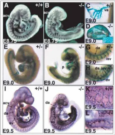
Figure 1.10. – Defective arterial and venous remodeling in Dll4−/− embryos ... 47
Figure 1.11. – Retinal vasculature in wild-type vs. Dll4+/- mice at postnatal P10 day ... 49
Figure 1.12. – Dll4 induction during tumor angiogenesis ... 50
Chapter II – RESEARCH DESIGN
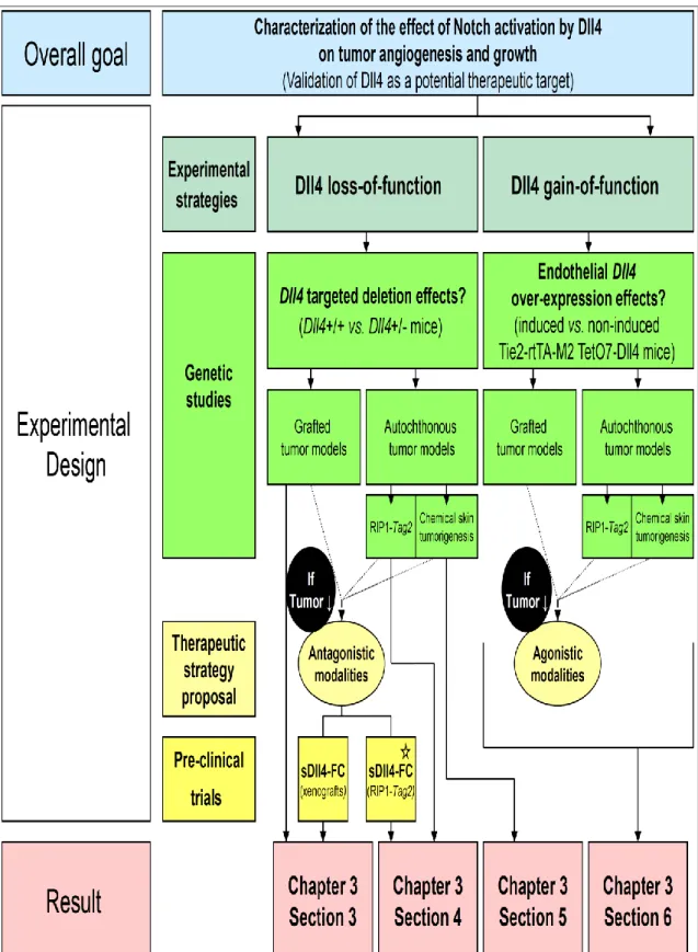
Figure 2.1. – Conceptual framework used for the investigation of Dll4 function in tumor angiogenesis and organization of the obtained results ... 70Figure 2.2. – Multi-step DMBA/TPA-induced mouse skin carcinogenesis ... 73
Figure 2.3. – Multi-step progression of tumors in RIP1-Tag2 mice and anti-angiogenic trials designed to target specific stages of carcinogenesis ... 74
XII
Chapter III – RESULTS
3. INHIBITION OF DLL4-MEDIATED SIGNALING INDUCES
PROLIFERATION OF IMMATURE VASSELS AND RESULTS IN
POOR TISSUE PERFUSION
Figure 3.1. – Dll4+/− mutant mice show defective increase in vascular proliferation ... 89
Figure 3.2. – Dll4 is activated in most but not all vessels in the tumor ... 90
Figure 3.3. - Biochemical properties of sDll4... 91
Figure 3.4. - sDll4 induces tubule formation in vitro ... 92
Figure 3.5. - sDll4 induces tubule formation in vitro ... 93
Figure 3.6. - sDll4 induces vessel response but lacks perfusion in murine Matrigel assay ... 94
Figure 3.7. - sDll4 inhibits the tumor growth in a murine tumor xenograft model ... 97
4. COMBINATION OF DLL4/NOTCH AND EPHRIN-B2/EPHB4
TARGETED THERAPY IS HIGHLY EFFECTIVE IN DISRUPTING
TUMOR ANGIOGENESIS
Figure 4.1. - Dll4 allelic deletion reduced tumor burden in RT2 mice due to increased nonproductive tumor angiogenesis ... 113Figure 4.2. -Differential gene expression in RT2 Dll4+/+ vs. RT2 Dll4+/- insulinomas ... 114
Figure 4.3. – sEphB4-Alb inhibited insulinoma growth and extended longevity in RT2 Dll4+/+ and Dll4+/- mice ... 117
Figure 4.4. – Combination therapy of sDll4-Fc and sEphB4Alb inhibits RT2 tumor growth with greater efficacy than either molecule alone ... 119
Figure 4.5 - Hepatic vascular lesions observed in endothelialspecific Dll4 knock-out mice are prevented by sEphB4-Alb administration ... 121
Figure 4.6. – Up-regulation of Rgs5 and PSENEN by sEphB4-Alb ... 125
5. DLL4 ALLELIC DELETION PROMOTES PRODUCTIVE
ANGIOGENESIS AND TUMOR GROWTH IN
CHEMICALLY-INDUCED MOUSE SKIN PAPILLOMAS
XIII Figure 5.1 - Dll4 allelic deletion promotes the onset and growth of chemically-induced skin tumors ... 138 Figure 5.2 – Partial Dll4/Notch inhibition due to haploid Dll4 deletion enhances less mature but productive angiogenesis in chemically-induced skin papillomas ... 140 Figure 5.3 – Dll4 allele deletion affects VEGF/VEGFR signaling as well as the regulators of pericyte recruitment in chemically-induced skin papillomas ... 141 Figure 5.4 – Dll4 deletion reduces sorafenib efficacy against chemically-induced skin papillomas ... 143
6. ENDOTHELIAL DLL4 OVEREXPRESSION REDUCES
VASCULAR RESPONSE AND TUMOR GROWTH IN GRAFTED
AND AUTOCHTHONOUS MOUSE MODELS
Figure 6.1. – Endothelial Dll4 overexpression restricts the growth and vascularization of subcutaneous LLC xenografts... 159 Figure 6.2. – Increased endothelial Dll4 expression suppresses chemical DMBA/TPA-induced skin tumor onset and development ... 161 Figure 6.3. – Endothelial Dll4 overexpression reduces vascular proliferation and promotes vessel maturation and perfusion in chemically-induced skin tumors ... 162 Figure 6.4. – Endothelial Dll4 overexpression and increased Dll4/Notch signaling modify the expression of major angiogenesis regulators and/or their receptors in chemically-induced skin papillomas ... 163 Figure 6.5. – Endothelial Dll4 overexpression negatively affects the RT2 insulinoma kinetics due to decreased vascular density although it enhances vascular maturation and competence ... 165
XIV
LIST OF PANELS
Chapter I – INTRODUCTION
Panel 1.1. – Angiogenesis regulators ... 7 Panel 1.2. – Diseases caused/characterized by abnormal or excessive angiogenesis ... 16 Panel 1.3. – Diseases caused/characterized by insufficient angiogenesis or vessel
regression ... 16 Panel 1.4. – Phenotypic characteristics of malignant cells/tissues ... 18 Panel 1.5. – Tumor mechanisms used to improve its blood supply ... 19 Panel 1.6. – Serious toxicities associated with bevacizumab ... 39
XV
LIST OF TABLES
Chapter I – INTRODUCTION
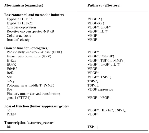
Table 1.1. – Angiogenic switch induction ... 22 Table 1.2. – Anti-angiogenic drugs approved for clinical use ... 33 Table 1.3. – Inclusive anti-angiogenic compounds: drugs with anti-angiogenic activity as a secondary function ... 34
Chapter III – RESULTS
4. COMBINATION OF DLL4/NOTCH AND EPHRIN-B2/EPHB4
TARGETED THERAPY IS HIGHLY EFFECTIVE IN DISRUPTING
TUMOR ANGIOGENESIS
Table 4.1.– Up-regulated genes in sEphB4-Alb treated RT2 mice ... 116
5. DLL4 ALLELIC DELETION PROMOTES PRODUCTIVE
ANGIOGENESIS AND TUMOR GROWTH IN
CHEMICALLY-INDUCED MOUSE SKIN PAPILLOMAS
XVI
ABBREVIATIONS
ADAM AIDS Alb AMD Ang1 Ang2 AP B16 Bcl-2 bFGF B-Raf BRCA1 BRCA2 CADASIL CD4 CD11 cDNA ChoK CIN c-KIT cmm COL-3 COX-2 Cre CSLA disintegrin and maetalloproteinase family Acquired immunodeficiency syndrome Albumin
Age-related macular degeneration Angiopoietin 1
Angiopoietin 2 Alkaline phosphatase
Mouse melanoma cell line 16
B-cell lymphoma 2 protein encoding gene Basic fibroblast growth factor
Murine sarcoma viral oncogene homolog B1 Breast cancer type 1 susceptibility protein Breast cancer type 2 susceptibility protein
Cerebral autosomal dominant arteriopathy with subcortical infarcts and leukoencephalopathy
Cluster of differentiation glycoprotein 4 Cluster of differentiation glycoprotein 11 Complementary deoxyribonucleic acid Chinese hamster cell line
Chromosomal instability
Mast/stem cell growth factor receptor Cubic millimeter
Incyclinide 3 Cyclooxygenase 2
Cre recombinase (topoisomerase) from P1 bacteriophage Cbf1/Su(H)/Lag1 DNA-binding protein
XVII D4BE D4OE DAPI DBP–MAF DCC Dll1 Dll3 Dll4 DMBA DMSO DNA DSL E ECM EDTA EFC-XV EFNB2 EGF EGFL7 EGFR EMT ENA78 EPC(s) EphA4 EphB1 EphB2 EphB3 EphB4 Ephrin-B2 Ephrin(s) Ephs Erb ERL Dll4 basic-expression mice Dll4 over-expressing mice
4′,6-diamidino-2-phenylindole dihydrochloride hydrate Vitamin-D-binding protein–macrophage-activating factor Deleted in colorectal cancer receptor
Delta-like ligand 1 Delta-like ligand 3 Delta-like ligand 4 7,12-dimethylbenz[a]anthracene Dimethyl sulfoxide Deoxyribonucleic acid
Delta, Serrate, Lag2 ligand family Embryonic day
Extra-cellular matrix
Ethylenediaminetetraacetic acid
Endostatin-like fragment from type XV collagen Human gene encoding Ephrin-B2
Epidermal growth factor
Epidermal growth factor ligand 7 Epidermal growth factor receptor Epithelial-to-mesenchymal transition Epithelial neutrophil-activating protein 78 Endothelial progenitor cell(s)
Erythropoietin-producing human hepatocellular carcinoma receptor A4 Erythropoietin-producing human hepatocellular carcinoma receptor B1 Erythropoietin-producing human hepatocellular carcinoma receptor B2 Erythropoietin-producing human hepatocellular carcinoma receptor B3 Erythropoietin-producing human hepatocellular carcinoma receptor B4 Eph family receptor interacting protein B2
Eph family receptor interacting protein(s)
Erythropoietin-producing human hepatocellular carcinoma receptors Erythroblastic leukemia viral oncogene
XVIII FACS Fc FGF FGF2 Flk-1 Flt-1 Flt-3 Flt-4 Fos GAPDH GCP-2 GIST Glu.Leu.Arg Gro H&E HDAC HER2 HERP HES HIF HP1 HPV HT29 HUVECs Id1 IFL IFN(s) IFN-α Ig IL(s) IL-4 IL-8 IMiDs
Fluorescence-activated cell sorter Immunoglobulin constant region Fibroblast growth factor
Fibroblast growth factor 2 Fetal liver kinase 1
Fms-like tyrosine kinase 1 Fms-like tyrosine kinase 3 Fms-like tyrosine kinase 4 Fibroblast oncogene
Glyceraldehyde 3-phosphate dehydrogenase encoding gene Granulocyte chemoattractant protein 2
Gastrointestinal stromal tumor
Glutamine-Leucine-Arginine sequence Growth-related oncogene protein Hematoxylin and eosin staining Histone deacetylase
Human Epidermal growth factor Receptor 2 HES-related repressor proteins
Hairy and enhancers of split transcription factors Hypoxia-inducible factor
Hypoxyprobe-1
Human papilloma virus
Human colon cancer cell line 29 Human umbilical vein endothelial cells DNA-binding protein inhibitor 1
Irinotecan, 5-fluorouracil and leucovorin combined chemotherapy Interferon(s) Interferon alpha Immunoglobulin Interleukin(s) Interleukin 4 Interleukin 8 Immunomodulatory drugs
XIX Jag1 Jag2 KDR KS LacZ LLC Mam MCF-7 MHC MIN MMP(s) MMP1 MMP2 MMP9 mRNA mTOR Myb Myc NG2 NICD NK NO p38 p53 PBS PBST PC(s) PC3 PCR PDGF PDGFB PDGFR-β PDZ Jagged ligand 1 Jagged ligand 2
Kinase domain-containing receptor Human Kaposi sarcoma cell line β-galactosidase reporter gene Lewis lung cancer cell line Mastermind co-activator
Michigan Cancer Foundation 7 breast cancer cell line Major histocompatibility complex
Microsatellite instability Matrix metalloproteinase(s) Matrix metalloproteinase 1 Matrix metalloproteinase 2 Matrix metalloproteinase 9 Messenger ribonucleic acid Mammalian target of rapamycin Myeloblastosis oncogene Myelocytomatosis oncogene
Neurogenin 2 chondroitin sulfate proteoglycan Notch intra-cellular domain
Natural killer cell Nitric oxide
Mitogen-activated protein kinase 38 Tumor protein 53 encoding gene Phosphate buffered saline
Tween-20 solution in PBS Pericyte(s)
Prostatic adenocarcinoma cell line 3 Polymerase chain reaction
Platelet-derived growth factor Platelet-derived growth factor B
Platelet-derived growth factor receptor β
XX PECAM PEDF PEX PFA PI3K PlGF PPAR-γ PTEN PTKs PTN PTTG1 PyMT Ras Rb RBC RET RIP ROBO 1 ROBO 2 ROBO 3 ROBO 4 ROS RT2 RT-PCR S180 SD sDll4 SEM Sema3A Sema3E Sema4A sEphB4
zonula occludens-1 protein
Platelet endothelial cell adhesion molecule Pigment epithelium-derived factor
Haemopexin C domain autolytic fragment of matrix metalloproteinase 2 Paraformaldehyde (PFA)
Phosphatidyl-inositol-3-kinase Placental growth factor
Peroxisome proliferator-activated receptor gama Phosphatase and tensin homolog encoding gene Receptor protein tyrosine kinases
Pleiotrophin
Pituitary tumor-derived transforming gene 1 Polyoma virus middle T oncogene
Rat sarcoma oncogene
Retinoblastoma protein encoding gene Red blood cell
Glial cell line–derived neurotrophic factor receptor Rat insulin promoter
Roundabout homologue 1 Roundabout homologue 2 Roundabout homologue 3 Roundabout homologue 4 Reactive oxygen species
RIP1-Tag2 transgenic mouse line
Reverse transcription polymerase chain reaction Mouse sarcoma 180 cell line
Standard deviation
Soluble Dll4 extra-cellular domain Standard error of the mean
Semaphorin 3A Semaphorin 3E Semaphorin 4A
XXI sFlt1 Src SSC SV40 T. f. VEGF Tag TGF-ß Tie-2 TIMP 1 TIMP 2 TIMP 3 TIMP 4 TIMPs TNF-α TPA TSP1 TSP2 UNC5 US FDA VEGF VEGFA VEGFA VEGFB VEGFC VEGFD VEGFE VEGFR1 VEGFR2 VEGFR3 VPF WT α-SMA β-gal
Soluble fms-related tyrosine kinase 1 Sarcoma oncogene
Squamous cell carcinoma Simian vacuolating virus 40
Trimeresurus flavoviridis vascular endothelial growth factor F
Large T antigen
Transforming growth factor beta
Tyrosine kinase with immunoglobulin-like and EGF-like domains 2 Tissue inhibitor of metalloproteinase 1
Tissue inhibitor of metalloproteinase 2 Tissue inhibitor of metalloproteinase 3 Tissue inhibitor of metalloproteinase 4 Tissue inhibitors of matrix metalloproteinase Tumor necrosis factor alpha
12-O-tetradecanoylphorbol-13-acetate Thrombospondin 1
Thrombospondin 2
Receptor of the uncoordinated-5 family United States Food and Drug Administration Vascular endothelial growth factor
Vascular endothelial growth factor A
Vascular endothelial growth factor A encoding gene Vascular endothelial growth factor B
Vascular endothelial growth factor C Vascular endothelial growth factor D Vascular endothelial growth factor E
Vascular endothelial growth factor receptor 1 Vascular endothelial growth factor receptor 2 Vascular endothelial growth factor receptor 3 Vascular permeability factor
Wild-type
Alpha smooth muscle actin β-galactosidase
CHAPTER I
INTRODUCTION
3
1
Introduction
In complex vertebrate organisms, the cardiovascular system, composed of the heart, blood vessels and blood itself, delivers oxygen and nutrients to all tissue cells and removes carbon-dioxide and metabolic waste from them (Aaronson et al., 2004). It also distributes water, electrolytes and hormones throughout the body, contributes to the infrastructure of the immune system, establishes hemostasis after injury, and participates in thermoregulation. Deregulated vascular formation and functions contribute to the pathogenesis of different congenital and later onset disorders, including inflammatory, ischemic, neoplastic, and degenerative pathologies (Carmeliet 2003). Thereby, significant efforts have been made over the last decades to better understand the mechanisms regulating vascular growth and homeostasis in both pre- and post-natal settings. Angiogenesis, the predominant mechanism of blood vessel proliferation under adult physiological and pathological conditions (Adams and Alitalo 2007), has been intensely under the focus of biomedical research resulting in the identification of rational bases for novel therapeutic strategies with revolutionary potential regarding the management of a range of human diseases.
This introducing chapter provides a brief overview of the angiogenic process and its regulation. The sites of physiological and pathological angiogenesis are subsequently presented while special attention is paid to the particularities of tumor angiogenesis. The successful attempts to translate laboratory bench results to the angiogenesis-targeting anti-cancer drug development are next summarized. Simultaneously, the major clinical problems arising from the use of currently approved angiogenesis-modulators are reviewed indicating the indisputable need to improve our understanding of tumor-driven angiogenesis. Finally, the attention is focused on Delta-like 4 (Dll4)/Notch signaling which crucial implication in embryonic vascular development (Duarte, Hirashima et al. 2004), together with increased Dll4 expression levels within human cancers (Mailhos, Modlich et al. 2001; Patel, Dobbie et al. 2006), directed the host laboratory and my professional interest to elucidate this signaling pathway role(s) in tumor vascularization.
4
1.1. OVERVIEW OF ANGIOGENESIS AND ITS REGULATION
1.1.1. Basic Mechanisms of Blood Vessel Formation
Growing tissues require an increased blood perfusion to guarantee adequate exchange of gases, nutrient supply and removal of waste metabolic products, which is achieved by the proliferation of blood vessels. This is evident during embryogenesis where hierarchically organized arteries, capillaries and veins develop from initial, undifferentiated vascular structures through dynamic growth and remodeling processes (Folkman 1995). Similar vascular expansive phenomena are also seen in the adults, in the female reproductive tract during the ovarian/menstrual cycles and in tissue regenerative processes such as wound healing (Klagsbrun and D'Amore 1991).
There are at least three different mechanisms that contribute to blood vessel formation and growth: vasculogenesis, angiogenesis and intussusceptions (Adams and Alitalo 2007). Vasculogenesis denominates the formation of blood vessels by the direct assembly of angiogenic progenitor cells while angiogenesis refers to the sprouting of novel capillary blood vessels from the pre-existing vasculature and subsequent stabilization of these sprouts by mural cells. Additionally, the vascular network can be enlarged by splitting a single pre-existing vessel in two new vascular segments through the insertion of a tissue pillar, a process called intussusception or splitting angiogenesis.
Initially, it was though that vasculogenesis occurred only in the embryos where the first vessels developed from endothelial progenitor cells (EPCs, i.e. angioblasts), while the adult vessel formation resulted exclusively from division of differentiated endothelial cells and angiogenic sprouting of pre-existing vessels. During early embryogenesis, mesodermal cells differentiate into EPCs and form aggregates, so-called blood islands, which subsequent fusion gives origin to the vasculogenic formation of primary capillary
plexi (Eichmann, Yuan et al. 2005). These primitive vascular structures are extended by
angiogenesis and remodelled into an embryonic vascular tree. Recent evidence suggests that the bone-marrow-derived circulating EPCs are capable of penetrating into the endothelium contributing to vasculogenic blood vessel growth in the adults (Rafii, Lyden et al. 2002). Nevertheless, angiogenesis is clearly the most common mechanism engaged in adults for vascular extension under both physiological and pathological conditions (Adams and Alitalo 2007).
5 1.1.2. The Angiogenic Cascade
Endothelial cells (ECs) are the basic and constant blood vessel components covering, in the form of a monolayer, their luminal surfaces. In favorable pro-angiogenic settings, principally induced by hypoxia, the angiogenic process is initiated by selective activation of individual ECs in differentiated capillaries (Adams and Alitalo 2007). While some ECs are activated others maintain the quiescent state. The selected ECs acquire invasive behavior, degrade the sub-endothelial basement membrane and depart from the vessel of origin following the gradients of pro-angiogenic stimuli. Known as the tip cells, they break down the extra-cellular matrix and provide directional migration (i.e., lead the growing sprout). Despite their highly migratory phenotype, tip cells remain in physically contact with their follower ECs, the stalk cells, which proliferate in response to pro-angiogenic factors constituting the endothelial component of the nascent vessel body and extending its length (Iruela-Arispe, 2008). Simultaneously with this endothelial proliferation, mesenchymal cells also proliferate, migrate through the cleaved extra-cellular matrix, incorporate in the new sprout, differentiate into mural cells, i.e. pericytes, and mature stabilizing the outgrowth (Papetti and Herman 2002). Both adhesive and repulsive interactions between tip cells from adjacent sprouts regulate their fusion and the establishment of novel vascular interconnections (Adams and Alitalo 2007). The vessel wall assembly, lumen acquisition, synthesis and organization of the basement membrane and specification of ECs to arterial or venous fate trigger the resolution of the endothelial activation (Iruela-Arispe, 2008). Once established blood flow improves tissue perfusion that reduces hypoxia-induced pro-angiogenic signals and enhances vascular maturation including the stabilization of EC-EC junctions, matrix deposition and tight pericyte attachment (Adams and Alitalo 2007).
Didactically, the described cellular phenomena can be divided in four consecutive phases representing together so-called angiogenic cascade. They are:
1. Stimulation of angiogenic response and selection of sprouting ECs, 2. Sprout outgrowth,
3. Sprout fusion and lumen formation, and 4. Vessel perfusion and maturation
6
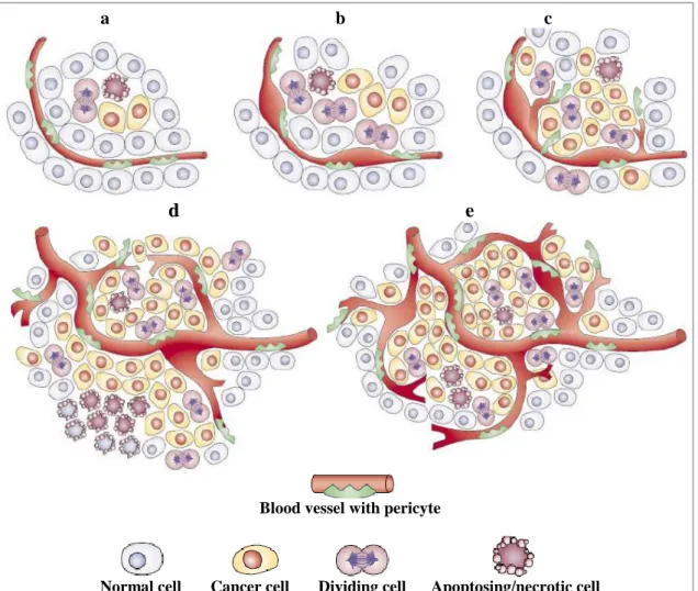
Figure 1.1. – Angiogenic cascade (reproduced from
Adams and Alitalo, 2007).
a) Angiogenic sprouting is controlled by the balance between pro-angiogenic signals (+), such as vascular endothelial growth factor (VEGF), and factors that promote quiescence (–), such as tight mural cell, i.e. pericyte (PC; yellow) contact, certain extra-cellular matrix (ECM) molecules or VEGF inhibitors. In conditions that favor angiogenesis, some endothelial cells (ECs) can sprout (green), whereas others fail to respond (grey). Sprouting requires the flipping of apical–basal polarity, the induction of motile and invasive activity, the modulation of cell–cell contacts and local matrix degradation. b) The growing EC sprout is guided by VEGF gradients. Other signals may include attractive (+) or repulsive (–) matrix cues and guidepost cells in the tissue environment. Release of platelet-derived growth factor B (PDGFB) by the tip cells promotes the recruitment of PCs to new sprouts. EC–EC junctions need to be maintained after lumen formation to prevent excessive leakage. c) Adhesive or repulsive interactions that occur when tip cells encounter each other regulate the fusion of adjacent sprouts and vessels. Lumen formation in stalk ECs involves the fusion of vacuoles but other mechanisms may also contribute. d) Fusion processes at the EC–EC interfaces establish a continuous lumen. Blood flow improves oxygen delivery and thereby reduces pro-angiogenic signals that are hypoxia-induced. Perfusion is also likely to promote maturation processes such as the stabilization of cell junctions, matrix deposition and tight PC attachment. Growth factor withdrawal can trigger sprout retraction and endothelial apoptosis. DLL4, Delta-like-4 ligand; EGFL7, Epidermal growth factor ligand-7; ROBO4,
Roundabout homologe-4; VEGF2, VEGF receptor-2.
7 1.1.3. Molecular Regulation of Angiogenesis
The angiogenic growth of blood vessels is regulated by different and numerous positively and negatively acting factors. They include soluble polypeptides, cell-cell interactions, cell-matrix interactions and hemodynamic effects (Papetti and Herman 2002). The principal angiogenesis regulators, classified as soluble mediators, membrane-bound molecules, and mechanical forces, are presented in Panel 1.1. Capillary blood vessels remain in the quiescent state when the balance between positive and negative regulatory factors is maintained stable while the shift to the side of pro-angiogenic stimuli triggers angiogenesis.
Panel 1.1. – Angiogenesis regulators (adapted from Papetti and Herman, 2001)
I - SOLUBLE MEDIATORS
Stimulatory Factors
Acidic fibroblast growth factor Angiogenin
Basic fibroblast growth factor Fibroblast growth factor 3 and 4 Hepatocyte growth factor Interleukin 8
Placental growth factor Platelet-derived growth factor Pleiotropin
Proliferin
Platelet-derived endothelia-cell growth factor Transforming growth factor αand β
Tumor necrosis factor α
Vascular endothelial growth factor
Inhibitory Factors
Angiostatin
Antithrombin III fragment Endostatin
Canstatin
Fragment of platelet factor 4 Interferon αand β Interferon-inducible protein 10 Maspin Prolactin fragment Thrombospondin 1 and 2 Tumstatin
Vascular endothelial growth inhibitor Vasostatin II - MEMBRANE-BOUND MOLECULS Stimulatory Effects Integrin α5β1 Integrin αvβ3 Integrin, αvβ5, Eph-4B/Ephrin-B2 Inhibitory Effects
Classe 3 semaphorins /Plexin D1 Netrin1/ uncoordinated-5B Slit?/Robo4
III - MECHANICAL FORCES (pro-angiogenic) Blood flow/Shear stress
8
1.1.3.1. Vascular Endothelial Growth Factor A and Stimulation of the Angiogenic Response
Current experimental evidence indicates that the most important pro-angiogenic factor is vascular endothelial growth factor A [VEGFA or VEGF; also known as vascular permeability factor (VPF)]. VEGFA is a member of a family of potent angiogenesis and/or lymphangiogenesis positive regulators that also includes VEGFB, VEGFC, VEGFD, placental growth factor (PlGF), Orf virus’ VEGFE, and
Trimeresurus flavoviridis (T. f.) svVEGFs (Lyttle, Fraser et al. 1994; Junqueira de
Azevedo, Farsky et al. 2001; Ferrara, Gerber et al. 2003). In humans and animals, VEGFA is secreted at low levels by various tissues, but in elevated concentrations, the growth factor is produced in the sites where angiogenesis is needed such as in the placenta, many embryonic/fetal tissues, in the proliferating endometrium, corpus luteum as well as in neoplastic tissues (Dvorak, Brown et al. 1995). VEGFA production is principally regulated by the tissue oxygen saturation (Shweiki, Itin et al. 1992). Under hypoxic conditions, VEGFA production is promoted by up-regulated hypoxia-inducible factor (HIF) that binds to the VEGFA promoter increasing the gene transcription (Kimura, Weisz et al. 2001).
VEGFA causes diverse effects on the endothelium. It specifically inhibits EC apoptosis (Gerber, McMurtrey et al. 1998) and stimulates EC proliferation (Connolly, Heuvelman et al. 1989), enhances EC migration (Dimmeler, Dernbach et al. 2000), and increases vascular permeability to circulating metabolites (Kitadai, Takahashi et al. 1999). Genetically targeted mouse embryos deficient in Vegfa virtually do not develop any vascular structure (Shalaby, Rossant et al. 1995; Ferrara 1999). Studies conducted in mice, on the neonatal corneas and healing bone grafts, indicated that the VEGFA roles are equally important for inducing pos-natal and adult capillary sprouting (Connolly, Heuvelman et al. 1989).
The endothelial responsiveness to VEGFA is regulated by several mechanisms including expression of alternatively spliced VEGFA isoforms. In the first place, alternative splicing of a single VEGFA gene can generate different variants of the growth factor composed of 121, 145, 165, 183, 189 or 206 amino acids (Tischer, Mitchell et al. 1991; Robinson and Stringer 2001). Despite the fact that all of these isoforms have the same biological activity, they can be either soluble (e.g. VEGFA-121 and the most commonly produced VEGFA-165) or immobilised, cell- or
matrix-9 associated due to their affinity for heparan sulfates (e.g. VEGFA-189 and VEGFA-206) (Houck, Leung et al. 1992). While immobilized VEGFA generates a gradient that is sensed by the tip cell providing directional migratory cues, the soluble VEGFA does not form the gradient, so ECs lose directionality becoming less migratory (Tischer, Mitchell et al. 1991; Ruhrberg, Gerhardt et al. 2002; Lee, Jilani et al. 2005). In addition, alternative VEGFA splicing can result in the production of “ b” isoforms that have anti-angiogenic properties (Ladomery, Harper et al. 2007). Thus, the balance between different VEGFA isoforms alters the EC behaviour in response to the growth factor.
The VEGFA effects depend also on the balance between the different VEGFA receptors. VEGFA binds to at least three tyrosine kinase receptors: Flt-1 (VEGFR1) (de Vries, Escobedo et al. 1992), KDR/Flk-1 (VEGFR2) (Klagsbrun and D'Amore 1991), and Flt-4 (VEGFR3) (Pajusola, Aprelikova et al. 1992). Binding of VEGFA to VEGFR2 leads to the activation of a cascade of downstream signaling pathways and generates the major signals for sprouting angiogenesis (Shibuya and Claesson-Welsh 2006). In contrast, pro-angiogenic VEGFA effects are minimized by VEGFR1, which has ten times higher affinity for the growth factor then VEGFR2, but very weak tyrosine-kinase activity, serving as a VEGFA trapper (Shibuya and Claesson-Welsh 2006). Besides, VEGFR1 appears in a soluble and catalytically inactive form (Shibuya 2006). Therefore, the VEGFR1/VEGFR-2 ratio importantly determines the VEGFA outcome.
Together with VEGFA, different other factors have been demonstrated to counteract pro-quiescent signals and positively influence the process of angiogenesis (Panel 1.1.). Nevertheless, VEGFA is thought to be the initiating angiogenesis stimulus.
1.1.3.2. Delta-like4/Notch Signaling and Acquisition of Tip vs. Stalk Endothelial Cell Identities
Stimulating EC survival, proliferation and migration, as presented above, VEGFA plays a fundamental role in the EC activation. Nevertheless, VEGFA signaling, on its own, does not provide an organized endothelial proliferation that is absolutely needed for hierarchical and thereby functional vascular network formation (Iruela-Arispe, 2008). A crucial element in angiogenesis regulation is to establish the leadership between equal ECs under favorable pro-angiogenic conditions and determine which
10
ECs will acquire the tip cells phenotype providing directional migration and which ECs will remain in the quiescent state within the pre-existing vessels or acquire the “follower”, stalk cells phenotype contributing to the body formation of a novel sprout. Interestingly, it has been documented that the tip and the stalk cells respond differently to VEGFA stimulation: while the tip cells predominantly migrate, the stalk cells mostly proliferate (Gerhardt and Betsholtz 2005).
Several studies in zebrafish (Leslie, Ariza-McNaughton et al. 2007; Siekmann and Lawson 2007) and mouse embryos (Duarte, Hirashima et al. 2004; Sainson, Aoto et al. 2005) as well as in mouse neonatal retinas (Claxton and Fruttiger 2004; Hellstrom, Phng et al. 2007; Lobov, Renard et al. 2007; Suchting, Freitas et al. 2007) have indicated that endothelial trans-membrane Notch receptors, activated by their Delta-like 4 (Dll4) ligand of adjacent ECs, trigger intra-cellular signaling that determines EC fate when exposed to pro-angiogenic stimuli. Most of the referred studies were conducted simultaneously with this PhD project. Since the very first of these findings from developmental and post-embryonic angiogenesis experiments directly inspired our research focusing the Dll4 implication in pathological (tumor-driven) angiogenesis in adult mice, the section “Dll4/Notch Signaling Pathway and its Involvement in Vascular
Biology” provides more extensive information. Briefly, targeted Dll4 deletion and
pharmacologically achieved Notch signaling blockade enhance ECs to acquire the tip
cell phenotype resulting in excessive angiogenic branching. Considering the fact that,
under VEGFA pressure, Dll4 has up-regulated expression in the tip cells, this implies that activated ECs use the Dll4/Notch system to signal to the neighboring quiescent ECs and suppress their activation. Mechanistically, Dll4/Notch signaling was found to repress VEGFR2 and increases the VEGFA trapper - VEGFR1 (Lobov, Renard et al. 2007; Suchting, Freitas et al. 2007). Thus, binding of Dll4 of already activated ECs (acquiring tip cells phenotype) to the Notch on adjacent cells seems to induce the acquisition of the stalk identities by enabling ECs to differentially sense the existent VEGFA gradient (Figure 1.2.).
Figure 1.2. – Selection of sprouting endothelial cells (adapted from Iruela-Arispe, 2008).
Departing form parental vessels, subset of activated ECs express Dll4, activate the Notch receptor in adjacent ECs reducing their sensibility to VEGFA (left). The outcome is the formation of a vascular sprout with highly migratory tip cells and proliferating stalk cell (right).
11 1.1.3.3. Guiding Cues for Sprout Outgrowth
Once determined the fate of ECs and established the leadership between them, organized EC proliferation and migration result in emerging sprout outgrowth. Expressing VEGFR2 in the tip cells, EC guidance is controlled by the special concentration gradient of matrix-anchored VEGFA isoforms that functions as a chemo-attractive force (Ruhrberg, Gerhardt et al. 2002; Gerhardt, Golding et al. 2003). In parallel, proliferating vascular sprouts generate the concentration gradient of platelet-derived growth factor B (PDGFB), which up-regulated expression in ECs promotes the recruitment of PDGF receptor β (PDGFR-β)-exhibiting pericytes and, thus, growing vessel stabilization (Gerhardt, Golding et al. 2003). In addition to VEGFA concentration gradient, the endothelial directional sprouting is regulated by attractive and repulsive tissue-derived navigational cues with pronounced analogy to growing nerve fibers (Carmeliet and Tessier-Lavigne 2005). Initially identified as axon guidance regulators, semaphorins, netrins, slit/robo proteins and ephrins have been evidenced to play prominent roles in angiogenic vessel growth (Huminiecki, Gorn et al. 2002; Gitler, Lu et al. 2004; Lu, Le Noble et al. 2004; Park, Crouse et al. 2004; Gu, Yoshida et al. 2005; Weinstein 2005; Wilson, Ii et al. 2006; Toyofuku and Kikutani 2007; Sawamiphak, Seidel et al. 2010; Wang, Nakayama et al. 2010)
Semaphorin family includes membrane-bound and secreted proteins that provide navigational signals during neuronal growth, but also, as recently documented, some of them influence vascular directionality (Weinstein 2005). Generally said, membrane-bound semaphorins signal by binding to plexins while secreted semaphorins signal through neuropilins (Bagri and Tessier-Lavigne 2002). Genetic targeting of Semaphorin4A (Sema4A) showed that it suppresses VEGF-induced EC migration and angiogenesis in vivo by interacting with Plexin D1 (Toyofuku and Kikutani 2007). In addition to Sema4A, at least two class 3 semaphorins, Sema3A and Sema 3E, can signal trough Plexin D1 serving as repulsive cues for ECs (Gitler, Lu et al. 2004; Gu, Yoshida et al. 2005).
The second group of ligands involved in both neuronal growth and vascular patterning are the netrins. In the nervous system, netrins function either as attractive cues, when they interact with the family of deleted in colorectal cancer (DCC) receptors, or as repulsive cues, when they signal through receptors of the
12
uncoordinated-5 (UNC5) family or UNC5–DCC heterodimers (Carmeliet and Tessier-Lavigne 2005). In mice and zebrafish, Netrin1-UNC5B signaling was found to result in retraction of the tip cell filopodia1 and inhibit endothelial sprouting (Lu, Le Noble et al. 2004; Park, Crouse et al. 2004). Nevertheless, netrins are also capable of promoting angiogenesis, probably, by using different receptors that remain to be identified (Wilson, Ii et al. 2006).
Slit proteins and roundabouts (Robo) represent an additional ligand/receptor family that contributes to neuronal patterning and seems to be involved in the regulation of EC sprouting. Slit/Robo signaling repels growing axons (Brose, Bland et al. 1999). From four Robo receptors identified in mammals (Robo1–4), Robo4, also known as
magic roundabout, is apparently a vascular-specific receptor, expressed at sites of active
angiogenesis, that inhibits EC migration (Huminiecki, Gorn et al. 2002). Robo1 is also actively involved in EC filopodia formation (Sheldon, Andre et al. 2009).
Finally, bidirectional signaling between Eph receptors and ephrin ligands consists one of the most prominent guidance cues for neuronal axons while ephrin-B2 reverse signaling regulates endothelial tip cell guidance (Egea and Klein 2007; Sawamiphak, Seidel et al. 2010; Wang, Nakayama et al. 2010). It is generally accepted that Ephs (erythropoietin-producing human hepatocellular carcinoma) and ephrins (Eph family receptor interacting proteins) function to translate the density of their “partner” on opposing membranes into precise cellular responses, resulting in cell-cell adhesion or cell-contact repulsion (Campbell and Robbins 2008). Encoded by the gene EFNB2, Ephrin-B2 activates several receptors of the Eph tyrosine kinase family, including EphB4 (‘forward’ signaling), and possesses intrinsic (‘reverse’) signaling activity (Himanen and Nikolov 2003). Ephrin-B2 is a well-known arterial EC marker while vascular expression of EphB4 is predominantly observed in venous endothelium (Adams and Alitalo, 2007). The involvement of Ephrin-B2 and EphB4 in mouse developmental angiogenesis was suggested by findings that knockout of either protein is embryonically lethal due to vascular arrest at the primitive capillary plexus stage (Adams, Wilkinson et al. 1999). At the time of writing this text, two publications appeared providing at least partial explanation regarding the mechanistic basis of
1 Filopodia are cellular processes that extend from the leading edge of migrating cells, attach to the
13 ephrin-B2 effects on EC behaviour (Sawamiphak, Seidel et al. 2010; Wang, Nakayama et al. 2010). In cultured vascular ECs, zebrafish and mice, Ephrin-B2 reverse signaling, involving PDZ interactions, increases the number of tip cells and their filopodial extensions, promoting motile behaviour and sprouting (Sawamiphak, Seidel et al. 2010; Wang, Nakayama et al. 2010). Ephrin-B2 reverse signaling was found to control VEGFR-2 internalization, which is necessary for activation, downstream signaling of the receptor and VEGFA-induced tip cell filopodial extension (Sawamiphak, Seidel et al. 2010). Simultaneously, in cultured lymphatic ECs, Ephrin-B2 was evidenced to induce the internalization of the VEGFC receptor VEGFR3, and, thus, positively regulate this principal pro-lymphangiogenic signaling axis (Wang, Nakayama et al. 2010). Together, these data suggest that Ephrin-B2 activation on tip cells directs VEGF-induced migration by stimulating the spatial activation of VEGFR endocytosis.
1.1.3.4. Transformation of Sprouts into Vessels – Sprout Fusion and Lumenization
The onset of blood flow requires the fusion of growing endothelial sprouts between themselves or with pre-existing capillaries as well as the formation of vascular lumen. Strong adhesive interactions among encountering tip cells and the firm EC–EC junctions enable the fusion of the sprout tips (Adams and Alitalo 2007). The repulsive interactions between tip cells of different sprouts probably also occur, preventing, for example, the fusion of incompatible vascular segments. Regarding the lumen formation, high-resolution time-lapse imaging of growing intersegmental vessels in zebrafish has indicated that pinocytosis2, vacuole formation, their intracellular fusion and subsequent intercellular fusion lead to the development of endothelial tubular structures (Kamei, Saunders et al. 2006). Additionally, there is evidence that Notch signaling contributes to lumen enlargement by increasing the ratio of stalk to tip cells (Lobov, Renard et al. 2007; Suchting, Freitas et al. 2007) while epidermal growth factor-like domain 7 (EGFL7; also known as vascular endothelial statin), a small EC-driven secreted factor, promotes vascular tubulogenesis by suppressing extra-cellular matrix adhesion (Parker, Schmidt et al. 2004). In contrast to immobilized VEGF, the soluble VEGF isoforms also favor increased vessel diameters (Tischer, Mitchell et al. 1991).
2 Pinocytosis is a process by which the extra-cellular or surrounding fluid is internalized into cells in the
14
1.1.3.5. Vessel Maturation and Termination of the Endothelial Activation
The establishment of blood flow and improved tissue perfusion suppress EC proliferation and promote the maturation of novel vascular connections since increased tissue oxygenation lowers the levels of VEGFA and other pro-angiogenic factors, shifting the balance to anti-angiogenic factor dominancy. Particularly important events during the terminal phase of angiogenic cascade are the basement membrane formation and the incorporation of pericytes into the newly formed endothelial tubes (Iruela-Arispe, 2008). As previously stated, the PDGFB expression in ECs promotes the recruitment of PDGFR-β-exhibiting pericytes (Gerhardt, Golding et al. 2003). Pericyte-mediated vascular stabilization involves inhibitors of extra-cellular matrix cleavage by matrix metalloproteinases (Iruela-Arispe, 2008). Remarkably, pericyte-derived tissue inhibitor of metalloproteinase-3 (TIMP-3), but also EC-derived TIMP-2, induce capillary stabilization upon endothelial-pericyte association, and prevent capillary regression (Davis and Saunders 2006). Nevertheless, capillaries may remain highly stable in the absence of pericytes (Iruela-Arispe, 2008). Therefore, much remains to be understood within this terminal angiogenesis step.
15 1.2. ADULT ANGIOGENESIS IN HEALTH AND DISEASE
Physiological angiogenesis ensures that normal, developing and growing or regenerating tissues receive an appropriate oxygen and nutrient supply. On the other hand, in various pathological conditions, normal angiogenesis mechanisms are usurped, directly provoking or enhancing the onset of disease.
In contrast to embryogenesis, EC turnover in the adult vasculature is quite minimal (Denekamp 1982). Angiogenesis is rarely observed in healthy adult organs with the exception of the female reproductive tract and during the repair of injured tissue. Each menstrual cycle, various tissues of the female reproductive system are subjected to rigorous vascular bed remodelling (Groothuis 2005). Cyclical changes of angiogenesis and vascular regression are observed in the growing follicles and corpus
luteum of the ovary, endometrium and the breast tissue. Beside the reproductive
function, successful adult tissue repair, including appropriate wound healing, also fundamentally depends on new blood vessel proliferation, with the important contribution of the accompanying inflammatory response that releases pro-angiogenic factors (Hunt et al., 2008). Although several regulators, both inducers and inhibitors, play critical roles, adult physiological angiogenesis, including the reparatory angiogenesis, is mainly triggered and mediated by VEGFA (Groothuis, 2005, Hunt et al., 2008).
Impaired vascular homeostasis, which results from either excessive or insufficient vessel growth, tremendously impacts human health contributing to the onset and/or progress of disorders from virtually all diagnostic categories (Panels 1.2. and 1.3.; Carmelitet, 2003). The best known pathologies characterized or caused by excessive vascular proliferation are cancer, psoriasis, rheumatoid arthritis and diabetic retinopathy, the leading cause of blindness in the developed world. Nevertheless, abnormal angiogenic response is also observed in common diseases such as infections, atherosclerosis, obesity and bronchial asthma. On the other hand, insufficient blood vessel growth and abnormal vascular regression lead to heath and brain ischemia, neurodegenerative lesions, arterial hypertension, pre-eclampsia, respiratory distress syndrome, osteoporosis and many other disorders. Apparently, there are not many other pathophysiological processes that threaten global human well-being such as pathological angiogenesis. Thereby, understanding it´s molecular basis might lead to
16
therapeutic improvements, particularly in the medical fields, such as oncology, where available treatment options usually do not provide a satisfactory outcome.
Panel 1.2. – Diseases caused/characterized by abnormal or excessive angiogenesis (adapted from Carmelitet, 2003)
Numerous organs – tumors, infectious diseases, autoimmune disorders
Blood vessels – vascular malformations, DiGeorge syndrome, cavernous hemangioma, atherosclerosis, transplant arteriopathy
Adipose tissue – obesity
Skin – psoriasis, warts, allergic dermatitis, scar keloids, pyogenic granulomas, blistering diseases, AIDS-associated Kaposi sarcoma
Eye – persistent hyperplastic vitreous syndrome, diabetic retinopathy, retinopathy of prematurity, choroidal neovascularization
Lung – primary pulmonary hypertension, asthma, nasal polyps
Gastrointestinal tract – inflammatory bowel and periodontal disease, ascites, peritoneal adhesions
Reproductive system – endometriosis, menorrhagia, ovarian cysts, ovarian hyperstimulation
Locomotor system – arthritis, synovitis, osteomyelitis, osteophytes
Panel 1.3. – Diseases caused/characterized by insufficient angiogenesis or vessel regression (adapted from Carmelitet, 2003)
Nervous system – Alzheimer disease, amyotrophic lateral sclerosis, diabetic neuropathy, stoke
Blood vessels – atherosclerosis, hypertension, diabetic vasculopathy, post-traumatic restenosis
Gastrointestinal tract –gastric and oral ulcerations, Crohn disease Skin – hair loss, skin purpura, teleangiectasia, venous lake formation Reproductive system – pre-eclampsia, menorrhagia
Lung – neonatal respiratory distress, pulmonary fibrosis, emphysema Kidney – nephropathy
17 1.3. OVERVIEW OF TUMOR ANGIOGENESIS
Tumor denominates a new growth of tissue, neoplasm, in which cell multiplication is uncontrolled and progressive, faster than that of normal tissue, continuing after cessation of the growth stimuli and having no useful physiologic purpose (Dorland 1953). Tumors can be either benign or malignant. While unregulated cell growth without invasion is a characteristic of benign tumors, cancers or malignant tumors, that involve carcinomas originating from epithelial tissues and sarcomas arising from mesenchymal tissues, are defined by two principal features – unrestrained cell proliferation and tissue invasion/metastasis (Fenton and Longo, 2008). Although the cancer incidence has been declining by about 2% each year since 1992 and the application of current treatment options (i.e., surgery, radiotherapy, chemotherapy and biological modalities) results in the cure of nearly two of three patients diagnosed with cancer, malignant neoplasms represent the second leading cause of death world-wide behind heart disease (Longo, 2008).
Cancer is not a single disease, but a group of genetic disorders that result from a series of somatic alterations in DNA, caused by random replication errors, exposure to carcinogens (e.g., radiation), and/or faulty DNA repair processes (Morin et al., 2008). Although most cancers appear sporadically, various histological types cluster in some families that carry a germline mutation in a cancer gene.
The phenotypic characteristics of malignant cells and tissues are summarized in Panel 1.4. One of the most prominent neoplasm features is its neovascularization. Such as normal tissues, both benign tumors and cancers require blood supply to promote their growth (Austen 1997). In 1971, Judah Folkman presented, for the first time, the concept that tumor growth depends on the formation of new blood vessels from the host pre-existing vasculature (Folkman 1971). The hypothesis arose from the observations that neither isolated animal organs nor the vitreous and aqueous humour of the human eye had supported the vascularization of malignant cell agglomerates and their enlargement more than 1-2 mm of diameter (Folkman, Cole et al. 1966; Lutty, Thompson et al. 1983). Subsequently, Folkman and co-workers identified a factor, named “tumor angiogenesis factor” (Folkman, Merler et al. 1971), postulating that the endothelial cells may be switched from a quiescent state to a rapid growth phase by diffusible chemical signals emitting from the tumor cells. Subsequent development of