SYNTHESIS AND CHARACTERIZATION OF NEW OLIGOSACCHARIDES WITH PREBIOTIC ACTIVITY
by
Maria Inês Pereira Montenegro
SYNTHESIS AND CHARACTERIZATION OF NEW OLIGOSACCHARIDES WITH PREBIOTIC ACTIVITY
Obtenção e Caracterização de Novos Oligossacáridos com Atividade Prebiótica
Thesis presented to Escola Superior de Biotecnologia of the Universidade Católica
Portuguesa to fulfil the requirements of Master of Science degree in Applied Microbiology
by
Maria Inês Pereira Montenegro
Place: CBQF/Escola Superior de Biotecnologia da Universidade Católica Portuguesa Supervision: Professora Maria Manuela Estevez Pintado
Co-Supervision: Alejandra Cardelle-Cobas and Beatriz Gullón
II
Resumo
A relevância da microbiota intestinal na manutenção da saúde do hospedeiro é bem conhecida e, nas últimas décadas, a consciencialização dos consumidores para a escolha de alimentos saudáveis tem vindo a aumentar. Existem diversas estratégias para estimular a proliferação de bactérias intestinais benéficas, incluindo o consumo de prebióticos. Atualmente, existe uma vasta gama de hidratos de carbono prebióticos no mercado, a maior parte isolados de polissacarídeos de plantas, de que são exemplo a inulina e frutooligossacarídeos (FOS). No entanto, existe um interesse crescente no desenvolvimento de novos prebióticos com funcionalidade adicional. Nesse sentido, o quitosano, sendo um polissacarídeo composto por unidades de glucosamina (GlcN) e N-acetil glucosamina (GlcNAc) unidas por ligações β (1→4), apresenta uma estrutura muito semelhante à dos atuais prebióticos glucooligossacarídeos. A diferença principal consiste na presença de grupos amina na sua estrutura, que lhe confere uma importante atividade antimicrobiana. A modificação química do quitosano por substituição dos seus grupos amina poderia eliminar este efeito antimicrobiano e converter o quitosano num novo e interessante ingrediente prebiótico. Assim, este trabalho teve por objetivo modificar quimicamente o quitosano para ser usado como ingrediente prebiótico na indústria alimentar.
Por forma a atingir este objetivo, foi levada a cabo a otimização da síntese dos derivados do quitosano por reacção de Maillard e hidrólise enzimática. Os derivados obtidos foram analisados quanto à distribuição de massa molar e caracterizados estruturalmente por Cromatografia de Exclusão Molecular (SEC), Espectroscopia de Infravermeho por Transformada de Fourier (FT-IR), Ressonância Magnética Nuclear (1H-NMR) e titulação coloidal. O potencial prebiótico do produto purificado foi avaliado em ensaios de fermentação
in vitro realizados em culturas puras e em inóculos humanos fecais.
Os resultados obtidos demostraram que os quitooligossacarídeos (COS) sintetizados, possuem potenciais efeitos prebióticos, que incluem alterações no padrão de produtos metabólicos gerados e nas contagens de Bifidobacterium, podendo, assim, contribuir para um ambiente intestinal saudável. Por fim, a avaliação da citotoxicidade dos derivados de COS foi realizada in vitro por citometria de fluxo. Os resultados obtidos demonstraram que os derivados sintetizados são moléculas biocompatíveis e que a substituição dos grupos amina diminuiu a citotoxicidade dos derivados quando comparados com COS não modificados. No entanto, estudos in vivo são recomendados para confirmar estes resultados in vitro.
III
Abstract
The importance of human intestinal microbiota in maintaining host health is well-known and in the past few decades, the consumer’s awareness for healthier foods has increased. There are several strategies to stimulate the proliferation of beneficial intestinal bacteria, including the consumption of prebiotics. Currently, there is a range of prebiotic carbohydrates on the market, most of them isolated from plant polysaccharides such as inulin and fructooligosaccharides (FOS) but there is an increasing interest in the development of new prebiotics, with added functionality. In this sense, chitosan, which is a polysaccharide composed of glucosamine (GlcN) and N-acetyl glucosamine (GlcNAc) units, linked by β (1→4) bonds, presents a structure very similar to prebiotic glucooligosaccharides. The main difference is the presence of amino groups, which are the cause of antimicrobial effect of chitosan. Chemical modification of chitosan by substitution of its amino groups could eliminate the antimicrobial effect and convert chitosan in a new interesting prebiotic ingredient. Thus, the objective of the present work is to chemically modify chitosan to be used in the food industry as prebiotic ingredient.
In order to achieve this objective, the optimization of the synthesis of chitosan derivatives with glucose by the Maillard reaction and enzymatic hydrolysis, was conducted and the obtained purified derivatives were assayed for molar mass distribution and structural characterization by Size Exclusion Chromatography (SEC), Fourier Transform Infrared Spectroscopy (FT-IR), Proton Nuclear Magnetic Resonance (1H-NMR) and colloid titration. The purified product was assayed for its prebiotic potential by means of in vitro fermentability assays performed with individual microbial strains and human faecal inocula.
The experimental data showed that the refined chitooligosaccharide (COS) derivatives obtained in this work had potential prebiotic effects, inducing changes in both the pattern of generated metabolic products and the count of Bifidobacterium, which might contribute to a healthy intestinal environment. Finally, the in vitro cytotoxicity of the COS derivatives synthesized was performed by flow cytometry. The results obtained demonstrated that the COS derivatives are biocompatible molecules. Also, the assay showed that the substitution of the amino groups decreased the cytotoxicity of the COS derivatives when compared to unmodified COS. Nevertheless, further studies are recommended, mainly in vivo tests, to eventually confirm these in vitro results.
IV
Acknowledgments
I would like to thank Prof. Manuela Pintado for conceding me the opportunity to develop this thesis under her supervision. Thank you very much for your kindness and support.
I would also like to express my profound gratitude to Alejandra Cardelle-Cobas and Beatriz Gullón for their unconditional support during all the stages of this thesis. Thank you for your patience and for the many things you have taught me.
My gratitude is extended to all my laboratory colleagues who were always willing to offer their help and made the days spent in the laboratory much more pleasant.
I would also like to thank the Department of Chemical Engineering, Faculty of Science, University of Vigo (Campus Ourense), specially to Dr. José Luis Alonso for giving me the opportunity to develop the in vitro fermentation assays with human faecal microbiota and the study of dynamics of the Bifidobacterium population by FISH in his laboratory. Thank you very much for your kindness.
I would not forget to thank the Department of Chemistry and Functionality of Carbohydrates and Derivatives, at the Institute of Food Research in Madrid (Centro de Investigación en Ciencias de la Alimentación, CIAL), specially to Dr. Nieves Corzo and Dr. Ana Ruiz. Thank you for your support and for providing all the facilities to carry out the spectroscopic analysis of the samples.
Moreover, I would like to express my gratitude to Prof. Alice Santos-Silva for conceding me the opportunity to develop the cytotoxicity assays in the Faculty of Pharmacy, University of Porto. In particular, I would like to thank João Fernandes for his help during the experiments and for all the hours dedicated to this work.
I must acknowledge with deep thanks my parents and all my family and friends for giving me the strength and advice I needed in the most difficult moments.
Finally and above all, I would like to express my infinite gratitude to my boyfriend, Carlos. Only through your love, patience, support and unwavering belief in me, I have been able to complete this journey. Thank you with all my heart and soul.
V
Contents
Resumo ... II Abstract ... III Acknowledgments ... IV List of Figures ... VIII List of Tables ... XI Abbreviations ... XII
1. Introduction ... 1
1.1. Chitosan derivatives ... 2
1.1.1. Modification of chitosan via sugar introduction. The Maillard reaction ... 5
1.1.2. Applications of chitosan derivatives. Food applications ... 6
1.2. Structural characterization of chitosan and its derivatives ... 9
1.2.1. Determination of the degree of acetylation and degree of substitution ... 9
1.2.2. Molecular weight determination ... 12
1.3. Functional foods ... 13
1.3.1. Gut microbiota ... 14
1.3.2. Prebiotics ... 17
1.3.2.1. Prebiotic oligosaccharides ... 18
1.3.2.1.1. Health benefits associated to prebiotics ... 20
1.3.2.1.2. In vitro and in vivo evaluation of prebiotic properties... 21
1.3.2.1.3. Molecular methods of bacterial identification ... 23
1.4. Objectives ... 24
2. Materials and Methods ... 25
2.1. Reagents ... 25
2.2. Microorganisms ... 25
2.3. Optimization of the synthesis of chitosan derivatives by Maillard reaction ... 25
VI
2.5. Analytical determinations ... 27
2.5.1. Characterization of chitosan, COS and derivatives ... 27
2.5.1.1. Determination of weight-average molecular weight ... 27
2.5.1.2. Determination of the extent of the Maillard reaction ... 27
2.5.1.3. Determination of degree of acetylation and the degree of substitution ... 27
2.5.1.3.1. Colloid titration method... 27
2.5.1.4. Fourier Transform Infrared Spectroscopy (FT-IR) ... 28
2.5.1.5. Proton Nuclear Magnetic Resonance (1H-NMR) ... 29
2.6. In vitro fermentation of COS derivatives ... 29
2.6.1. Growth with pure cultures ... 29
2.6.2. Growth with mixed cultures (human faecal inocula) ... 29
2.6.2.1. Faecal inocula ... 29
2.6.2.2. Fermentation media ... 30
2.6.2.3. Determination of fermentation products in batch cultures ... 31
2.6.2.4. Fluorescent in situ hybridisation (FISH) assays ... 31
2.7. Evaluation of the cytotoxic effect of COS derivatives by flow cytometry ... 32
2.8. Statistical analysis ... 33
3. Results and Discussion ... 34
3.1. Optimization of the synthesis of chitosan/COS derivatives by Maillard reaction ... 34
3.1.1. Effect of the molecular weight of chitosan, glucose concentration, temperature and reaction time on the Maillard reaction ... 34
3.1.2. Synthesis of COS-Glc derivative under optimal conditions ... 41
3.2. Characterization of LMWC-Glc derivative obtained by Maillard reaction ... 42
3.2.1. SEC-HPLC analysis ... 43
3.2.2. Colloid titration analysis ... 44
3.2.3. FT-IR analysis ... 44
3.2.4. 1H-NMR analysis ... 45
3.3. Enzymatic hydrolysis of chitosan/chitosan derivatives ... 47
VII
3.4.1. SEC-HPLC analysis ... 49
3.4.2. Colloid titration analysis ... 50
3.4.3. FT-IR analysis ... 50
3.4.4. 1H-NMR analysis ... 51
3.5. In vitro fermentation studies of chitosan derivatives ... 52
3.5.1. Effect of COS derivatives on bacterial growth in pure cultures ... 53
3.5.2. Modulation of the intestinal microbiota by COS-Glc derivatives ... 57
3.5.2.1. Dynamics of the Bifidobacterium population ... 58
3.5.2.2. Short chain fatty acids production in faecal cultures ... 59
3.6. Evaluation of the cytotoxic effect of COS derivatives by flow cytometry ... 64
4. Conclusions ... 67
5. Future Work ... 68
VIII
List of Figures
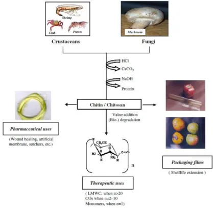
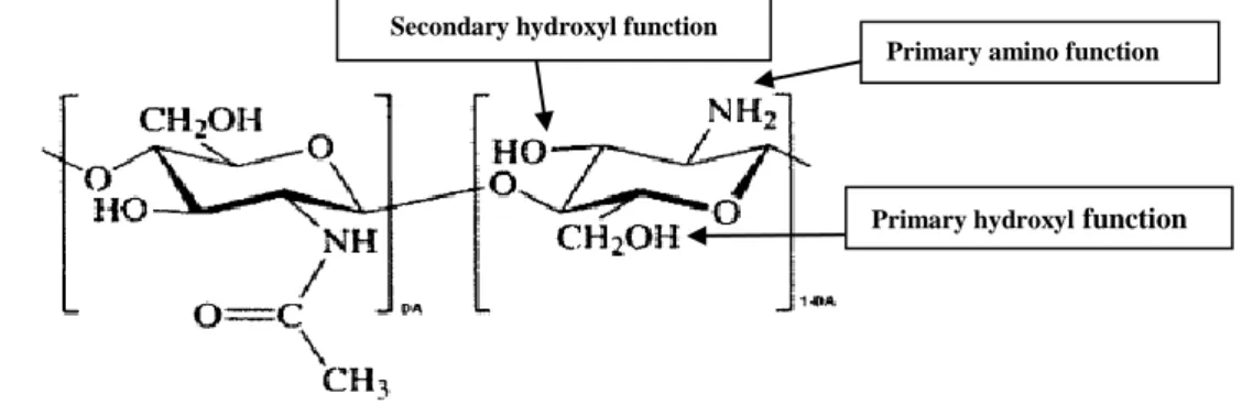
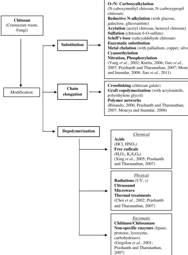
Figure 1.1. Production and value use of chitin/chitosan (Prashanth and Tharanathan, 2007). .. 1 Figure 1.2. Chemical structure of partially acetylated chitosan and its main reactive functional groups; DA=Degree of Acetylation (Rinaudo, 2006). ... 2 Figure 1.3. Methods of chitosan modification described in the literature. ... 4 Figure 1.4. Basic gut anatomy (Gibson and Roberfroid, 2008; Roberfroid et al., 2010). ... 15 Figure 1.5. Schematic representation of an adult microbiota (Roberfroid et al., 2010). The major phyla and genera are located on a logarithmic scale as number of CFU/g of faeces. Genera on the left site (pink) are likely to be potentially harmful whereas those on the right site (blue) are potentially beneficial to health. Those that are located both on the left site and the right site either (black) contain species that are potentially harmful and species that are potentially beneficial to health or contain genera/species that still need to be classified (white). Indeed many of these have only recently been identified in the gut microbiota and their roles are still largely unknown. ... 16 Figure 3.1. SEC-HPLC profiles of LMWC (A) and HMWC (B). ... 35 Figure 3.2. Absorbance (294 and 420 nm) and fluorescence measurements (excitation wavelength of 350/emission wavelength of 420 nm) for the samples withdrawn during the reaction of LMWC (A) and HMWC (B) with 0.5% (w/v) of Glc at 40 ºC during 72 h. Error bars indicate standard deviations. ... 35 Figure 3.3. Absorbance (294 and 420 nm) and fluorescence measurements (excitation wavelength of 350/emission wavelength of 420 nm) for the samples withdrawn during the reaction of LMWC with 0.5% (w/v) (A), 1% (w/v) (B) and 2% (w/v) (C) of Glc at 40 ºC during 72 h. Error bars indicate standard deviations. ... 37 Figure 3.4. Absorbance (294 and 420 nm) and fluorescence measurements (excitation wavelength of 350/emission wavelength of 420 nm) for the samples withdrawn during the reaction of LMWC with 1% and 2% (w/v) of Glc at 40 ºC (A, D), 60 ºC (B, E) and 80 ºC (C, F) during 72, 52 and 24 h respectively. Error bars indicate standard deviations. ... 39 Figure 3.5. Absorbance (294 and 420 nm) and fluorescence measurements (excitation wavelength of 350/emission wavelength of 420 nm) for the samples analyzed during the reaction of 2% (w/v) of LMWC with 2% (w/v) of Glc at 60 ºC during 52 h. Error bars indicate standard deviations. ... 41 Figure 3.6. Absorbance (294 and 420 nm) and fluorescence measurements (excitation wavelength of 350/emission wavelength of 420 nm) for the samples analyzed during the reaction of 2% (w/v) of COS with 2% (w/v) of Glc at 60 ºC and 32 h. Error bars indicate standard deviations. ... 42
IX
Figure 3.7. SEC-HPLC chromatograms obtained from the analysis of chitosan samples before (A) and after (B) reaction carried out with 2% (w/v) of LMWC, 2% (w/v) of Glc at 60ºC and 32h of reaction. ... 43 Figure 3.8. FT-IR spectra obtained for the LMWC-Glc derivative obtained by Maillard reaction under the optimal conditions (A) and comparison between LMWC and LMWC-Glc IR spectra (B) (region 800-1400 cm-1). ... 45 Figure 3.9. 1H-NMR spectrum of LMWC (A) and LMWC-Glc derivative (B). HOD: Signal corresponding to solvent. ... 46 Figure 3.10. SEC-HPLC profiles obtained from the analysis of LMWC and LMWC-Glc before (A and C, respectively) and after enzymatic reaction (B and D) at a concentration of 2% (w/v) with Pectinex Ultra SP-L (120 UE) at 40 ºC during 16 h of reaction. ... 48 Figure 3.11. SEC-HPLC profiles obtained from the analysis of COS-Glc1 (A) and COS-Glc2 (B). ... 49 Figure 3.12. FT-IR profiles obtained for unmodified chitooligosaccharides (COS) and modified chitooligosaccharides (COS-Glc1 and COS-Glc2). ... 50 Figure 3.13. 1H-NMR spectrum of COS (A) and COS derivatives, Glc1 (B) and COS-Glc2 (C). HOD: Signal corresponding to solvent. ... 52 Figure 3.14. Growth curves of L. brevis L24, L. casei L01, L. acidophilus LA5, L. acidophilus LA10, L. paracasei L26 and L. plantarum, in media containing MRS broth (with and without Glc as carbon source), supplemented with 0.5% (w/v) of COS-Glc1. ... 54 Figure 3.15. Growth curves of L. brevis L24, L. casei L01, L. acidophilus LA5, L. acidophilus LA10, L. paracasei L26 and L. plantarum, in media containing MRS broth (with and without Glc as carbon source), supplemented with 0.5% (w/v) of COS-Glc2. ... 55 Figure 3.16. Growth curves of B. animalis Bb12 and B. lactis B94 in media containing MRS broth (with and without Glc as carbon source), supplemented with 0.5% (w/v) of COS-Glc1 or 0.5% (w/v) of COS-Glc2. ... 57 Figure 3.17. Increment in total bacteria and Bifidobacterium counts determined by FISH in faecal cultures from three donors using COS, and the derivatives COS-Glc1 and COS-Glc2 as a carbon source. Data obtained after 48 h of incubation. Error bars indicate standard deviations. Means within different letters are significantly different (p < 0.05). Control does not include carbohydrate source added. Initial Log CFU/mL Bifidobacterium counts: donor 1=7.33±0.08, donor 2=7.38±0.06, donor 3=7.43±0.06. ... 58 Figure 3.18. Non-viable (Annexin V+/7AAD- plus Annexin V+/7AAD+) lymphocytes following treatment with the COS-Glc1, COS-Glc2 and COS (average ± standard deviation). ... 65
X
Figure 3.19. Necrotic (Annexin V+/7AAD+) and apoptotic (Annexin V+/7AAD-) lymphocytes following treatment with COS-Glc1, COS-Glc2 and COS (average ± standard deviation). ... 66
XI
List of Tables
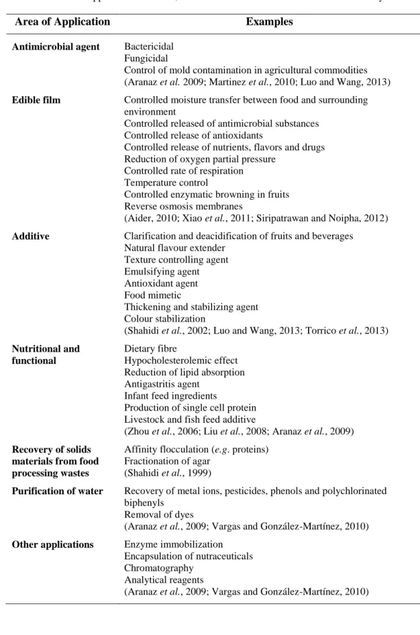
Table 1.1. Applications of chitin, chitosan and their derivatives in the food industry. ... 8 Table 1.2. Summary of the advantages and disadvantages of the different methods for the determination of DA of chitosan and its derivatives (Kasaai, 2009). ... 10 Table 1.3. Principal techniques used to establish the Mw of chitosan and its derivatives. ... 13 Table 1.4. Main classes of functional foods (Spence, 2006; Siró et al., 2008; Kaur and Das, 2011). ... 14 Table 1.5. Main health benefits of prebiotic oligosaccharides (Mussato and Mancilha, 2007). ... 20 Table 1.6. Principal molecular methodologies of bacterial identification (Gibson et al., 2004; Roberfroid, 2007). ... 23 Table 3.1. Average molecular weights (Mw ± SD) obtained for the analysis of the samples of LMWC and LMWC-Glc by SEC-HPLC before and after reaction of hydrolysis with Pectinex Ultra SP-L. ... 48 Table 3.2. Concentrations (mM) of the three major SCFAs values of faecal cultures from three donors. A= Acetate, P= Propionate, B= Butyrate. ... 63
XII
Abbreviations
CFU Colony Forming Unit
COS Chitooligosaccharides
COS-Glc1 Hydrolyzed LMWC-Glc derivative
COS-Glc2 Hydrolyzed LMWC subjected to the Maillard reaction
DA Degree of Acetylation
DD Degree of Deacetylation
DP Degree of Polymerization
DS Degree of Substitution
DSC Differential Scanning Calorimetry
FBS Fetal Bovine Serum
FDA Food and Drug Administration
FI Faecal Inoculum
FISH Fluorescent In Situ Hybridization
FOS Fructooligosaccharides
FT-IR Fourier Transform Infrared Spectroscopy
GC Gas Chromatography
Glc Glucose
GlcN Glucosamine
GlcNAc N-acetyl glucosamine
GPC-UV Gel Permeation Chromatography-Ultraviolet
GRAS Generally Recognized As Safe
XIII
HPLC High Performance Liquid Chromatography
1
H-NMR Proton Nuclear Magnetic Resonance
IR Infrared
LMWC Low Molecular Weight Chitosan
LMWC-Glc LMWC subjected to the Maillard reaction
LS Light-Scattering
MRS Man-Rogosa-Sharpe
Mw Molecular Weight
NDOs Non-Digestible Oligosaccharides
NIR Near-Infrared
NMR Nuclear Magnetic Resonance
OD Optical Density
PCR Polymerase Chain Reaction
PVSK Potassium Polyvinyl Sulfate
PS Phosphatidylserine
RI Refractive Index
RPS Reduced Physiological Salt Solution
SCFAs Short Chain Fatty Acids
SEC Size Exclusion Chromatography
TSB Trypticase Soya Broth
UV Ultraviolet
1
1. Introduction
Chitin is a linear polymer of N-acetyl glucosamine (GlcNAc) units linked by β (1→4) bonds and is mainly obtained as waste biomass from the seafood processing industry, although other sources such as fungi are also increasing. It is the second most abundant polymer in nature after cellulose and is the primary structural component of the shells of crustaceans, insects and fungal cell walls (Aranaz et al., 2009; Luo and Wang, 2013; Ruiz-Matute et al., 2013).
The deacetylated form of chitin is chitosan which is composed of units of glucosamine (GlcN) and GlcNAc linked by β (1→4) bonds. It is the only natural polysaccharide that presents cationic character due to the presence of free amino groups, usually responsible for biological activity. At low pH these groups are protonated and can interact with negatively charged compounds such as proteins, anionic polysaccharides (alginates, carragenates, pectins, among others), fatty acids, bile acids and phospholipids (Agulló et al., 2003; Guo et
al., 2005). This fact combined with its biocompatibility, biodegradability and non-toxicity has
made it prone to multiple applications namely in fields such as technology, food, cosmetics, medicine, biotechnology, agriculture and the paper industry (see Figure 1.1) (Prashanth and Tharanathan, 2007; Aranaz et al., 2009).
2
Despite this broad spectrum of applications, it is already recognized that the physicochemical properties of chitosan, such as solubility, molecular weight (Mw) and viscosity, can restrict its applicability in certain fields. In particular, chitosan is only soluble in acid aqueous solutions with a pH below its pKa (6.0-6.5) being insoluble in water and most organic solvents (Ruiz-Matute et al., 2013). The poor solubility of chitosan is the major limiting factor to its use and consequently, the interest in developing new strategies to modify the structure of chitosan in order to obtain novel derivatives with different physicochemical properties has increased in the last years (Luo and Wang, 2013; Ruiz-Matute et al., 2013). Moreover, the Mw and, consequently, the viscosity of chitosan, is considered a limiting factor in its application in certain fields. In fact, chitosan with high Mw, due to its high viscosity, pose important problems in terms of manipulation, which restricts its applicability in a commercial context (Chung et al., 2005; Aranaz et al., 2009).
Due to the presence of hydroxyl and amino groups on its backbones (see Figure 1.2), chitosan offers scope for manipulation enabling the production of a vast range of derivatives with application in different fields (Prashanth and Tharanathan, 2007; Luo and Wang, 2013).
Figure 1.2. Chemical structure of partially acetylated chitosan and its main reactive functional groups;
DA=Degree of Acetylation (Rinaudo, 2006).
1.1. Chitosan derivatives
Chemical derivatives of chitosan have received increasing interest over the past decades due to their associated chemical, biological, and functional advantages over unmodified chitosan. These include, but are not limited to, improved biocompatibility, better solubility in aqueous solutions over a wider range of pH, gelling properties, possibility to revert the net charge from polycationic to polyanionic, design of hydrophobic derivatives with amphiphilic character and capacity to harness self-assembling nanostructures and chemical conjugates
Primary amino function Secondary hydroxyl function
3
with an assortment of bioactive and therapeutic molecules (Sarmento et al., 2011). Several chemical modifications such as oligomerization, alkylation, acylation, quaternization, hydroxyalkylation, carboxyalkylation, thiolation, sulfation, phosphorylation, enzymatic modifications and graft copolymerization along with many assorted modifications have been carried out. Figure 1.3 summarizes the different methods that have been used to modify the structure of chitosan as well as some examples of derivatives already synthesized. The chemical modification affords a wide range of derivatives with modified properties for specific applications in diversified areas mainly pharmaceutical, biomedical and biotechnological.
The chemical modification of chitosan is often applied to obtain low molecular weight chitosan (LMWC), oligosaccharides and/or monomers. Among the various methods already described, the enzymatic methods are gaining importance because they allow a regioselective depolymerization under mild conditions (Prashanth and Tharanathan, 2007). In the case of enzymatic degradation of chitosan, LMWC with high water solubility was produced using chitinase, chitosanase, glucanase, lipase and some proteases (Pantaleone et al., 1992; Kumar
et al., 2004). There are also non-specific enzymes (Muzzarelli, 1997), including lysozyme,
cellulase, lipase, amylase, papain and pectinase (Nordtveit et al., 1996; Grigolon et al., 2001; Ruiz-Matute et al., 2013) that are capable of depolymerizing chitosan.
In recent years, assorted modifications including chitosan hybrids with sugars, cyclodextrins, dendrimers, and crown ethers have also emerged as interesting multifunctional macromolecules. The versatility in possible modifications and applications of chitosan derivatives presents a great challenge to scientific community and to industry and open opportunities for new research studies (Mourya and Inamdar, 2008).
4
Figure 1.3. Methods of chitosan modification described in the literature. O-/N- Carboxyalkylation
(N-caboxymethyl chitosan, N-carboxypropil chitosan)
Reductive N-alkylation (with glucose,
galactose, glucosamine)
Acylation (acetyl chitosan, benzoyl chitosan) Sulfation (chitosan 6-O-sulfate)
Schiff’s base (salicyaldehyde chitosan) Enzymatic substitution
Metal chelation (with palladium, copper, silver) Cyanoethylation
Nitration, Phosphorylation
(Yang et al., 2002; Kurita, 2006; Guo et al., 2007; Prashanth and Tharanathan, 2007; Mourya and Inamdar, 2008; Jiao et al., 2011)
Chitosan
(Crustacean waste, Fungi)
Substitution
Crosslinking (chitosan galate)
Graft copolymerization (with acrylonitrile,
polyethylene glycol)
Polymer networks
(Rinaudo, 2006; Prashanth and Tharanathan, 2007; Mourya and Inamdar, 2008)
Chain elongation Modification Depolymerization Chemical Acids (HCl, HNO2) Free radicals (H2O2, K2S2O8)
(Xing et al., 2005; Prashanth and Tharanathan, 2007) Physical Radiations (UV, ) Ultrasound Microwave Thermal treatments
(Choi et al., 2002; Prashanth and Tharanathan, 2007)
Enzymatic Chitinase/Chitosanase Non-specific enzymes (lipase,
protease, lysozyme, carbohydrases) (Grigolon et al., 2001; Prashanth and Tharanathan, 2007)
5
1.1.1. Modification of chitosan via sugar introduction. The Maillard reaction
The introduction of hydrophilic residues in the chitosan molecule, through the formation of covalent bonds with the reactive amino groups, is one of the most described strategies to increase the solubility of chitosan. Because of its hydrophilic nature, carbohydrates and particularly mono- and disaccharides, have been the most preferred compounds to carry out these modifications. It has been established that substitutions of 3-20% of the amino groups leads to a drastic increase in solubility, whereas the remaining free amino groups ensure the biological activity of the derivatives (Il’ina and Varlamov, 2007). The preparation of glycosylated derivatives of chitosan, involves, among other reactions, the Maillard reaction, reductive alkylation (the Maillard reaction in the presence of sodium borohydride) and amide formation.
Amide formation. Derivatives of chitosan with lactobionic acid through amide formation
generate branched derivatives with modified characteristics. These derivatives have been widely used as drug delivery systems for low molecular drugs, peptides and genes (Gao et al., 2003; Li et al., 2011a; Zhang et al., 2011), as effective synthetic extracellular matrices for the attachment of hepatocytes (Chung et al., 2002; Park et al., 2003; Mi et al., 2007) and as stabilizer for obtaining iron oxide nanoparticles for the preparation of multifunction nanoprobes (Bahadur et al., 2009). Recently, Ruiz-Matute et al. (2013) showed that these derivatives are very promising compounds to be applied as an additive in the food industry (for example to bind fat and cholesterol or avoid hardening of foods).
Reductive alkylation. By reductive alkylation it is possible to obtain branched chitosan
with modified functional properties or even to induce some new chemical and biological properties. To expand the range of solubility, derivatives of chitosan have been prepared with mono-, di-, tri- and polysaccharides (using cianoborohydride for the reduction), which presented an excellent solubility in water (Yalpani and Hall, 1984; Yang et al., 2002). Using sodium borohydride, N-alkyl derivatives of chitosan have been prepared with better fungicidal and insecticidal properties than native chitosan (Rabea et al., 2006). Cianoborohydride has been used for other applications of chitosan than the food sector, in which the presence of trace amounts of cyanide does not represent a problem. The use of sodium borohydride allows the formation of derivatives in which the presence of boron in trace amounts can be admitted in food ingredients at similar levels to those present in several foods and not exceeding the maximum recommended amounts (Li and Zhang, 2007).
6
Maillard reaction. The Maillard reaction takes place by condensation of the carbonyl
group of reducing sugars, aldehydes or ketones with amino groups of amino acids, proteins or nitrogenous compounds (Chang et al., 2011). It is well known that this reaction occurs during the heating, storage, and processing of foods. It influences the food quality by affecting factors such as colour, flavour, taste, and nutrition (Kanatt et al., 2008). It is characterized by the mildness of the reaction, easy operation and controllability (Chung et al., 2005).
Owing to the presence of a large number of amino groups in the chitosan molecule, it can be involved in the Maillard reaction. In fact, several authors have carried out this modification with the main objective of improving chitosan solubility and increase its applicability at neutral pH values. Indeed, Tanaka et al. (1993) obtained chitosan-glucose derivatives by Maillard reaction with improved functional properties. Depending on the stage of the Maillard reaction, changes in certain properties were observed. In particular, water binding capacity and antimicrobial activity decreased to some extent in the initial stages, while acidic dye binding capacity increased considerably as the reaction progressed. Other properties such as fat binding capacity and iron chelating were not affected by the reaction (Tanaka et al., 1993). In addition, Chung et al. (2005, 2006) prepared derivatives of chitosan with different carbohydrates (glucose, fructose, maltose and GlcN) by the Maillard reaction. In these studies, the derivatives formed with GlcN were those showing the higher solubility and the best metal chelating and antibacterial properties. Derivatives obtained by Maillard reaction from chitosan and xylose showed a bactericidal effect higher than native chitosan confirming their application to preserve refrigerated fresh pasta (Huang et al., 2007). More recently, Kanatt et al. (2008), obtained derivatives of chitosan with glucose by Maillard reaction that presented excellent antioxidant and antimicrobial properties and could be considered as promising food preservatives.
1.1.2. Applications of chitosan derivatives. Food applications
As it was mentioned before, chitosan is the only natural polysaccharide that presents cationic character due to the presence of free amino groups. Indeed, these groups are responsible for various unique properties such as biocompatibility, biodegradability, non-toxicity, and characteristic physicochemical and biological activities, which open opportunity for multiple applications. The broad fields of application of chitosan include medicine, biotechnology, pharmaceutical, cosmetics, foods and agriculture (Kurita, 2006; Prashanth and Tharanathan, 2007; Aranaz et al., 2009).
7
Regarding the food industry, conversion of processing waste and by-products into valuable products and alternative specialty materials has been identified as a timely challenge for food research and development associated with numerous applications of chitosan polymers. In that sense, these biopolymers offer a wide range of unique applications including production of value-added food products (Rinaudo, 2006; Aranaz et al., 2009), preservation of foods from microbial deterioration assuring extension of shelf-life (No et al., 2002; Bautista-Baños
et al., 2006; Vargas and González-Martínez, 2010; Jiang et al., 2012), biodegradable edible
films and coatings (Hernández-Muñoz et al., 2006; Santos et al., 2006; Ribeiro et al., 2007; Aranaz et al., 2009), recovery of valuable compounds from food processing waste and by-products (Shahidi et al., 1999; Casal et al., 2006), purification of water (No and Meyers, 2000; Crini, 2005; Guibal et al., 2006), separation and purification of proteins (Chen and Chuang, 2013) and clarification and deacidification of fruit juices (Soto-Perlata et al., 1999; Chatterjee et al., 2004; Rungsardthong et al., 2006).
Another important property attributed to chitosan and its derivatives is their scavenging capacity against different radical species, conferring them antioxidant properties (Prashanth and Tharanathan, 2007; Aranaz et al., 2009; Vargas and González-Martínez, 2010). The origin of the scavenging ability of chitosan and derivatives is related with the presence of active hydroxyl and amino groups in the polymer chains. The hydroxyl groups in the polysaccharide units can react with free radicals and the amino groups can react with free radicals to form additional stable macroradicals (Yen et al., 2008). Regarding the effect of the Mw of chitosan on its antioxidant properties, Xing et al. (2007) showed through an in vitro study that LMWC had stronger scavenging activity effect on oxygen and hydroxyl groups than high molecular weight chitosan (HMWC). The same effect was observed by Feng et al. (2007) by reducing chitosan Mw by means of irradiation treatments.
Associated to the antioxidant property, chitosan and chitosan derivatives can be used to decrease oxidation process, in particular to prevent oxidative reaction in food or beverage products (Vargas and González-Martínez, 2010). The antioxidant activity of chitosan solutions was tested in a cooked cod fish model system, demonstrating a reduction in peroxides index and thiobarbituric reactive substances, with increasing efficiency according chitosan concentration and decrease in the Mw (Shahidi et al., 2002). Table 1.1 shows the main applications of chitin, chitosan and their derivatives in the food industry.
8
Table 1.1. Applications of chitin, chitosan and their derivatives in the food industry.
Area of Application Examples
Antimicrobial agent Bactericidal Fungicidal
Control of mold contamination in agricultural commodities (Aranaz et al. 2009; Martinez et al., 2010; Luo and Wang, 2013)
Edible film Controlled moisture transfer between food and surrounding environment
Controlled released of antimicrobial substances Controlled release of antioxidants
Controlled release of nutrients, flavors and drugs Reduction of oxygen partial pressure
Controlled rate of respiration Temperature control
Controlled enzymatic browning in fruits Reverse osmosis membranes
(Aider, 2010; Xiao et al., 2011; Siripatrawan and Noipha, 2012)
Additive Clarification and deacidification of fruits and beverages Natural flavour extender
Texture controlling agent Emulsifying agent Antioxidant agent Food mimetic
Thickening and stabilizing agent Colour stabilization
(Shahidi et al., 2002; Luo and Wang, 2013; Torrico et al., 2013)
Nutritional and functional
Dietary fibre
Hypocholesterolemic effect Reduction of lipid absorption Antigastritis agent
Infant feed ingredients
Production of single cell protein Livestock and fish feed additive
(Zhou et al., 2006; Liu et al., 2008; Aranaz et al., 2009)
Recovery of solids materials from food processing wastes
Affinity flocculation (e.g. proteins) Fractionation of agar
(Shahidi et al., 1999)
Purification of water Recovery of metal ions, pesticides, phenols and polychlorinated biphenyls
Removal of dyes
(Aranaz et al., 2009; Vargas and González-Martínez, 2010)
Other applications Enzyme immobilization Encapsulation of nutraceuticals Chromatography
Analytical reagents
9
1.2. Structural characterization of chitosan and its derivatives
The content and sequence of GlcN and GlcNAc units in the chitosan molecule will determine the physicochemical and the biological properties of the polymer. It is known that heterogeneous conditions during deacetylation of chitin to obtain chitosan, provide a block-wise distribution, whereas under homogeneous conditions a random distribution of acetyl groups appears in chitosan. Thus, the degree of acetylation (DA) and the Mw (specifically the weight-average molecular weight) are the two major parameters that affect the properties and the applications of chitosan (Rinaudo, 2006; Aranaz et al., 2009). In the case of the derivatives obtained by substitution, the degree of substitution (DS) is also an important parameter to take into account (Ruiz-Matute et al., 2013). Additionally, due to its rigid and specific crystalline structures, possible through intra- and intermolecular hydrogen bonding, chitosan has the ability to exist in nature in different polymorphic forms, whose properties vary considerably.
1.2.1. Determination of the degree of acetylation and degree of substitution
The DA corresponds to the ratio between the acetylated and not acetylated GlcN units into the molecule of chitosan (Kasaai, 2008; 2009). For the chitosan derivatives, the DS can be obtained by the difference between DA before and after substitution reaction (Ruiz-Matute et
al., 2013). Several methods have been reported to determine the DA of chitosan. These
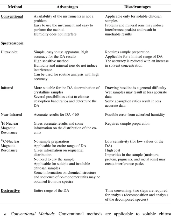
methods can be classified in three groups: (a) conventional, (b) spectroscopic and (c) destructive (Rinaudo, 2006; Kasaai, 2009). Table 1.2 shows the main advantages and disadvantages of these methods, which will be briefly explained below.
10
Table 1.2. Summary of the advantages and disadvantages of the different methods for the determination of DA
of chitosan and its derivatives (Kasaai, 2009).
Method Advantages Disadvantages
Conventional Availability of the instruments is not a problem
Easy to use the instrument and easy to perform the method
Humidity does not interfere
Applicable only for soluble chitosan samples
Proteins and mineral ions may induce interference peak(s) and result in unreliable results
Spectroscopic
Ultraviolet Simple, easy to use apparatus, high accuracy for the DA results High sensitive method
Humidity and mineral ions do not induce interference
Can be used for routine analysis with high accuracy
Requires sample preparation
Applicable for a limited range of DA The accuracy is reduced with an increase in solvent concentration
Infrared More suitable for the DA determination of crystalline samples
Several possibilities exist to choose absorption band ratios and determine the DA
Drawing baseline is a general difficulty Wet samples may result in less accurate data
Some absorption ratios result in less accurate data
Near-Infrared Accurate results for DA ≤ 60 Possible error from adsorbed humidity
1H-Nuclear
Magnetic Resonance
Gives accurate results and some information on the distribution of the co-units
Requires sample preparation
13C-Nuclear
Magnetic Resonance
No sample preparation
Applicable for entire range of DA Gives information on sequential distribution
No need to dry the sample
Applicable for soluble and insoluble chitosan samples
Some information on chemical structure and sequence of co-monomer units may be obtained from the spectra
Low sensitivity (for low values of the DA)
High cost
Impurities in the sample (moisture, protein, pigments, and metal ions) may create interference peaks
Destructive Entire range of the DA Time consuming: two steps are required for analysis (decomposition and analysis of the decomposed species)
a. Conventional Methods. Conventional methods are applicable to soluble chitosan
samples as well as those with low acetyl contents. These methods include titration with an
acid or alkali, colloid titration, conductometry, potenciometric titration, the ninhydrin assay
and the adsorption of free amino groups of chitosan by picric acid (Kasaai, 2009). The use of these different methods by diverse research groups has given contradictory data and
11
conclusions for low acetyl contents of chitosans. This is because the amino groups of chitosan (with low acetyl contents) are accessible for titration in the conventional methods (Nanjo et
al., 1991; Varum et al., 1995).
b. Spectroscopic Methods. The spectroscopic methods not only give information about the
DA of chitosan and its derivatives but also on their chemical structure, sequence and morphology. These methods include Nuclear Magnetic Resonance (NMR) spectroscopy (1 H-NMR, 13C-NMR and 15N-NMR), Infrared (IR) spectroscopy, Ultraviolet (UV) spectrometry and Gel Permeation Chromatography with Ultraviolet detection (GPC-UV) (Kasaai, 2009).
A chitosan sample produces a larger absorbance in UV technique in comparison with near-infrared (NIR), IR and NMR methods. In the latter methods, in order to obtain sufficient absorbance, more amounts of the sample or greater concentrations are needed. Thus, determination of the DA by UV spectrophotometry results in greater accuracy when compared to NIR, IR and NMR methods. In fact, the IR technique is mainly used for qualitative analysis and comparison studies. It has been used for quantitative analysis of crystalline samples, since these samples created sharper signals and higher resolution compared to amorphous samples (Kasaai, 2009). 1H-NMR technique has usually been employed as a standard method to calibrate other methods (Varum et al., 1991a; Shigemasa et
al., 1996; Brugnerotto et al., 2001). Among various conditions proposed for determining the
DA of chitosan by 1H-NMR (Brugnerotto et al., 2001; Terry and Joyce, 2004; Bautista-Baños
et al., 2006), the procedures proposed by Hirai et al. (1991) and Varum et al. (1991a) have
been widely accepted. 15N-NMR and 13C-NMR do not need a dried sample. However, these two techniques are not appropriate for chitin/chitosan having low DA values since they resulted in underestimated values (Domard, 1987; Raymond et al., 1993; Deserieres et al., 1996; Heux et al., 2000).1H-NMR and 13C-NMR spectroscopy may also provide information on the sequential distribution of free amino and N-acetyl groups (Varum et al., 1991a; Varum
et al., 1991b).
c. Destructive Methods. Destructive methods (Elemental analysis, Thermal analysis by
Differential Scanning Calorimetry (DSC), acid or enzymatic hydrolysis followed by High Performance Liquid Chromatography (HPLC) or spectrophotometry analysis, and pyrolysis-Gas Chromatography (GC) analysis) can be used for the entire range of the DA. In the HPLC and GC analyses of chitin/chitosan samples in the presence of other carbohydrates and polysaccharides, overestimated DA values may be obtained. This is due to the formation of
12
additional acetic acid from the impurities. Excess amounts of oxalic acid had an adverse effect on the experimental results obtained from the pyrolysis-GC method (Sato et al., 1998).The impurities do not create any difficulties for the DA analysis by HPLC and GC if the impurities are clearly separated in different peaks from those corresponding to the sample. The error variation in the elemental analysis method is relatively large (Roberts, 1992). This is because the presence of organic materials or polysaccharides other than chitin/chitosan (as impurities) significantly changes the ratio of Nitrogen (N)/Carbon (C).
1.2.2. Molecular weight determination
The physicochemical, biological and rheological properties of chitosan vary significantly as a function of its weight-average Mw and Mw distribution (Beri et al., 1993; Denuzière et
al., 1995). It is therefore important to have precise and accurate values of the Mw of chitosan
(Nguyen et al., 2009).
It is well known that the determination of the Mw of polyelectrolytes is complex (Terbojevich et al., 1993). In the case of chitosan, this situation is exacerbated due to the marked tendency of this polymer to form resilient aggregates in solution (Anthonsen et al., 1994; Philippova et al., 2001; Liu and Yao, 2002; Schatz et al., 2003). In order to determine the Mw of chitosan and its derivatives, viscosimetry (Knaul et al., 1998; Kumar, 2000; Aranaz et al., 2009), Size Exclusion Chromatography (SEC) (also called gel filtration chromatography) (Aranaz et al., 2009; Nguyen et al., 2009) and Light-Scattering (LS) (Kumar, 2000; Aranaz et al., 2009) have been used. Table 1.3 describes the principles of each method as well as their advantages and disadvantages.
13
Table 1.3. Principal techniques used to establish the Mw of chitosan and its derivatives.
Method Description Advantages/Disadvantages
Viscosimetry Based on the use of the Mark–Houwink equation: [η]= K x M a, where [η] is the intrinsic viscosity, M is the Mw, K and a are constants depending on the polymer and the solvent system used as well as the temperature (Rinaudo, 2006)
Simple and rapid method
Not greatly affected by the presence of negligible amounts of very high Mw polymer
Requires calibration curves (Terbojevich and Cosani, 1997)
Size Exclusion Chromatography
(SEC)
A liquid mobile phase is passed through a column (stationary phase) at a fixed flow rate. The molecules with different molecular sizes are separated into distinct chromatographic bands (Wu, 1995)
Provides the number-average Mw and the weight-average Mw in a single measurement Requires calibration curves
The ionic strength of the SEC mobile phase can affect precision and variability of chitosan SEC analyses (Nguyen et al., 2009)
Light-Scattering (LS)
Based on the interaction of electromagnetic waves with matter by measuring the changes in the number (intensity), the direction (momentum) and the frequency (energy) of each type of photon in the incident and the emerging light beam (Chu, 1970)
LS measurements are difficult to perform Sometimes the data are not easy to interpret, in the presence of aggregation and/or association (Terbojevich and Cosani, 1997)
1.3. Functional foods
In the last decades, consumer demands in the field of food production have changed considerably. In fact, nowadays foods are not intended to only satisfy hunger and to provide necessary nutrients to the normal body function, but also to prevent nutrition-related diseases and improve physical and mental well-being (Roberfroid, 2000b; Menrad, 2003). In this view, functional foods play an outstanding role (Roberfroid, 2000a, 2000b; Kotilainen et al., 2006).
The term "functional food" was first used in Japan, in the 1980s, for food products fortified with special constituents that possess advantageous physiological effects (Hardy, 2000; Stanton et al., 2005; Kaur and Das, 2011). Presently, there is no universally accepted definition for functional foods, which are more accurately viewed as a concept than a well-defined group of food products (Siró et al., 2008; Kaur and Das, 2011). Indeed, a large number of definitions exist worldwide for functional foods (Doyon and Labrecque, 2008; Siró
et al., 2008).
In a review paper published in 2008 (Doyon and Labrecque, 2008) a study of over a hundred definitions for functional foods was carried out. In this study, the following working definition for functional foods was proposed:"A functional food is, or appears similar to, a
14
normal quantities. It has proven health benefits that reduce the risk of specific chronic diseases or beneficially affect target functions beyond its basic nutritional functions".
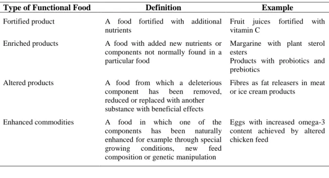
According to Spence (2006), Siró et al. (2008) and Kaur and Das (2011), it is possible to classify the functional foods in different classes, as it is shown in Table 1.4.
Currently, the market of functional foods is dominated by gut health products (Siró et al., 2008). Indeed, it is now well established that the gut microbiota has a profound influence on health. In this view, there is presently a great deal of interest in the use of functional foods to manipulate the composition of the gut microbiota in order to improve health (Wang, 2009; Roberfroid et al., 2010).
Table 1.4. Main classes of functional foods (Spence, 2006; Siró et al., 2008; Kaur and Das, 2011).
Type of Functional Food Definition Example
Fortified product A food fortified with additional nutrients
Fruit juices fortified with vitamin C
Enriched products A food with added new nutrients or components not normally found in a particular food
Margarine with plant sterol esters
Products with probiotics and prebiotics
Altered products A food from which a deleterious component has been removed, reduced or replaced with another substance with beneficial effects
Fibres as fat releasers in meat or ice cream products
Enhanced commodities A food in which one of the components has been naturally enhanced for example through special growing conditions, new feed composition or genetic manipulation
Eggs with increased omega-3 content achieved by altered chicken feed
1.3.1. Gut microbiota
The human gastrointestinal tract is the natural habitat for a complex bacterial community (Roberfroid et al., 2010). It is recognized that its colonization starts immediately after birth (Guarner and Malagelada, 2003) and then a pattern that resembles the adult microbiota is established (Gibson and Roberfroid, 1995).
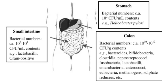
Moreover, it is known that factors such as pH, nutrient availability, host health, bacterial adhesion, transit time, among others, influence the numbers and diversity of bacteria present in the different regions of the gastrointestinal tract (Kerckhoffs et al., 2006). Figure 1.4 illustrates the basic gut anatomy and shows that different types of microbial community in terms of both species diversity and numbers colonize the different regions within the gut. The conditions of the stomach reduce the microbial load to approximately 102 colony forming unit
15
(CFU) (per mL) while in the small intestine it reaches 102-104 CFU/mL. The colon is the region with the highest microbial load namely 1010-1012 CFU/g. Furthermore, within the colon the distal region is the area of highest colonization with more than 500 different species and up to 100 billion microbial inhabitants per gram of contents (Gibson and Roberfroid, 2008; Roberfroid et al., 2010).
Figure 1.4. Basic gut anatomy (Gibson and Roberfroid, 2008; Roberfroid et al., 2010).
Apart from the knowledge on the complexity of the gut microbiota, it is also known that certain bacteria are associated with toxin formation and even pathogenicity when they become dominant. These potentially harmful bacteria belong to species within groups such as clostridia and bacteroides. On the other hand, potentially healthy bacterial groups are characterized, besides absence of toxin production, by a beneficial metabolism towards the host through the formation of short chain fatty acids (SCFAs) or vitamin synthesis. They may also inhibit pathogens through a multiplicity of mechanisms (competition for colonization sites and nutrients, production of bacteriocins and acids and consequently reduction of in situ pH). Acknowledged examples of beneficial bacteria present in the gut microbiota are bifidobacteria and lactobacilli (Roberfroid et al., 2010). Other groups like streptococci, enterococci, eubacteria and bacteroides have also been considered as potentially beneficial to health, however some of them include potentially harmful species, in particular enterococcus. With regard to some of the most recently identified genera in the major phyla (Firmicutes,
Actinobacteria and Bacteroidetes), classification as potentially beneficial or potentially
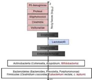
harmful to health still remains to be ascertained (Roberfroid et al., 2010). Figure 1.5 illustrates the main bacteria present in the adult gut.
Stomach
Bacterial numbers: c.a. 102 CFU/mL contents
e.g., Helicobacter pylori Small intestine Bacterial numbers: ca. 102-104 CFU/mL contents e.g., lactobacilli, Gram-positive cocci Colon
Bacterial numbers: c.a. 1010-1012 CFU/g contents
e.g., bacteroides, bifidobacteria,
clostridia, peptostreptococci, fusobacteria, lactobacilli, enterobacteria, enterococci, eubacteria, methanogens, sulphate reducers, etc.
16
Figure 1.5. Schematic representation of an adult microbiota (Roberfroid et al., 2010). The major phyla and
genera are located on a logarithmic scale as number of CFU/g of faeces. Genera on the left site (pink) are likely to be potentially harmful whereas those on the right site (blue) are potentially beneficial to health. Those that are
located both on the left site and the right site either (black) contain species that are potentially harmful and species that are potentially beneficial to health or contain genera/species that still need to be classified (white).
Indeed many of these have only recently been identified in the gut microbiota and their roles are still largely unknown.
It has become clear that the gut microbiota plays important nutritional and physiopathological roles which include: prevention of gut colonization by potentially pathogenic microorganisms by competing with invading pathogens for ecological niches and metabolic substrates; important sources of energy for the cells of the gut wall through the fermentation of carbohydrates to SCFAs; modulation of the immune system; modulation of gene expression and cell differentiation in the gut wall (Gibson and Roberfroid, 2008; Roberfroid et al., 2010).
It is already recognized that products causing a selective modification in the composition and/or activity of gut microbiota can induce beneficial effects in human health and well-being. In this view, there is currently a great deal of interest in the use of food ingredients to manipulate the composition of the microbiota, particularly in the colon, in order to improve health (Wang, 2009).
There are three methods of manipulating the composition of the colonic microbiota, namely by the use of probiotics, prebiotics or the combination of both (symbiotic) (Roberfroid
et al., 2010).
The concept of probiotics was introduced long before that of prebiotics. A probiotic has been defined as "Live microorganisms which when administered in adequate amounts confer a health benefit on the host" (FAO/WHO, 2002). In the case of probiotics, the microorganism is introduced into the host intestinal microbiota and causes a selective modification of its
17
composition. It is the probiotic by itself that, by implanting into the gut microbiota, is responsible for the resulting effects (Gibson and Roberfroid, 2008). The most common probiotics currently used belong to the genera Bifidobacterium and Lactobacillus. The intake of probiotics has been associated with a considerable number of health benefits, namely reduction of diarrhoea, reduction of gastrointestinal tract symptoms (constipation, bloating, etc.), stimulation of the immune system, prevention of cancer, among others (Simmering and Blaut, 2001; Parvez et al., 2006).
Another approach consists in the use of prebiotics. In this case, the effect is essentially indirect because the prebiotic selectively feeds one or a limited number of microorganisms thus causing a selective modification of the host’s colonic microbiota. Therefore, it is not the prebiotic by itself but rather the changes induced in the composition of gut microbiota that are responsible for its effects (Gibson and Roberfroid, 2008).
1.3.2. Prebiotics
The concept of prebiotic was first defined by Gibson and Roberfroid in 1995 as "A non-digestible food ingredient that beneficially affects the host by selectively stimulating the growth and/or activity of one or a limited number of bacteria in the colon, and thus improving host health". The most recent definition of prebiotic is the following: "A dietary prebiotic is
a selectively fermented ingredient that results in specific changes, in the composition and/or activity of the gastrointestinal microbiota, thus conferring benefit(s) upon host health" (ISAPP, 2008).
To be classified as a prebiotic, a food ingredient must fulfil three requirements: 1) resistance to gastric acidity; 2) fermentation by intestinal microbiota; and 3) selective stimulation of the growth and/or activity of those intestinal bacteria that contribute to health and well-being (Roberfroid, 2007).
In this view, any dietary component that reaches the colon intact (or partly so) is a potential candidate for prebiotic attribute. However, it is the latter of the three mentioned criteria which is crucial but still the most difficult to fulfil (Roberfroid et al., 2010). Several dietary carbohydrates (polydextrose, glucooligosaccharides, lactose, hemicellulose, resistant starch, resistant dextrins, β-glucans, sugar alcohols such as lactitol) and also other compounds (for example peptides and yeast extract) showed some fermentation selectivity when tested in laboratory systems. Nevertheless, the ultimate test for prebiotic activity (i.e. human trials) is lacking for the majority of these compounds (Roberfroid et al., 2010).
18
Therefore, there is much interest in the development of novel prebiotics with validated bioactivity, with well-established relationship structure-activity, addressed to reach the distal regions of the colon unaltered and promote the growth of specific bacteria. These may conceivably possess desired attributes not present in the current generation of molecules (Rastall and Maitin, 2002). In particular, prebiotic oligosaccharides have been a target of intensive research as ingredients with potential health-promoting properties (Korhonen, 2002).
1.3.2.1. Prebiotic oligosaccharides
Oligosaccharides are carbohydrates with a low degree of polymerization (DP) and consequently low Mw. They have been defined as including from 2 to 20 monosaccharide units (Roberfroid and Slavin, 2000). However, according to IUB-IUPAC (Joint Commission on Biochemical Nomenclature, 1982) the dividing point between oligo- and polysaccharides is 10. Moreover, oligosaccharides have been classified according to their physiological properties as digestible and digestible (Cummings et al., 1997). The concept of non-digestible oligosaccharides (NDOs) originates from the observation that the anomeric C atom (C1 or C2) of the monosaccharide units of some dietary oligosaccharides has a configuration that makes their osidic bounds non-digestible to the hydrolytic activity of the human digestive enzymes (Roberfroid and Slavin, 2000).
NDOs can be found as natural components in milk, honey, fruits and vegetables. Generally, the concentration of NDOs in these types of foods range between 0.3 and 6% of fresh weight (Mussatto and Mancilha, 2007). In what concerns the caloric value, it has been estimated to be 1.5-2.0 Kcal/g, which is approximately 40-50% of those of digestible carbohydrates such as sucrose (Mussatto and Mancilha, 2007).
Among all the food ingredients, NDOs are the most relevant prebiotic candidates. Due to their chemical structure, they are resistant to gastrointestinal absorption and hydrolysis by digestive enzymes being considered as colonic ingredients. Moreover, some oligosaccharides can selectively stimulate the growth and/or activity of certain intestinal bacteria with positive health outcomes which make them prebiotic ingredients. Among all the recognized NDOs there are currently three that fulfil the criteria mentioned before and are thus considered as prebiotics: inulin-type fructans, galactooligosaccharides and lactulose (Gibson and Roberfroid, 2008). In fact, these are the only compounds that have, until now, proved their
19
prebiotic effects by their ability to change the gut microbiota composition after a short feeding period at reasonably low doses (Roberfroid et al., 2010).
Oligosaccharides obtained from chitosan by partial hydrolysis - i.e. chitooligosaccharides (COS), as well as LMWC have been claimed to exhibit prebiotic effects (Lee et al., 2002; Fernandes et al., 2012). In fact, these compounds possess a structure similar to prebiotic glucooligosacharides. However, the presence of the amino groups in their molecule confers an important antimicrobial activity (Kong et al., 2010; Hafdani and Sadeghinia, 2011), which can cause a decrease in the bacterial host population (Vernazza et al., 2005) with negative health outcomes. Chemical modification of chitosan and COS by substitution of their amino groups could eliminate this antimicrobial effect and convert them into new prebiotic ingredients.
Another important aspect to consider is related to the biocompatibility and toxicity of these potential prebiotic ingredients. In fact, any substance that is intentionally added to food (food additive), must be subjected to premarket review and approval by Food and Drug Administration (FDA). Indeed, it must be demonstrated that the substance is safe under the conditions of its intended use, so the ingredient must be Generally Recognized As Safe (GRAS). Some studies carried out with COS reported toxic effects upon human cells (Xu et
al., 2008; Fernandes et al., 2011) associated to its amino groups. In this view, the
modification of COS through the substitution of the amino groups may reduce their cytotoxicity allowing their safe use as prebiotic compounds. However, prior to its use, it is important to evaluate the cytotoxicity of the modified COS when compared with the unmodified COS to guarantee its safety as prebiotic ingredients. Most of the studies on COS cytotoxicity use the MTT colorimetric assay (Fernandes et al., 2011) which is based on the capacity of mitochondrial enzymes of viable cells to transform the MTT tetrazolium salt into MTT formazan (Mosmann, 1983). Flow cytometry is another method widely used to evaluate the cytotoxicity. This method can provide rapid, quantitative and objective evaluation of cell viability, and may further provide enumeration of apoptotic or necrotic cells. It has become the method of choice to assay for apoptosis and necrosis in a variety of cell systems. The double-staining Annexin V/7-AAD assay discriminates cells that are undergoing early or late apoptosis and necrosis (Baudouin et al., 2007).
Prebiotics have received much attention in the last years and intensive research has been conducted in this field. In particular, prebiotic oligosaccharides derived from food industry by-products have been a target of great interest as ingredients with potential health-promoting
20
properties (Korhonen, 2002). This type of products are regarded as an alternative to synthetic polymers combining the production of manufactured products with the protection of environment, cost reductions and waste material recycling (Ruiz-Matute et al., 2013). Since chitosan and COS are by-products of the seafood industry, they represent an alternative with great potential in the development of novel prebiotic ingredients with health-promoting properties.
1.3.2.1.1. Health benefits associated to prebiotics
The fermentation of prebiotic oligosaccharides by colonic bacteria may result in several health benefits (see Table 1.5).
Table 1.5. Main health benefits of prebiotic oligosaccharides (Mussato and Mancilha, 2007).
Health Benefit Description
Modification of the colonic microbiota
Stimulation of the growth and proliferation of beneficial bacteria (Lactobacillus and Bifidobacterium). The production of SCFAs by these microorganisms results in a pH decrease and thus inhibition of the growth of pathogenic bacteria
Nutrient production Production of vitamins of the B complex (B1, B2, B6 and B12, nicotinic and folic acid)
Constipation relief and effects on intestinal motility
The ingestion of prebiotic oligosaccharides has demonstrated to prevent constipation. The end products of the fermentation, the SCFAs, are efficiently absorbed and used by the human colonic epithelial cells, stimulating their growth as well as salt and water absorption. The increased humidity of the faecal bolus improves the intestinal motility
Protective effect against infection in the
gastrointestinal,
respiratory and urogenital tracts
These effects are related to the pH decrease due to the production of SCFAs, the production of bacteriocins by several species of Lactobacillus and Bifidobacterium and the competition for nutrients and adhesion sites in the epithelial surface
Increase in absorption of minerals
The increased absorption of iron, calcium, and magnesium is related to the binding/sequestering capacity of the prebiotic oligosaccharides. Thus, these minerals are not absorbed in the small intestine and reach the colon, where they are released from the carbohydrate matrix and absorbed
Beneficial effect on carbohydrates and lipids metabolism
The ingestion of prebiotic oligosaccharides decreases the blood concentration of cholesterol, triglycerides and phospholipids, reducing the risk of diabetes and obesity. Some strains of Lactobacillus acidophilus assimilate the cholesterol present in the medium, while others appear to inhibit the absorption of cholesterol through the intestinal wall. Also, changes in lipid metabolism were suggested to be a consequence of a metabolic adaptation of the liver induced by SCFAs
Reduction of cancer risk The anticarcinogenic effect appears to be related to the increase of beneficial bacteria and SCFAs production during the fermentation. It has been demonstrated that the intake of certain prebiotic disaccharides reduces faecal physiological parameters (pH, ammonia, p-cresol and indole) considered to be risk factors for cancer development