FCUP
Diversity and biotechnological potential of epiphytic bacteria of macroalgae 3
Agradecimentos
Quero dirigir as minha primeiras palavras à minha Orientadora Prof. Dr. Olga Maria Oliveira da Silva, por me “acolher” num momento crucial do meu percurso académico e me mostrar que a microbiologia realmente é interessante. Antes de tudo, obrigada pela paciência, apoio, motivação e carinho prestados durante todo este ano. Com a professora aprendi que para “fazer ciência” não precisamos de estar num “regime militar” e que a podemos fazer com um sorrisso diário no rosto. Saio desta faculdade com consciência que melhorei muito as minhas competências ciêntificas por ter realizado esta Tese.
Em segundo lugar quero expressar toda a minha gratidão à minha “Co-Orientadora” Patrícia Graça, por todos os ensinamentos científicos, paciência e acima por toda a amizade desenvolvida este ano. Esta tese só foi possivel graças ao seu suporte, carinho e cumplicidade.
À Dra. Claudia Serra agradeço a colaboração dispensada na realização da técnica do DGGE e ao Dr. José Catita a disponibilização do microscópio de varrimento.
Agradeço também a todos meus colegas do LEMUP, que me acompanharam neste percurso e contribuiram de alguma forma para a realização desta tese.
Aos meus queridos “Sapos”, obrigada por terem partilhado comigo os melhores momentos que durante estes cinco anos passei nesta faculdade e por me apoiarem nos bons e maus momentos. “One Sapo Very Sapo”.
A todas as meninas do 304 da RUCA que por lá passaram durante a minha longa estadia e com as quais partilhei longas conversas, jantares, festas e momentos para sempre guardados na memória.
Quero agradecer ao “GANG”, os amigos de sempre e para todo o sempre, que me apoiam diáriamente e incentivam para ultrapassar todas as “pedras” que aparecem no caminho.
Um especial agradecimento à minha querida Cató por toda a paciência, dedicação, carinho e grande amizade que teve durante estes longos cinco anos. Começamos, acabamos e começaremos uma nova jornada juntas.
Ao Pipe, por me ajudar em todos os momentos da minha vida e por me apoiar e motivar quando mais precisei.
Por fim, quero agradecer à minha família que durante estes anos, com muito carinho, me ajudaram, aconcelharam e encaminharam para conseguir terminar esta etapa e seguir os meus sonhos.
Resumo
Os oceanos são um ecossistema com uma elevada diversidade de espécies, contendo milhões de microrganismos, muitos deles ainda por descobrir. Com os avanços tecnológicos, novas espécies microbianas têm sido descritas e demonstraram ser de extrema relevância para o ambiente e qualidade de vida humana. As bactérias são os principais intervenientes nos processos ecológicos e biogeoquímicos que ocorrem no ecossistema marinho. São encontradas na superficie de vários macroorganismos formando complexas associações denominadas biofilmes, com importância ecológica.
As superfícies das macroalgas são comummente colonizadas por uma diversa comunidade bacteriana que tende a ser específica e constante em diferentes espécies de macroalgas. As complexas interações entre as macroalgas e as bactérias revelam-se cruciais para o derevelam-senvolvimento de ambos os organismos. Novas espécies e géneros de bactérias têm sido encontradas no biofilme da superficie das macroalgas por métodos dependentes e independentes de cultivo.
Para além disso, as bactérias comunicam entre si e com o hospedeiro através de interações complexas mediadas pela produção de pequenas moleculas que podem ter potencial bioactivo. O conhecimento do funcionamento destes processos tem permitido descobrir compostos bioactivos com potencial biotecnológico.
Neste sentido, o objetivo principal deste estudo é analisar a diversidade bacteriana que se encontra no biofilme de três diferentes espécies de macroalgas: Ulva sp., Porphyra dioica e Sargassum muticum através de diferentes métodos de cultivo e moleculares. A observação da superficie das macroalgas por microscopia óptica e eletrónica de varrimento permitiu visualizar esta diversidade. Através do isolamento de bactérias em culturas puras, através de amostras das macroalgas amostradas no Outono, obtiveram-se 245 isolados (41% da Ulva sp., 25 % da P. dioica e 34 % do S. muticum). Até ao momento, apenas 86 culturas foram identificadas com base no gene do rRNA 16S, tendo-se obtido bactérias que pertencem ao filo das Gammaproteobacteria, Alphaproteobacteria, Bacteroidetes, Planctomycetes, Firmicutes e Actinobacteria. Vibrio foi o género mais abundante nos isolamentos.
A partir de macroalgas colhidas no Outono e Primavera, foram obtidos isolados de planctomycetes os quais são filogenéticamente afiliados a Rhodopirellula baltica.
Ambos os métodos de pirosequenciação (Sequenciação de nova geração) e de Eletroforese em Gel de Gradiente de Desnaturação (DGGE) permitiram verificar que
as macroalgas e a água do mar envolvente não apresentaram diferenças significativas entre as suas comunidades bacterias, e que o mesmo ocorreu em termos sazonais.
O estudo do potencial biotecnológico das bactérias isoladas foi realizado pela análise dos genes que codificam para policetídeo-sintases (PKS) e para sintetases de peptídeos não ribossomais. A amplificação destes genes ocorreu em cerca de 30% dos isolados mostrando um nivel apreciável de potencial bioactivo.
Com o objectivo de identificar bactérias com genes responsáveis pela comunicação interbacteriana, o gene luxS foi estudado. Apesar de várias tentativas, até ao momento não foi conseguida a sua amplificação inviabilizando o estudo do “quorum-sensing”.
Na generalidade, os resultados obtidos demonstraram que esta diversidade é a que está normalmente associada à comunidade epifítica das macroalgas.
Palavras-chave: Macroalgas, biofilmes, bactérias, diversidade, potencial bioactivo,
Abstract
Oceans are ecosystems that encompass a high diversity of species and include millions of microorganisms, many of them still undiscovered. Through the technological advances, novel microbial species have been discovered and show to be extremely important for environment and human well-being.
Bacteria are the main key players involved in ecological and biogeochemical processes on the marine ecosystem and appear in complex interactions called biofilms at the surface of macroorganism. Macroalgae surfaces are normaly colonized by a diversity of bacterial communities that shows to be stable and specific for each macroalgae species. Furthermore, the interactions between macroalgae and bacteria are essential in the development of both organisms. Novel bacteria genera and species have been found associated to the biofilm of macroalagae surface by independent culture-dependent and culture-independent techniques. Moreover, bacteria communicate with others and the host through complex interactions that are mediated by the production of small molecules usually with bioactive potential. The knowledge of the functioning of these processes allowed the discovery of the secondary metabolites with biotechnological potential.
Thereafter, the main goal of this study was to analyze the bacterial diversity present in the biofilm associated with three different species of macroalgae: Ulva sp, Porphyra dioica and Sargassum muticum, and compare this diversity through different culture and molecular methods. Optical and scanning electron microscopy allowed the observation of this diversity.
Through cultivation of pure bacterial cultures obtained from the macroalgae in Autumm, 245 isolates were obtained (41% from Ulva sp., 25 % from P. dioica and 34 % from S. muticum). Until now, 86 isolates were identify based on the 16S rRNA gene and Gammaproteobacteria, Alphaproteobacteria, Bacteroidetes, Planctomycetes, Firmicutes and Actinobacteria were obtained. Vibrio was the most abundant genus in the isolations. From macroalgae sampled in Autumn and Spring, planctomycetes isolates phylogeneticaly affiliated to the R. baltica were obtained.
Both pyrosequencing and Denaturing Gradient Gel Electrophoresis (DGGE) methods showed that no significant differences existed among the bacterial communities from macroalgae and surrounding seawater and that the same result was obtained in terms of sazonality.
The study of the biotechnological potential of the isolated bacteria was done based on the analysis of the Polyketide synthases (PKS) and Nonribosomal peptide
sinthethases (NRPS) genes. The amplification of these genes ocorred in about 30% of the isolates, showing a considerable level of bioactive potential.
For the identification of bacteria that produce genes responsible for the bacterial communication, the presence of luxS gene was study. However, after several attempts, no amplification was obtained until now, making impossible the study of the quorum-sensing.
In general, our results showed that this diversity is the one usually associated to the epiphytic community of macroalgae.
Keywords: Macroalgae, biofilm, bacteria, diversity, bioactive potential, DGGE
Table of contents
Agradecimentos ... I Resumo ... II Abstract ... IV Table of contents ... VI Table Index ... VIII Figure Index ... IX Abbreviations ... XII
1. Introduction ... 1
1.1. Marine microorganisms – Overview ... 1
1.2. Marine bacterial diversity ... 3
1.2.1. The phyla Planctomycetes ... 4
1.3. Microbial biofilms ... 4
1.4. Bacterial communities associated with macroalgae ... 5
1.5. Chemical interactions between macroalgae and bacteria ... 6
1.5.1. Quorum sensing - signalling... 6
1.5.2. Biotechnological potential – secondary metabolites ... 7
1.6. Genomic studies ... 8
1.6.1. Denaturing gradient gel electrophoresis (DGGE) ... 8
1.6.2. Next generation sequencing - Pyrosequencing ... 9
2. Objectives ... 11
3. Material and Methods ... 12
3.1. Biological material ... 12
3.2. Isolation of microorganisms ... 13
3.3. Molecular Analysis ... 14
3.3.1. Community DNA extraction and amplification ... 14
3.3.3. Biotechnologic potential – search of polyketide synthase and
nonribosomal syntethase genes ... 15
3.3.4. Quorum sensing analysis – luxS gene ... 15
3.3.5. DGGE fingerprinting – seasonal variation in the epibacterial community ... 16
3.3.6. Pyrosequencing analysis ... 17
4. Results and Discussion ... 19
4.1. Macroalgae associated biodiversity ... 19
4.1.1. Bacterial Isolation Studies ... 19
4.1.2. Seasonal phylogenetic analysis of Plantomycetes ... 26
4.1.3. Bacterial diversity by Pyrosequencing ... 29
4.2. Analysis of bacterial seasonal variation ... 38
4.3. Search of potential PKS-I and NRPS genes ... 43
4.4. Quorum sensing ... 45
5. Conclusion ... 46
6. Future perspectives ... 47
Table Index
Table 1 – Some bacterial species found in marine ecosystems. ... 3
Table 2 – Designation of each abbreviation used in samples for all seasons. ... 12
Table 3 – LuxS primers used (forward and reverse). ... 16
Table 4 – Good`s coverage estimations per sample. ... 29
Table 5 – Alpha diversity parameters per sample. ... 31
Table 6 – Abundance of the OTUs found in the samples. Genera level identification is presented where possible. Data represented are relative abundance (%). ... 36
Table 7 – Parameters analyzed from seawater. ... 38
Table 9 – R and p values obtained in ANOSIM (Bray-Curtis measure) based on the DGGE profiles when macroalgae and seawater were constrained. ... 42
Table 10 – Alpha diversity parameters by species, seawater and seasonality in pyrosequencing analysis. ... 42
Table 11 – Alpha diversity parameters by species, seawater and seasonality in DGGE analysis... 43
Figure Index
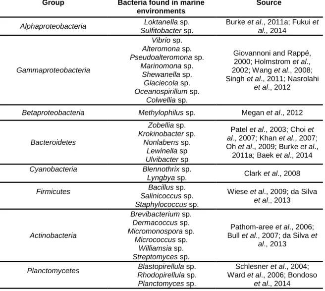
Figure 1 – Schematic representation of the pyrosequencing enzyme system. If the
added dNTP forms a base pair with the template, Polymerase incorporates it into the growing DNA strand and pyrophospate (PPi) is released. ATP Sulfurylase converts the PPi into ATP which serves as substrate for the light producing enzyme Luciferase. The produced light is detected as evidence of that nucleotide incorporation has taken place (Adapted from Ahmadian et al., 2006). ... 10
Figure 2 – Different species of macroalgae used in this study. A – Ulva sp., B – Porphyra dioica and C – Sargassum muticum. ... 13 Figure 3 – Microbial biofilm obtained through optical microscopy (OM) in each
macroalgae. A – Ulva sp., B – Porphyra dioica and C – Sargassum muticum. ... 19
Figure 4 – Observation of the microbial biofilm, by scanning electron microscopy
(SEM), associated to the macroalgae. A – Ulva sp., B – Porphyra dioica, C and D – Sargassum muticum. ... 20 Figure 5 – Number of colonies in percentage obtained from Ulva sp. (UAP), P. dioica
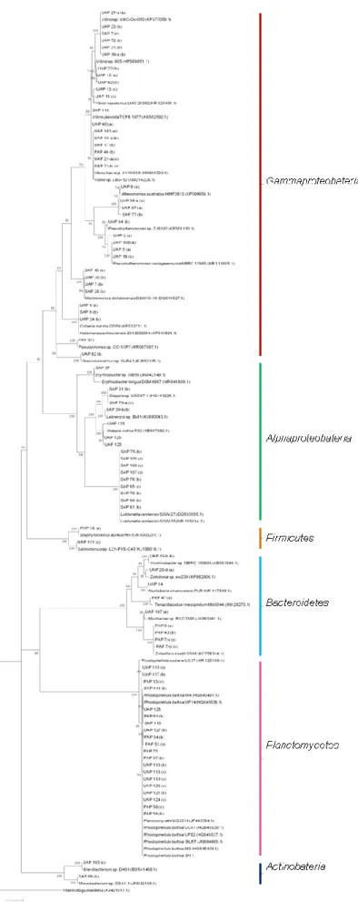
(PAP) and S. muticum (SAP) sampled in Autumn in Porto for all media... 21 Figure 6 - Phylogenetic 16SrRNA gene tree generated by maximum-likelihood analysis based in General Time Reversible model and Gamma distributed with Invariant sites (G+I) indicating the relationship of the bacteria isolated from the three macroalgae that demonstrated bioactive potential. Thermatoga maritima was used as out-group. The numbers beside nodes are the percentages for bootstrap analyses; only values above 50% are shown. Bar – 0.05 substitutions per 100 nucleotides. (a) Presence of NRPS gene; (b) Presence of PKS-I gene; (c) Presence of both genes. ... 22
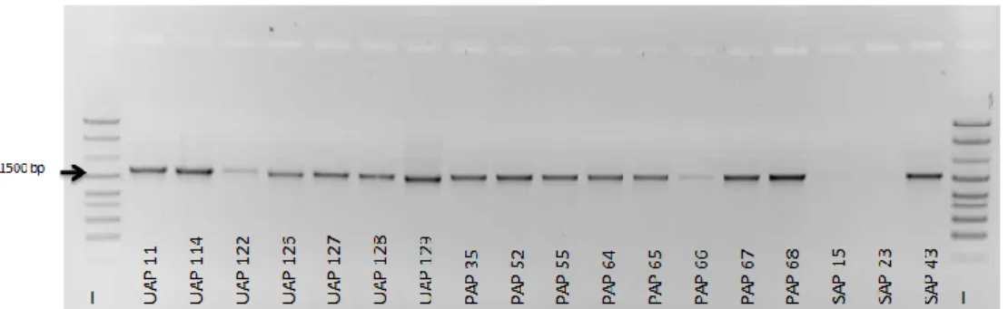
Figure 7 – Electrophoretic gel showing the 16S rRNA gene amplification in some
bacteria isolated from Ulva sp., P. dioica and S. muticum. L – Ladder (GeneRulerTM DNA Ladder Mix). For isolates designations see Table 2. ... 23
Figure 8 – Number of isolates obtained in each taxonomic group associated to the
different macroalgae. ... 23
Figure 10 – Electrophoretic gel showing the 16S rRNA gene amplification of bacteria
isolated in Spring from each macroalgae (Ulva sp., P. dioica and S. muticum). L – Ladder (GeneRulerTM DNA Ladder Mix). For isolates designations see Table 2. ... 26
Figure 11 – Phylogenetic 16S rRNA gene tree generated by maximum-likelihood
analysis based in General Time Reversible model and Gamma distributed with Invariant sites (G+I) indicating the relationship of the Planctomycetes isolated in Autumn and Spring seasons from the three macroalgae. Blastopirellula marina was used as out-group. ... 27
Figure 12 – OM image of a planctomycetes isolate obtained from Ulva sp. in Spring.
The characteristic rosette formation of Rhodopirellula spp. is well evident. ... 28
Figure 13 - Portion of Ulva in M13 medium showing the growth of isolates USpP 9 and
USpP 10. ... 28
Figure 14 – Good`s coverage rarefaction and observed species curves of the
samples. ... 29
Figure 15 – Proportion of reads of the samples assigned at the phylum level. Data
represented are means of the phyla with abundance higher than 0.5%. *Bacteria_Cyanobacteria_Chloroplast ... 31
Figure 16 – Bacterial groups obseved in pyrosequencing without the values relative to
chloroplast contamination. A – Macroalgae and seawater; B – Ulva sp.; C – P.dioica; D – S. muticum. ... 32
Figure 17 – Proportion of reads of the samples assigned at the genera level. ... 35 Figure 18 – Seasonal comparison of bacterial communities of Ulva sp. (lanes 1, 2, 8,
9, 15, 16, 22, 23), P. dioica (lane 3, 4, 10, 11, 17, 18, 24, 25), S. muticum (5, 6, 12, 13, 19, 20, 26, 27) and seawater (7, 14, 21, 28) through the analysis of 16S rRNA gene DGGE gel profiles. L – Ladder. ... 39
Figure 19 – (A) Dendrogram of DGGE profiles of macroalgae and seawater samples,
based on Bray-Curtis similarity. (B) Non-metric multidimensional analysis scaling (nMDS) plot (stress: 0.257) based on Bray-Curtis similarity. ... 40
Figure 20 - (A) Dendogram obtained by pyrosequencing based on Bray-Curtis
similarity. (B) Distance-based ReDundancy Analysis (dbRDA) of beta rarefaction metrics of the samples constrained by species and seawater: Bray-Curtis dissimilarity.
(C) Dendrogram of DGGE profile of macroalgae and seawater samples in Autumn and Winter, based on Bray-Curtis similarity. (D) Non-metric multidimensional analysis scaling (nMDS) plot (stress: 0.1645) based on Bray-Curtis similarity. ... 41
Figure 22 – Example of an electrophoresis gel of PKS-I and NRPS amplifications
(arrow) in some strains representative of all samples. L – Ladder (GeneRulerTM DNA Ladder Mix). ... 43
Figure 23 – Percentage of potential PKS-I and NRPS genes search in the bacterial
Abbreviations
® registered trademark ™ trademark symbol ºC degree Celsius μL microlitre μM micromolar Μm micrometre % percent sign16S rRNA 16S ribosomal ribonucleic Acid
ATP Adenosine triphosphate
BLAST Basic Local Alignment Search Tool
Bp base-pair
CFU Colony-forming unit
DGGE Denaturing Gradient Gel Electrophoresis
DNA deoxyribonucleic acid
dNTPs deoxyribonucleotide triphosphate
e.g exempli gratia
EPS Extracellular polymeric Substance
G gram
M Molar
MA Marine agar medium
MF Medium F mL milliliter Mg milligram MgCl2 Magnesium Chloride mM milliMolar Min minutes
mS/cm milliSiemens per centimetre
Ng nanogram
NGS Next-generation sequencing
NRPS Nonribosomal peptide sinthethases
OM Optical microscopy
OTU Operational Taxonomic Unit
PAST Paleontological statistics software package
pH Potential of Hidrogen
PKS Polyketide synthases
Ppi Inorganic pyrophosphate
Ppt Parts per thousand
QS Quorum sensing
RNA Ribonucleic acid
rRNA Ribosomal ribonucleic acid
S Seconds
SEM Scanning electron microscopy
sp. Specie
ST Starch marine medium
TAE Tris-acetate
1. Introduction
1.1. Marine microorganisms – Overview
The world oceans are the largest ecosystem on earth (Rumney, 1968). It covers over 70% of the earth's surface and contains a rich diversity of microorganisms estimated in several millions different species (Whitman et al., 1998). The total number of prokaryotic cells in the oceans is 1029 (Whitman et al., 1998) with 106 bacterial taxa suggested (Pedrós-Alió, 2006). Per mL of seawater it is possible to find a range of millions of viruses and bacteria, thousands of fungi and microalgae, and hundreds of microscopic larvae and spores (Harder, 2009). In recent years the interest in understanding the function of marine ecosystems has been accelerated because of its increasing impact on human life. However, and in general, marine microbes have not been studied as extensively as their terrestrial counterparts (National Research Council (US) Committee on Molecular Marine Biology, 1994). The impossibility to observe in great detail microorganisms by direct methods allied to difficulties in sampling some marine inaccessible areas delayed the increasing knowledge of marine microbial species (DeLong and Pace., 2001; Brandt et al., 2014). In spite of their importance, knowledge of marine microbial diversity is insufficient in terms of quantity and quality of the microorganisms present in the oceans and their ecological interactions and functions. Only in the late 19th century with the development of pure-culture techniques it was possible to enlarge our knowledge on microbial species and their characteristics. However, this approach only provides limited information about the organisms that can grow under determined conditions restricting the knowledge of microbial diversity (Pace, 1997). It is estimated that only 1% of marine bacteria can be isolated by cultivation methods (Amann et al., 1995). The development and application of novel and powerful molecular tools allowed increasing greatly the analysis of microbial diversity and of the community dynamics in their environment. In the early 90’s the analyses of ribosomal RNA (rRNA) sequences permitted to organize all living organisms into three major Domains: Bacteria and Archaea (both forming the “Prokaryotes”) and Eukarya (Woese et al., 1990). As the molecular markers rRNA genes have a highly conserved nature that anneal with “universal” PCR primers, they allowed the demonstration of the evolutionary relationships between all organisms and the reorganization of the phylogenetic tree (Woese et al., 1990; Pace, 1997). The 16S rRNA gene has been used for the study of bacterial phylogeny and taxonomy (e.g. Fox et al., 1977; Woese and Fox, 1977; Weisburg et al., 1991; Coenye and Vandamme, 2003; Woo et al., 2008). This genetic marker is considered a universal bacterial
identification tool as it is present in all bacteria, often existing as a multigene family, or operon; its function do not change over time (Woese,1987) and the size of the gene (approximately 1500 bp) is sufficiently large for informatic purposes as it contains statistically relevant sequence information (Patel, 2001). The 16S rRNA molecule possesses approximately 50 functional domains. The number of domains is important because the introduction of selected changes in one domain does not greatly affect sequences in other domains and these changes have lower impact on phylogenetic relationships (Woese, 1987). The discovery of 16S rRNA gene became an important milestone for detection, classification and reclassification of numerous bacterial species and genera and also for the discovery of uncultivable bacteria (Janda and Abbott, 2007; Woo et al., 2008).
In the sea world, prokaryotes encompass the majority of the genetic diversity (Glockner et al., 2012) and represent the second most abundant group after the viruses (Breitbart, 2012). The ubiquity of Bacteria and Archaea is favoured by their widespread dispersal capacity, metabolic flexibility and versatility combined with the fact that these organisms have the capacity to resist to extreme and antagonistic conditions (Schlegel and Jannasch, 2006). They play an essential role in ecology and biochemistry cycles, energy flow and nutrition (Arrigo, 2005; Thakur et al., 2008). For this reason, during evolutionary times these organisms have been playing an important role in the chemistry of the oceans and the atmosphere (Redfield, 1958) and are responsible for 50% of the global primary production occurring in the marine environment (Odeyemi, 2013). In bacteria, prochlorophytes and cyanobacteria are the main organisms that contribute to phytoplankton biomass and are responsible for energy flow and microbial food webs cycling in oligotrophic oceanic ecosystems (Sherr and Sherr, 1991; 1994). In the oceans, bacteria had to adapt to various, sometimes harsh, environments which implicated the development of diverse metabolisms and surviving strategies. Additionally, bacteria have the capacity to interact in different ways with marine macroorganisms, like macroalgae, sponges, anemones and other invertebrate animals (Bewley and Faulkner, 1998; Goecke et al., 2010; Barott et al., 2011; Graça et al., 2015), and are capable of colonizing their surface forming biofilms (Weinberger, 2007). Furthermore, surviving strategies led bacteria to produce bioactive compounds which may be used for many applications (Proksch et al., 2002; Graça et al., 2015). For example, actinomycetes have been an important bacterial group in the research of new products with antibiotic and anticancer potential (Goodfellow and Haynes, 1984; Jensen et al., 1991; Kim et al., 2005; Kim et al., 2006) and also in the recycling of organic matter (Srinivasan et al., 1991). So, bacteria are, thus, environmentally relevant as key players in all major biogeochemical cycles, energy fluxes, processes in
marine ecosystems and also to improve the human well-being by their biotechnological potential.
1.2. Marine bacterial diversity
As a liter of seawater contains in general 109 bacterial cells (Curtis et al., 2002), it is evident that the marine environment is the shelter of an enormous bacterial diversity (Giovannoni and Stingl, 2005; Zinger et al., 2011). This varies due to changes in water temperature, salinity, nutrients and other physicochemical parameters (Alavandi, 1990) and is also controlled by biological interactions. The interest to determine bacterial marine diversity has been important to understand communities’ structure, distribution and function but also to identify their ecological importance for the support of other organisms and ecosystems. The marine environment encompasses about 20 recognized culturable and well-studied major phyla of Bacteria (Ciccarelli et al., 2006). Some of these divisions are represented in Table 1.
Table 1 – Some bacterial species found in marine ecosystems.
Group Bacteria found in marine
environments
Source
Alphaproteobacteria Loktanella sp.
Sulfitobacter sp.
Burke et al., 2011a; Fukui et
al., 2014 Gammaproteobacteria Vibrio sp. Alteromona sp. Pseudoalteromona sp. Marinomona sp. Shewanella sp. Glaciecola sp. Oceanospirillum sp. Colwellia sp.
Giovannoni and Rappé, 2000; Holmstrom et al., 2002; Wang et al., 2008; Singh et al., 2011; Nasrolahi
et al., 2012
Betaproteobacteria Methylophilus sp. Megan et al., 2012
Bacteroidetes Zobellia sp. Krokinobacter sp. Nonlabens sp. Lewinella sp Ulvibacter sp
Patel et al., 2003; Choi et
al., 2007; Khan et al., 2007;
Oh et al., 2009; Burke et al., 2011a; Baek et al., 2014
Cyanobacteria Blennothrix sp.
Lyngbya sp. Clark et al., 2008
Firmicutes Bacillus sp.
Salinicoccus sp. Staphylococcus sp.
Wiese et al., 2009; da Silva
et al., 2013 Actinobacteria Brevibacterium sp. Dermacoccus sp. Micromonospora sp. Micrococcus sp. Williamsia sp. Streptomyces sp. Pathom-aree et al., 2006; Bull et al., 2007; da Silva et
al., 2013
Planctomycetes Blastopirellula sp.
Rhodopirellula sp. Planctomyces sp.
Schlesner et al., 2004; Ward et al., 2006; Bondoso
For the measurement of the bacterial community structure by culture-independent methods, some indices have been used. Particularly the Shannon index, the evenness index, derived from it, and Simpson’s dominance index (Shannon and Weaver, 1963; Dunbar et al., 1999; McCaig et al., 1999; Cho and Kim, 2000). The Shannon index (H’) has the advantage to consider both the richness, i.e. the number of different species in the sample, and also, the dominance of some species. This index is suggested as one of the most appropriated methods to study diversity among different communities (Hill et al., 2003).
1.2.1. The phylum Planctomycetes
The phylum Planctomycetes is a group of bacteria that exhibit unusual characteristics some of which are shared with eukaryotic cells (Lage, 2013). Normally, they appear in low abundance in samples (Rusch et al., 2007). They were found in a variety of ecosystems demonstrating a cosmopolitan distribution (Chouari et al., 2003; Buckley et al., 2006; Bondoso et al., 2011; Fuchsman et al., 2012; Sheng et al., 2012) and have the capacity to adapt to the extreme environments such as acidic habits, extreme saline habitats, thermophilic habitats, etc (Giovannoni et al., 1987; Drees et al., 2006; Byrne et al., 2009; Li et al., 2010; Bernhard et al., 2012, Ivanova and Dedysh, 2012; Urbieta et al., 2012; Tang et al., 2013). Furthermore, planctomycetes also appear associated with eukaryotic hosts, such as macroalgae where they are common inhabitants in the biofilm of the epibacterial community (Longford et al., 2007; Bengtsson et al., 2010; Burke et al., 2011a; Lachnit et al., 2011; Lage and Bondoso, 2011; de Oliveira et al., 2012; Hollants et al., 2013; Bondoso et al., 2014b; Miranda et al., 2013). These recently confirmed associations allowed the discovery of novel planctomycetes taxa (Fukunaga et al., 2009; Bondoso et al., 2014a, 2015).
1.3. Microbial biofilms
In aquatic environments microorganisms are capable of living using different strategies. In addition to the capacity to live as individual cells in aqueous suspensions, microorganisms can colonize surfaces by the formation of the so-called biofilms. The attachment in solid surfaces and consequently the formation of biofilms is favoured by humid or aqueous environments (Weinberger, 2007). A biofilm can be defined as an assemblage of microbial cells that is irreversibly associated with a surface of living or non-living materials enclosed in an extracellular polymeric substance (EPS) matrix (Donlan, 2002) mainly composed of high-molecular weight polysaccharides. EPS matrix may account for 50% to 90% of the total organic carbon and controls the
physical properties of biofilms. The community of microbial cells can be structured and enhanced or reduced by this substance (Decho, 2000). The association of one organism growing on the surface of another is referred to as epibiosis (Bengtsson, 2011) and advantages and disadvantages may occur for both organisms. Algae, corals and sponges are some examples of eukaryotes that harbour complex epiphytic microbial communities and are susceptible to biofilm formation (Olsan and Kellogg, 2010). A number of observed host-associated microbial communities tend to be very host-specific, generally stable and normally distinct from planktonic communities in the surrounding water column (Erwin et al., 2011). These communities are composed by a variety of organisms such as bacteria, fungi, unicellular algae such as diatoms, and protozoa (Marshall and Bowden, 2000; Maki, 2002; Goecke et al., 2010). Bacteria are one of the first colonizers of the surface usually due to its high abundance in seawater (Dang and Lovell, 2000). Biofilms represent a common adaptation, perhaps even a life stage or style, of bacteria (Costerton et al., 1995) and the bacterial number observed in biofilms is significantly higher than in the planktonic life form (Robinson et al., 2010). An important feature of these epiphytic associations is that, the microbial assemblages are involved in important metabolic transformations modulating this association. In some cases, the microbial biofilm can influence the host promoting its metabolic functions such as nutrition or reproduction (Goecke et al., 2010; Kazamla et al., 2012).
1.4. Bacterial communities associated with macroalgae
In aquatic environments where macroalgae live, diseases, parasitism, epibiosis and biofouling are quite common phenomena (Harder, 2009). Thus, macroalgae are important key components in marine ecosystems providing microhabitats for many organisms. The occurrence of the macroalgae in the photic zone makes them susceptible to epibiosis because the conditions are optimal for microbial growth, namely of phyto- and zoo- species, allowing them to survive without environmental stress (Nys et al., 1995; Potin et al., 2002; Burke et al., 2011a). Bacteria are considered the primary colonizers of algal surfaces with a high complexity of interactions, followed by diatoms and fungi (Qian et al., 2007, Lam et al., 2008; Burke et al., 2011a).
Macroalgae produce organic material, such as organic carbon, providing a rich habitat in nutrients for bacteria and other microorganisms (Lane and Kubanek, 2008). The symbiotic association can protect the macroalgae from UV radiation (Koch and Brandt, 2003) and promote nitrogen fixation (Thevanatan et al., 2000). Additionally, bacteria may supply vitamins and/or growth regulators to the algae (Croft et al., 2006;
Kazamla et al., 2012). It was proved that some molecules produced by bacteria determine zoospores settlement in Ulva species (Joint et al., 2000) or their liberation in Acrochaetium (Weinberger et al., 2007) and Gracilaria species (Singh, 2013). On the other hand, when these associations are harmful for macroalgae, bacteria may provoke diseases, tissue necrosis, growth and photosynthesis reduction (Armstrong et al., 2000; Vairappan et al., 2001). Bacterial community can also affect the host by the production of a variety of toxins, digestive enzymes, inhibitors and waste products (Ivanova et al., 2002). However, the macroalgae developed, overtime, strategies to defend their surfaces against bacteria (Wahl, 2008; Steinberg and de Nys, 2002).
The first studies about bacteria associated to macroalgae surface dates back to before 1875 (Johansen et al., 1999). Even though the studies about these associations are still scarce, the utilization of the classical molecular tools associated with new tools designed as next-generation sequencing (NGS) techniques, have enabled to increase our knowledge about bacterial-algal interactions (Lachnit et al., 2009). Recently, a variety of symbiotic, pathological and opportunistic interactions between macroalgae and bacteria are being discovered (Goecke et al., 2010). Novel described bacterial species and genera were isolated from some common algae such as Ulva, Porphyra, Fucus and Saccharina (Goecke et al., 2013).
Regarding the diversity of bacteria found in macroalgae surfaces, some studies showed that different macroalgae species in the same location maintain different bacterial communities (Lachnit et al., 2009; Nylund et al., 2010), and the same macroalgal species from different locations have high similarities in the composition of the associated microbial communities (Staufenberger et al., 2008; Lachnit et al., 2009; Sneed and Pohnert, 2011).
1.5. Chemical interactions between macroalgae and bacteria
1.5.1. Quorum sensing – signalling
The cooperative activities and physiological processes between bacteria are regulated through a mechanism called quorum sensing (QS), in which bacterial cells communicate with each other by releasing, sensing and responding to small diffusible signal molecules. Cell-to-cell communication signals have important effects in the formation and organization of bacterial population density that contribute to the structure of biofilms, stress resistance and production of secondary metabolites (Davies et al., 1998; Parsek and Greenberg, 2000; Sauer et al., 2002; Paul and Ritson-Williams, 2008; Dobretsov et al., 2009). These signals are produced and excreted with
feedback regulation, and the detection of these signals eventually leads to global changes in bacterial gene expression (Waters and Bassler, 2005).
The QS in the bacteria have been generally divided into at least three classes: LuxI/LuxR–type quorum sensing in Gram-negative bacteria, which uses acyl-homoserine lactones (AHL) as signal molecules; oligopeptide-two-component-type quorum sensing in Gram-positive bacteria, which uses small peptides as signal molecules; and luxS-encoded autoinducer 2 (AI-2) quorum sensing in both Gram-negative and Gram-positive bacteria (Miller and Bassler, 2001; Federle and Bassler, 2003; Waters and Bassler, 2005).
In marine bacteria, AI-2 group of signalling molecules are important in the metabolic transformation carried out by LuxS enzyme that regulates the genes responsible for bioluminescence, formation of biofilms, virulence, and antibiotic production (Chen et al., 2002; Miller et al., 2002; Henke and Bassler, 2004; Miller et al., 2004; Bodor et al., 2008). This enzyme was found in Vibrio harveyi and since then, the LuxS has been identified in over 50 different bacterial species (Schauder et al., 2001; Winzer et al., 2002; Xavier and Bassler, 2003; Vendeville et al., 2005). As referred by Atkinson and Williams (2009), the “cross-kingdom sensing of QS signal molecules may therefore constitute an adaptive survival strategy, enabling bacteria and eukaryotes to monitor their surroundings and adjust their behaviour in response to environmental challenge and population flux”.
1.5.2. Biotechnological potential – secondary metabolites
During the last decades, cultivable microorganisms have been used as a “pool” of natural product drug discovery and have provided unique compounds with chemical structures that have direct application in curing diseases. The discovery of novel metabolites has been focused on marine microorganisms because the enzymes produced by these organisms are more potent biochemically and stable than those derived from plants and animals (Bull and Ward, 2000; Kin, 2006). As referred by Fusetani in 2000, the search of new drugs from marine organisms resulted in the isolation of approximately 10000 metabolites. Nowadays these organisms are viewed as sources for therapeutic agents used for treatment of cardiac, respiratory and gastrointestinal diseases (Laport et al., 2009). Furthermore, some evidences indicate that many bioactive compounds previously reported in marine animals and plants were in fact produced or metabolised by the associated microorganisms (Unson and Faulkner, 1993; Schupp et al., 1999; Davidson et al., 2001; Luesch et al., 2001; Proksch et al., 2002; Penesyan et al., 2010). In their competition for survival, bacteria
associated with other organisms produce antimicrobial and antifouling substances (Laport et al., 2009) demonstrating to be a rich source of bioactive compounds (Bull and Stach, 2007; Egan et al., 2008; Graça et al., 2013, 2015). Streptomyces, Alteromonas, Pseudoalteromonas, Roseobacter and Actinomyces are examples of the many genera of bacteria that produced bioactive compounds (Okazaki et al., 1975; Fenical, 1993; Wagner-Dobler et al., 2002).
Secondary metabolites are the substances that mediate many of these host-microbe associations in the ocean environment (Lane et al., 2010). Moreover, the competition for space, nutrients and defence strategies are responsible for the production of many natural products by microorganisms (Armstrong et al., 2001). Polyketide synthases (PKS) and nonribosomal peptide synthetases (NRPS) are usually involved in the production of bioactive secondary metabolites (Donadio et al., 2007; Foerstner et al., 2008). PKSs type I are large, multifunctional enzyme complexes where intermediates translate along modules. Other PKSs have been described showing that the diversity of these systems is greater than previously recognized (Shen, 2003). The NRPS are involved with the production of small peptides. PKSs and NRPS share a similar model of biosynthesis. Both are created on modular enzymatic assembly lines, with their structural diversity governed by optional enzymes within the enzyme complexes (Walsh, 2004).
1.6. Genomic studies
Many studies of microbial communities are done, at the genetic level, without the isolation and culture of microorganisms, avoiding thus the limitation imposed by this method (Amman et al., 1995). For this purpose, the new molecular techniques developed have the aim to analyze the microbial structure and diversity of environmental samples and monitor changes in microbial communities (Muyzer and Smalla, 1998; Lyautey et al., 2005).
1.6.1. Denaturing gradient gel electrophoresis (DGGE)
The first study applied to the field of microbial ecology was developed by Muyzer and colleagues (1993). DGGE technique is usually used for detecting the phylogenetic “fingerprint” of diverse organisms by the separation of the double-stranded DNA PCR products of similar length but with different sequence composition. This separation is possible due to the denaturing gradient (urea and formamide) used in the polyacrylamide gel that cause partial denaturation of the DNA templates. In the 5’-end of one of the primers it is attached a GC-clamp (a GC-rich sequence usually
30-50 nucleotides) to stop the complete dissociation of the double-stranded DNA (Muyzer and Smalla, 1998; Muhling et al., 2008; Pollet et al., 2011). After DNA separation in the gel, the analysis consists in the evaluation of the number, precise position and intensity of the bands which give an estimation of the number and relative abundance of numerically dominant ribotypes in the sample. Profiles are analyzed on the basis of their banding pattern (presence/absence and intensity of bands) allowing a rapid comparison of the different bacterial communities (Zoetendal et al., 2001). The individual bands in the gel can also be excised, reamplified and sequenced to obtain phylogenetic information (Thakur et al., 2008). In the study of marine bacteria, DGGE fingerprinting has been applied for many purposes, such as picoeukaryotic assemblages (Schauer et al., 2000 D ez et al., 2001), epiphytic bacteria of coral species (Morrow et al., 2012), sponges (Li et al., 2007; Thiel et al., 2007) and macroalgae (Ohkubo et al., 2006; Bondoso et al., 2014). Furthermore, DGGE approach allows to study seasonal and spatial variations of bacterial communities (Murray et al., 1998; Riemann et al., 1999; Riemann and Middelboe, 2002; Kan et al., 2006) and has revealed that bacterial communities in association with algae diverge from planctonic communities (Burke et al., 2011a; Goecke et al., 2013).
1.6.2. Next generation sequencing - Pyrosequencing
Pyrosequencing is one of the next generation sequencing techniques that permits to increase in a great detail the knowledge of microbial communities, without isolation and culture methods. This approach, based on sequencing by-synthesis principle, consists on the luminometric detection of inorganic pyrophosphate (PPi) through the action of four enzymes (DNA Polymerase I, ATP sulfurylase, Luciferase and Apyrase) in a cascade of reactions that incorporate nucleotides and produce a detectable light signal (Fig. 1). When a nucleotide is introduced in the DNA-strand, pyrophosphate is released, ATP is generated which is used for the conversion of the luciferin to oxyluciferin with generation of visible light in amounts proportional to the produced ATP (Gharizadeh et al., 2007; Ahmadian et al., 2006). The emerging of this technique allows the reducing of some limitations associated with Sanger sequencing, commonly used since 1977 (Sanger et al., 1977; Gharizadeh et al., 2007).
Pyrosequencing includes advantages, comparatively to the other sequencing methods, in terms of accuracy, flexibility, parallel processing, automatization and it does depend of labeled primers, labeled nucleotides, and gel electrophoresis (Ronaghi, 2001). Moreover, it allows to sequence short DNA sequences (at least 20 bases), and provides numerous applications such as sequencing of whole genomes, determination
of the known as well as unknown polymorphic positions, comparisons of multiple strains of bacteria and quick detection of point mutations responsible for antibiotic resistance (Ahmadian et al., 2006; Moder et al., 2007).
In this way, the application of pyrosequencing technology has revolutionized the understanding about communities’ structure of bacteria in marine environment and increases the discovery of novel species which are not possible to be isolated by cultivation dependent methods.
Figure 1 - Schematic representation of the pyrosequencing enzyme system. If the added dNTP forms a base pair with
the template, Polymerase incorporates it into the growing DNA strand and pyrophospate (PPi) is released. ATP Sulfurylase converts the PPi into ATP which serves as substrate for the light producing enzyme Luciferase. The produced light is detected as evidence of that nucleotide incorporation has taken place (Adapted from Ahmadian et al., 2006).
2. Objectives
The main aim of the present work is the study of the epiphytic bacterial diversity associated with Ulva sp., Porphyra dioica, Sargassum muticum through culture- dependent and independent methods. Culture-dependent methods include isolation, cultivation in pure culture and identification through the analysis of the 16S rRNA gene. Seasonal variations of bacterial communities of macroalgae and surrounding water were analyzed by two independent methods: Denaturing Gradient Gel Electrophoresis (DGGE) and Next Generation Sequencing (NGS) technology. Another aim was to explore the biotechnological potential of the isolated bacteria through the search of polyketide synthase and nonrinosomal synthetase genes, which are important in the production of secondary metabolites. Potential ecological interaction between macroalgae and the different isolated bacteria was assessed through the analysis of luxS gene responsible for quorum sensing signalling.
3. Material and Methods
3.1. Biological material
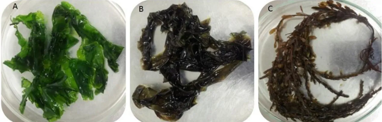
The specimens of macroalgae were harvested at low tide from the intertidal rock platform at Luz beach, in the coast of Porto, Atlantic Ocean, Portugal (41º18’North, 8º44’West). To study the variation of microorganism’s community associated to the surface of macroalgae, samples of the four seasons were collected during one year period. Specimens of each macroalgae, Ulva sp., Porphyra dioica and Sargassum muticum (Fig. 2) were removed and transferred into separate sterile plastic bags. One litter of the surrounding seawater was also sampled in a sterile bottle. Samples were immediately transported to the LEMUP (Laboratory of Microbial Ecophysiology of University of Porto) laboratory. Description and designation of the samples are referred in Table 2.
Microscopic observation of the macroalgae biofilm was assessed in an AxioCam MRc optical microscope and in a Phenom ProX scanning electron microscope.
Table 2 – Designation of each abbreviation used in samples for all seasons.
Samples Description (Species; Season; Study area)
UAP Ulva sp.; Autumn; Porto
PAP Porphyra dioica; Autumn; Porto
SAP Sargassum muticum.; Autumn; Porto
H2OAP Sea water; Autumn; Porto
UWP Ulva sp.; Winter; Porto
PWP Porphyra dioica; Winter; Porto
SWP Sargassum muticum.; Winter; Porto
H2OWP Sea water; Winter; Porto
USpP Ulva sp.; Spring; Porto
PSpP Porphyra dioica; Spring; Porto
SSpP Sargassum muticum.; Spring; Porto
H2OSpP Sea water; Spring; Porto
USP Ulva sp.; Summer; Porto
PSP Porphyra dioica; Summer; Porto
SSP Sargassum muticum.; Summer; Porto
Figure 2 – Different species of macroalgae used in this study. A – Ulva sp., B – Porphyra dioica and C – Sargassum
muticum.
3.2. Isolation of microorganisms
Isolation of the microorganisms was done from the samples collected in autumn. In the laboratory, the samples were manipulated under aseptic conditions, inside a flow chamber. Portions from each macroalgae were washed three times with sterile sea water, to remove non-associated bacterial cells and other small particles. One gram wet weight of each sample was macerated in 10 ml sterile natural sea water with sterile glass-beads and serially diluted (100, 10-2, 10-4 and 10-6). Thereafter, from each dilution, 100 µl were spread in eight selected isolation marine agar media, referred below. The cultures were incubated in the dark at 25ºC and presence of growth was checked daily. Distinct colony morphotypes were identified (colour, size, texture and shape) in a dissecting microscopy and transferred to the respective media for isolation. The pure bacterial cultures were cryopreserved in seawater supplemented with 20% glycerol at -80ºC.
The marine media used for bacteria isolation in this study were Marine Agar (MA) (Becton Dickinson), Medium F (MF) (Li et al., 2007), modified M13 Medium (Lage and Bondoso, 2011), Actinomycetes Medium (Zhang and Zhang, 2011), Bacillus Medium (HiCrome™), BG11 agar (HIMEDIA®) for the cultivation and maintenance of cyanobacteria, Starch Marine (HIMEDIA®), modified Myxobacterial Medium (Zhang et al., 2013) without cycloheximide, and for isolation of cellulose-degrading myxobacteria we used slices of filter paper which were placed on top of the same medium. For quantification of the Colony Forming Units (CFU), three different plates with MA medium for each dilution of each macroalgae were used.
Moreover, for the isolation of Planctomycetes, portions of macroalgae were placed in different plates containing modified M13 medium that was supplemented with streptomycin, ampicillin and pevaryl, and incubated in the dark at 25ºC according to Lage and Bondoso (2011). This procedure was repeated in all seasons.
Seawater samples were filtered through a 0.22 µm pore filter for DGGE and pyrosequencing analyses (described below), and temperature, salinity, conductivity and pH were measured.
3.3. Molecular Analysis
3.3.1. Community DNA extraction and amplification
The genomic DNA of bacteria isolated from the specimens were extracted using the E.Z.N.A. bacterial KIT from OMEGA, according to the manufacturer’s instructions. The taxonomic identification of bacteria isolated was based on the analysis of the 16S rRNA gene. This gene was amplified from the extracted DNA with the universal primers, 27F and 1492r (Lane, 1991) in 50 µl of PCR mixture (1 x Green GoTaq® Flexi Buffer; 1.5 mM MgCl2; 1 unit of GoTaq® Flexi DNA Polymerase; 200 µM of each
deoxynucleoside triphosphate (dNTPs); 2 µM of each primer). Two µl of DNA template were used for the PCR reaction. The PCR program was performed in a MyCycler™Thermo Cycler (Bio-Rad) and amplification conditions comprised initial denaturing step of 5 minutes at 95 ºC; 30 cycles of 1 minute at 94 ºC; 1 minute at 52 ºC, 90 seconds at 72 ºC and a final extension of 5 minutes at 72 ºC. PCR products were visualized after electrophoresis in a 1.2% agarose gel stained with Roti Safe (Roth) in 1 x Tris Acetate and EDTA (TAE) buffer (OMEGA)
.
3.3.2. Identification and phylogeny analysis
Amplification products were purified using illustra GFX PCR DNA and Gel Band Purification Kit (GE Healthcare) and sequenced for the 16S rRNA gene at Macrogen. The sequences were edited and checked manually using CHROMAS 2 (Goodstadt and Ponting, 2001) correcting possible errors in chromatograms. The corrected sequences were assembled and consensus of the strains was constructed in ProSeq v2.91 and Vector NTI 11.5.3.
Alignment of all consensus sequences was performed using MEGA 6 (Molecular Evolutionary Genetics Analysis) software, that permits to infer overtime the molecular evolutionary between genes, genomes and species (Tamura et al., 2013). The construction of the phylogenetic tree was performed using calculation methods (maximum likelihood – ML) in MEGA 6, applying General Time Reversible model and Gamma distributed with Invariant sites (G+I). The aligned sequences were compared in GenBank using a Basic Local Alignment Search Tool (BLAST). Different phylotypes
were considered based on a 97% 16S rRNA gene threshold (Stackebrandt and Goebel, 1994).
3.3.3. Biotechnologic potential - search of polyketide synthase
and nonribosomal peptide syntethase genes
The presence of the genes PKS-I and NRPS involved in the production of secondary metabolites was screened in all bacteria isolated from the specimens of macroalgae. Amplification of the extracted DNA was achived with MDPQQRf and HGTGTr (Kim et al., 2005) and DKf and MTr (Neilan et al., 1999) primers, specific for PKS-I and NRPS genes respectively, in 25 µl of PCR mixture (1 x Green GoTaq® Flexi Buffer; 1.7 mM MgCl2; 0.8 unit of GoTaq® DNA Polymerase; 0.2 mM of each dNTPs;
0.1mM of each primer and 2 µl DNA template. The same PCR program was used for the amplification of two genes in a MyCycler™Thermo Cycler (Bio-Rad). The amplification conditions consisted of an initial denaturing step of 5 min at 95 ºC; 11 cycles of 1 min at 95 ºC; 30 s at 60 ºC and 1 min at 72 ºC, with the annealing temperature reduced by 2 ºC per cycle, followed by 30 cycles of 95 ºC for 1 min, 40 ºC for 30 s and 72 ºC for 1 min with a final extension of 10 min at 72 ºC. The PCR products were visualized by electrophoresis for the presence of approximate 700 bp and 1000 bp size amplicons, for PKS-I and NRPS respectively, in a 1.2% agarose gel in 1 x TAE buffer.
3.3.4. Quorum sensing analysis - LuxS gene
The search of the luxS gene was based on Bodor et al., (2008), who used marine, Gram positive and negative bacteria, to analyze the potential for luxS signaling. PCR based on the amplification of luxS gene was performed using LuxS_degfor3 and LuxS_degrev4 primers (Table 3) with a highly degenerate 5’ end and a specific 3’ end. PCR mixture (50 µl) contained: 1 x Green GoTaq® Flexi Buffer; 3 mM MgCl2; 1.25 unit
of GoTaq® DNA polymerase; 1 µM of each primer; 200 µM dNTPs and 2 µl DNA template. The PCR program was conducted in a MyCycler™Thermo Cycler (Bio-Rad) and the conditions consisted in a predenaturing at 94 ºC for 1 min; 30 cycles of denaturing at 94 ºC for 10 s; annealing at 48 ºC for 30 s; elongation step at 68 ºC for 40 s; and a final extension at 68 ºC for 1 min 30s. PCR products were visualized after electrophoresis in 1.2 % agarose gel in 1 x TAE buffer.
Table 3 – LuxS primers used (forward and reverse).
3.3.5. DGGE fingerprinting of seasonal variation in the epiphytic
community
For the analysis of DGGE bacterial profiles an extraction kit was used, the PowerSoil® DNA Isolation Kit, that allows the isolation of microbial genomic DNA from soil and other environmental samples. In this case, the samples used were the homogenized from each macroalgae and the filtrate from sea water sampled.
To compare the profiles of bacterial communities of all seasons (macroalgae and seawater), PCR based on the analysis of 16S rRNA gene were performed using encoding primers GC-358F (5′-CCT ACG GGA GGC AGC AG-3′ with a GC-clamp at the 5´ end) and 907r (5′-CCG TCA ATT CMT TTG AGT TT-3′) in 50 µl of PCR mixture: 1x Green GoTaq® Flexi Buffer, 1.5 mM MgCl2, 0.2 µM of each dNTPS, 0.5 µM of each
primer, 0.75 units of GoTaq® Flexi DNA Polymerase and 5 µl of DNA as template. These primers are specific for bacteria and the DNA fragments produced have around 550 bp.
PCR amplification was performed in a MyCycler™Thermo Cycler (Bio-Rad), that consisted in a initial denaturing step of 94 ºC for 5 minutes; 10 cycles of 1 minute at 94 ºC; 1 minute at decreasing temperature with each cycle starting at 65 ºC and ending at 55 ºC, 3 minutes at 72 ºC; 20 cycles of 1 minute at 94 ºC; 1 minute at 55 ºC; 3 minutes at 72 ºC and a final extension of 10 minutes at 72 ºC. PCR products were separated by electrophoresis on 1.2% agarose gel in 1 x TAE buffer.
The concentrations of the samples were calculated with the Thermo Scientific™ µDrop™ Plate that use a photometric measurement to quantify the nucleic acids in a sample. For quantification 2 µl of sample and standard (Green GoTaq® Flexi Buffer) were used.
After this procedure, about 600 ng of PCR products from each mixture were loaded in a DGGE gel and run at 60 °C at constant voltage of 120 volts for 16 hours in a DGGEK-2401-220 system (CBS Scientific Company). The 6% acrylamide gel with a linear gradient of denaturing conditions (100% denaturant agent is 7 M urea and 40% deionized formamide) ranged from 40 to 60%, 40 to 70% and 40 to 80% (based on Díez et al., 2001).
Name Sequence (5’-3’) Length Degeneracy
Ann.
Temp. Start position
LuxS_degfor3 CATTATTAGATAGCTTTACADTNGAYCAYA 30 bp 48 48 ºC 4
The ladder was prepared with a mixture of PCR products of bacterial communities associated with macroalgae. Gels were stained with SYBR® Gold Nucleic Acid Gel Stain during 1 hour in 1 × TAE buffer and visualized by UV light in a ChemiDoc system (Bio-Rad).
The digitalized DGGE gel was analyzed with the QuantityOne software (Bio-Rad). This software performs an optical intensity profile through each lane, detects the bands and identifies the bands occupying the same position in the different lanes of the gel.
Matrix reports were statistical analysed using PAST 2.17 (Hammer et al., 2001) and cluster analysis, non-metric multidimensional scaling (nMDS), similarity percentage analysis (SIMPER), analysis of similarity (ANOSIM), all based in Bray-Curtis coefficient (Bray and Curtis, 1957) were performed. Furthermore, diversity index (Shannon-Weiner and CHAO1) was also analysed.
3.3.6. Pyrosequencing analysis
Pyrosequencing sequencing and analysis were conducted at Plymouth University. Autumn and winter samples used in this analysis were the same that were extracted for DGGE analysis. PCR amplification of the 16S rRNA V1-V2 region was conducted using the primers 338R (5’-GCW GCC WCC CGT AGG WGT-3’) and 27F (5’-AGA GTT TGA TCM TGG CTC AG-3’) according to Newton and Roeselers (2012). Each PCR reaction contained 1 µl of each primer, 25 µl of MyTaqTM, 23 µl of molecular grade water and 1 µl of DNA template. Thermal cycling was ferformed using a TC-512 thermal cycler in the following conditions: initial denaturating step at 94 °C for 7 min, 10 cycles at 94 °C for 30 s, touchdown of 1 °C per cycle from 62 to 53 °C for 30 s and 72 °C for 30 s. A further 25 cycles were performed at 94 °C for 30 s, 53 °C for 30 s and 72 °C for 30 s before a final extension for 7 min at 72 °C.
PCR products were cleaned using PCR purification columns (QIAquick PCR Purification Kit; Qiagen) prior to high-throughput sequencing. To ensure that there was sufficient DNA present, some PCR reactions were made in duplicate (UWP, PWP, SWP) and pooled into a single sample prior to cleaning.
The amplified and purified DNA from the samples was quantified using a Qubit® 2.0 Fluorometer (Invitrogen). Prior to sequencing, the amplicons were assessed for fragment concentration using an Ion Library Quantitation Kit (LifeTechnologiesTM) and concentrations were then adjusted to 26 pM.
Amplicons were attached to Ion Sphere Particles using Ion PGMTM Template OT2 400 kit (LifeTechnologiesTM) according to the manufacturer’s instructions.
Multiplexed sequencing was conducted using Ion XpressTMBarcode Adapters (LifeTechnologiesTM) and a 318TM chip (LifeTechnologiesTM) on an Ion Torrent Personal Genome Machine (LifeTechnologiesTM) at the Systems Biology Centre in Plymouth University. Sequences were binned by sample and filtered within the Ion Personal Genome Machine (PGM) software to remove low quality reads.
Data were then exported as FastQ files. Taxonomic analyses of sequence reads were performance after the removal of low quality scores (Q score < 20) with FASTX-Toolkit (Hannon Lab). Sequences were concatenated and sorted by sequence similarity into a single fasta file. Sequences were denoised and analyzed with QIIME (Caporaso et al., 2010b). Briefly, OTU (Operational Taxonomic Unit) mapping was performed using the USEARH quality filter pipeline (Edgar, 2010), to remove putatively erroneous reads (chimeras), then OTU picking was achieved with a minimum pair wise identity of 97%.
The most abundant sequence in each OTU were selected to assign a taxonomic classification based on the Greengenes database (DeSantis et al., 2006) using the RDP classifier (Wang et al., 2007), clustering the sequences at 97% similarity with a 0.80 confidence threshold. Sequences were filtered to remove outliers and filter positions with gaps (0.95) and singletons. PyNast was used to create a multiple alignment of the representative sequences for each OTU (Caporaso et al., 2010a) with minimum sequence length threshold of 150 bp and 95% identification.
Alpha diversity metrics were calculated on rarefied OTU tables with QIIME to assess sampling depth coverage using Chao1, Good’s coverage, observed species, Phylogenetic Diversity and Shannon’s diversity index. QIIME was also used to calculate beta diversity metrics among samples using weighted and unweighted Unifrac distances (Lozupone et al., 2007) and BrayCurtis similarity (Bray and Curtis, 1957).
To determine if significant differences among seasonality or macroalgae species and seawater in high-throughput data non-parametric tests were performed on the OTU relative abundance data and alpha diversity metrics. Additionally, Vegan and APE packages of R were used to analyze the beta diversity of the communities from the eukaryotic species and seawater and the effect of seasonality. Statistical significance was accepted at the p < 0.05 level.
4. Results and Discussion
4.1.
Macroalgae associated biodiversity
4.1.1. Bacterial isolation studies
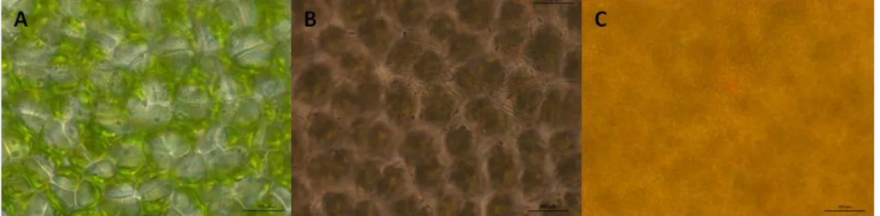
The characterization of the bacterial community associated with the three macroalgae under study, Ulva sp., Porphyra dioica and Sargassum muticum was done in the samples collected in Autumn through cultural methods. These algae are representatives of the three lineages of macroalgae, Chlorophyta, Rhodophyta and Heterokontophyta, that are commonly found in the North Coast of Portugal. Ulva and P. dioica are native inhabitants in this area (Oliveira, 1990; Pereira et al., 2001) but S. muticum is an invasive species (Vaz-Pinto et al., 2014). The three macroalgae were observed by optical and electron microscopy (OM and EM) to assess the biofilm on their surfaces (Figs 3 and 4).
Figure 3 - Microbial biofilm obtained through optical microscopy (OM) in each macroalgae. A – Ulva sp., B – Porphyra dioica and C – Sargassum muticum.
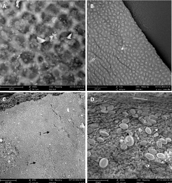
Microbial biofilm is well visible by OM in Ulva sp and P. dioica surfaces but impossible to visualize in S. muticum thalus due to its width (Fig. 3). By EM the biofilm was clearly visible in the three macroalgae. S. muticum, however, showed zones without (Fig. 4C-1) and with (Fig. 4C-2) epibacterial community. In the kelp Laminaria hyperborea was also observed a highly variable density and distribution of the microbial cells on its surface (Bengtsson et al., 2010). S. muticum also revealed the presence of high abundance of diatoms (Fig. 4D-3).
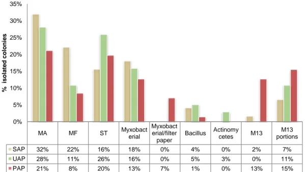
A total of 245 isolates were obtained, 100 (41 %) from Ulva sp., 61 (25 %) from P. dioica and 84 (34 %) from S. muticum. Bacteria were obtained in all media assayed except in BG11 agar and the highest number of colonies was obtained for the three macroalgae species in the non-selective MA medium (Fig. 5). As previously referred, MA is a medium allowing the growth of a high number of isolates of heterotrophic marine bacteria (Webster et al., 2001, Radwan et al., 2010; Graça et al., 2015). On the contrary, more selective media, like the Actinomycetes medium, allowed low levels of
isolation. Curiously, Joint et al., (2010) on an isolation study of bacterial groups from seawater of the English Channel obtained a very high number of isolates in Actinomycete medium (mainly Gammaproteobacteria and Actinobacteria) and a much lower number in MA. Seawater is a much lower organic carbon content environment when compared to biofilms. It is thus expected that a medium like MA that contains organic carbon in concentrations much higher than those found in most natural environments like seawater (Toledo et al., 2006), favours the growth of typically fast growing heterotrophic marine bacteria present in biofilms. The planctomycetes selective M13 medium also allowed a quite high percentage of isolation (around 8 %) which may be due to the quite high percentage of planctomycetes known to exist on macroalgae surfaces (Bengtsson and Øvreås, 2010; Lachnit et al., 2011).
Figure 4 – Observation of the microbial biofilm, by scanning electron microscopy (SEM), associated to the macroalgae. A – Ulva sp., B – Porphyra dioica, C (1 – Without epibacterial community; 2 – With epibacterial community) and D (3 – diatoms) – Sargassum muticum.
A
B
C
D
1
2
In MA medium, cell concentration associated with each macroalgae was 9.03x10³ CFU per 1 g of wet biomass for Ulva sp., 2.28x10⁴ CFU per 1 g of wet biomass for P. dioica and 1.86 x 10⁴ CFU per 1 g of wet biomass for S. muticum. Ulva sp. revealed the lowest number of associated bacteria. Beleneva and Zhukova (2006) obtained higher bacterial cell concentrations (from 1.37×10⁵ to 6.14×10⁵ cells per 1 g of wet biomass) on healthy brown (Desmarestia viridis and Chordaria flagelliphormis) and red algae (Gracilaria verrucosa and Camphylaephora hyphaeoides) grown on Y-K-agar (Youschimizu and Kimura, 1976). This medium has higher organic concentration then MA.
Figure 5 – Number of colonies in percentage obtained from Ulva sp. (UAP), P. dioica (PAP) and S. muticum (SAP)
sampled in Autumn in Porto for all media.
The phylogenetic identification of the bacterial isolates was, in general, only performed for the bacteria that demonstrated bioactive potential through the search of the PKS-I and/or NRPS genes due to financial constraints. Only 86 bacteria were identified by the analysis of the 16S rRNA gene sequence from the 245 microorganisms isolated (Fig. 6). Figure 7 shows the amplification of the 16S rRNA gene for different isolates.
MA MF ST Myxobacterial
Myxobact erial/filter paper
Bacillus Actinomycetes M13 portions M13
SAP 32% 22% 16% 18% 0% 4% 0% 2% 7% UAP 28% 11% 26% 16% 0% 5% 3% 0% 11% PAP 21% 8% 20% 13% 7% 1% 0% 13% 15% 0% 5% 10% 15% 20% 25% 30% 35% % is ol a te d c ol oni e s
Figure 6 ─ Phylogenetic 16SrRNA gene tree generated by maximum-likelihood analysis based in General Time Reversible model and Gamma distributed with Invariant sites (G+I) indicating the relationship of the bacteria isolated from the three macroalgae that demonstrated bioactive potential. Thermatoga maritima was used as out-group. The numbers beside nodes are the percentages for bootstrap analyses; only values above 50% are shown. Bar – 0.05 substitutions per 100 nucleotides. (a) Presence of NRPS gene; (b) Presence of PKS-I gene; (c) Presence of both genes.