RESUMO.- [Aspectos da espermatogênese e morfologia microscópica testicular em Emas, Rhea americana (Linnaeus, 1758).] O objetivo do estudo foi estudar a morfologia microscópica do parênquima testicular de emas (Rhea americana). Foram utilizados 54 machos adultos criados em cativeiro de 2,5±0,5 anos de idade. Durante o
abate comercial foram coletadas amostras de testículos em novembro/2005, dezembro/2006 e maio/2007, para efeitos de comparação. As amostras foram processadas e para microscopia ótica de rotina para análise. Foram medidas diâmetro total de túbulos seminíferos (ST), lúmen, espessura do epitélio e a proporção volumétrica dos componentes do parênquima. O ST apresentou forma circular nas seções transversais. Em novembro/2005 e dezembro/2006, se observaram os tipos de células germinativas e espermatozoides no lúmen. Em maio/2007, as amostras de epitélio se observaram escassas meioses e imagens de
mitose e era difícil de ver qualquer espermatozoide, em
muitos dos túbulos o lúmen era inexistentes ou diminuído de tamanho. Em dezembro/2006 e maio/2007, as médias
Aspects of spermatogenesis and microscopic testicular morphology
in Greater Rhea,
Rhea americana
(Linnaeus, 1758)
1Gustavo E. Freneau2*, Saulo F.M. Carvalho2, Simone M.T. Saboia-Morais3
and Breno N. Freneau2
ABSTRACT.- Freneau G.E., Carvalho S.F.M., Saboia-Morais S.M.T. & Freneau B.N. 2016. As-pects of spermatogenesis and microscopic testicular morphology in Greater Rhea, Rhea americana (Linnaeus, 1758). Pesquisa Veterinária Brasileira 36(10):1045-1052. Laboratório de Andrologia e Tecnologia do Sêmen, Escola de Veterinária e Zootecnia, Universidade Federal de Goiás, Campus Samambaia, Av. Esperança s/n, Campus Universitário, Goiânia, GO 74690-900, Brazil. E-mail: gfreneau@gmail.com
The purpose of this study was to study the microscopic morphology of the testicular parenchyma of Rhea americana birds. Fifty-four 2.5±0.5 year-old male adults bred in captivity. were used. During commercial slaughter, samples of testis were collected in November/2005, December/2006 and May/2007, in order to compare possible differences. The samples underwent optical microscopy analysis and measurements of seminiferous tubule (ST) total diameters, lumen, epithelium thickness and the relative volume of parenchyma. The ST had circular form in transverse cross sections. November/2005 and December/2006 samples had many types of germinative cells and spermatozoa in lumen, but in May/2007 the samples of epithelium were poor regarding meiotic and mitotic
pictures, and it was difficult to find any spermatozoon; in many tubules the lumen was
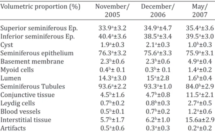
inexistent or diminished. In December/2006 and May/2007 the averages were: tubule diameter 110.3 and 5.3mµ, lumen 52.4 and 4.5mµ, epithelium thickness 57.8 and 0.7mµ respectively. The volumetric proportions were: seminiferous epithelium 75.6 and 75.9, cysts in epithelium 2.1 and 1.0, ST 93.3 and 84.0, interstitium 6.2 and 15.6 respectively. The sperm reserves were: 19.7±2 and 0±0 x109 sperm cells in December 2006 and May 2007 respectively. Microscopic measures of seminiferous tubules, spermatic cells and diameter
of the nuclei were presented. These data confirm reproductive seasonality, with breeding
season in spring-summer with sperm production. A great variation n parenchyma, when compared breeding was noticeable.
INDEX TERMS: Spermatogenesis, testicular morphology, greater rhea, Rhea americana, ratites, reproduction, seminiferous tubules.
1 Received on June 10, 2015.
Accepted for publication on June 2, 2016.
2 Laboratório de Andrologia e Tecnologia do Sêmen, Escola de Veterinária e Zootecnia, Universidade Federal de Goiás (UFG), Campus Samambaia, Av. Esperança s/n, Campus Universitário, Goiânia, GO 74690-900, Brazil. *Corresponding author: gfreneau@gmail.com, gfreneau@ufg.br
das características estudadas foram: diâmetro dos túbulos 110,3 e 5,3 mµ, lúmen 52,4 e 4,5mµ, espessura do epitélio 57,8 e 0,7mµ, respectivamente. As proporções volumétricas foram: epitélio seminífero 75,6 e 75,9, cistos no epitélio 2,1 e 1,0, túbulos seminíferos 93,3 e 84,0, interstício 6,2 e 15,6, respectivamente. Foram apresentadas medidas microscópicas de túbulos seminíferos, diâmetro dos núcleos
das espermátides. Estes dados confirmam a sazonalidade
reprodutiva, com época de reprodução na primavera - verão, com a produção de esperma. Foi perceptível uma grande variação nas medidas do parênquima testicular, quando se comparou a estação reprodutiva.
TERMOS DE INDEXAÇÃO: Espermatogênese, morfologia microscópica testicular, Emas, Rhea americana, ratitas,
reprodução, túbulos seminíferos, parênquima testicular.
INTRODUCTION
The seminiferous tubules of Galliformes and Columbiformes
are constituted by a stratified epithelial tissue with
germinative cells. These species have already been studied, as well as the turkey and the garganey (Soley 1993, Soley & Roberts 1994, Soley 1997). The contribution was not made for the study of microscopic tissues alone, but also for the study of reproduction of these species (Baraldi Artoni et al. 1997).
Avian reproduction occurs during determined periods of the year, it is synchronized with seasonal variations and it is often dependent of photoperiod (Nicholls et al. 1988). In the domestic Quail and other bird species, there are studies correlating the effects of photoperiod with gonadal development, or with gonadal activity during the stages of testicular cycle, taking into account the variations of testicular weight during these stages (absolute or relative), (Tanaka et al. 1964, Hamner 1966, Schwab 1970, Brillard & Reviers 1981, Hess & França 2008).
In Rhea Americana were reported seasonality pattern in sex hormones (Valdez et al. 2014) and macroscopic morphology of testicles and epididymis (Carvalho et al. 2015). There is no report about spermatogenesis in this South American bird.
The purpose of this research was to evaluate spermatogenesis aspects of the Greater Rhea, with the description of microscopic testicular morphology, volumetric proportions of the testicular parenchyma, seminiferous tubule measurements, spermatic reserve and measures of cellular nuclei of seminiferous tubule cells, for three samples of distinct periods of the year.
MATERIALS AND METHODS
Fifty-four sexually mature Greater Rheas were utilized; with an age of 2.5±0.5 years and body weight of 30.12±1.87 kg, slaughtered in a slaughterhouse credentialed by the Brazilian Federal Inspection Service and located in Santa Maria/RS: latitude -29° 41’, longitude +53° 48’, average altitude of 151m, humid subtropical climate, average annual temperature of 26.37°C, and annual photoperiod ranging from 10.0 to 14.2 hours/day. Climate data can be reviewed on Table 1, it was collected from the Santa Maria Meteorological Station, National Meteorology Institute (INMET). The samples were collected in November/2005 (n=14),
December 2006 (n=20) and May 2007 (n=20), according to the slaughterhouse activity. The birds came from a commercial breeding ranch, located in the same county, that possessed a commercial breeder license (Registry no. 65. 2515) from Instituto Brasileiro de Meio Ambiente (IBAMA). It was informed that the animals were fed with a balanced diet and water ad libitum. For the research proposal, IBAMA provided the Research License 058/2005.
After the slaughtering process, the testicles were collected and separated from the epididymes. Tissue samples were taken from cranial, medial and caudal regions of the testicles. The samples were fixated in Bouin solution for 12 hours, after being washed and placed in 70% alcohol until dehydration. The tissue samples were dehydrated in serial alcohol, diafanized in xilol, embedded in paraffin and sliced into sections of 4 µm. The sections were stained in hematoxylin and eosin (HE) (Luna 1968). After the processing of the tissue, the structure of the testicular parenchyma was described, starting from the capsule to the lumen. The morphology of the seminiferous tubules was studied from the albuginea to the epithelium, including the shape and cellular types. Total diameter of the tubular lumen and epithelial height of the seminiferous tubules were estimated from the averages of ten tubules of a single section, with five blocks per animal. The measure of the tubular and lumen diameters was performed through vertical and horizontal measures of the tubules. The result from averaging these last measures was considered as the diameter of the seminiferous tubule (Weibel et al. 1966), with the support of a binocular light microscope, 40x objective and
software Axio Vision® 3.0.6 sp4 (Carl Zeiss, GmbH). Circular or
almost circular shaped tubules were solely utilized.
The volumetric proportions of the components of the testicular parenchyma were measured with the support of an integrating eyepiece with reticules Zeiss Kpl 8x/18, containing a grid with five parallel lines and 25 equidistant points, coupled by a 40x objective. Carried out next was the examination of 40 fields, randomized by horizontal scan for each animal. Counted points were expressed through percentages. The same five blocks from each animal were used. The volumetric proportions of structures were calculated through the Pi/Pt formula, in which Pi is the number of counted points and Pt is the total number of points (Weibel et al. 1966). The volumetric proportions of the following structures were estimated: seminiferous epithelium, lumen, cyst, myoid cells, basement membrane, conjunctive tissue, Leydig cells, blood vessels and artifacts. With possession of this data, the total volumetric proportion of the epithelium was calculated, which was the sum of the superior and inferior epithelium and the cyst. The volumetric proportion of the seminiferous tubule was also calculated, which was total epithelium plus the basement membrane, the myoid cells and the lumen. Lastly, the interstitial or intertubular tissue was calculated as the sum of the conjunctive tissue, Leydig cells and blood vessels.
The width, the length and the diameter of the cellular nuclei were measured, and in this manner, it was computed the nuclei of
Table 1. Means, maximum and minimum values of humidity and radiation during collection, November/2005,
December/2006 and May 2007, Santa Maria, RS
Months/Year Temperature °C Humidity Precipitation Sunny
mean max min (%) (mm) (hs)
November/2005 22.2 29.4 16.0 67.1 57.2 251.7 December/2006 25.5 32.2 20.0 68.7 84.2 262.8 May/2007 13.8 20.2 9.4 82.4 102.8 173.5 Source: Meteorological Station of Santa Maria, National Meteorology
different types of spermatogenic cells and the nucleoli of Sertoli cells. Ten transverse sections from the seminiferous tubules of each animal testicle were used. The nuclei of the following cellular types were measured: spermatogonia, young primary spermatocytes (preleptotene and leptotene stages), old primary spermatocytes (pachytene stage), round spermatids and Sertoli cells.
The calculation of the testicular sperm reserve was made through the hemocytometer method (Neubauer). Through this method, spermatic cells (spermatozoid heads and/or elongated spermatids) were counted from diluted homogenized fragments from the testicular parenchyma. The homogenization of fragments from the testicular parenchyma was done with a blender, along with a diluent of STM solution (saline + Triton x-100®, Merk, USA + Thimerosal), prepared as follows: NaCl at
0.9%, 100 mL; Triton x-100®, 0.05mL and 100ppm of Thimerosal.
The concentration of Triton x-100 in the solution was 0.5%. This method is based on the principle that, along spermiogenesis, the plasmatic and nuclear membranes, as well as the nuclear material of spermatids, acquire strong resistance to physical destruction provoked by homogenization, making it possible to isolate and count the numbers of this cellular type without the interference of other cellular types of the testicular parenchyma, which end up destroyed in the process (Grant & Hjerten 1977).
The testicular spermatic reserve was estimated for both testicles. After the sampling, the testicles were weighted and the following measurements were recorded: length, width, thickness and volume. Once the sampling of testicular parenchyma for histological analysis was finished, the testicles were wrapped in aluminium foil and placed in a Styrofoam container with ice, transported and then stored at -20oC until the moment of processing.
Samples of the testicular parenchyma were weighted and placed on a Petri dish with 20mL of STM, with the help of a tweezers and a scissor. The samples were fragmented and grinded until reduced to a mass of pasty consistency. Afterwards, this mass was transferred to a blender cup with the assistance of successive rinses, with STM, of the dish and the instrumental, in order to remove as much material adhered to them as possible. The volume of STM used for cleansing of the instrument was added to total volume. The material inside STM solution was homogenized in a blender at a maximum speed of approximately 18,000 rpm and during a period of 3 minutes. This initial homogenized material was moved to a graduated beaker, and the final volume acquired was recorded. At this point, the blender cup and the instrumental were also rinsed and the volume was added up, in order to observe the relationship “fragment weight x total volume of STM used”. From this initial homogenized material, a section of 1.0mL was removed and placed in a beaker of 10mL along with 9.0mL of STM. This new homogenized fragment, diluted at 1/10, was agitated sufficiently to suspend the appearance of possible sediments, and a Pasteur pipet was used to remove enough of it to fill two hemocytometer chambers (Neubauer), 3 minutes was the waiting period for material sedimentation. The counted cells were spermatozoid heads and/or elongated spermatids (cells resistant to homogenization). The cell counts were made for the two chambers and also posteriorly repeated. From the number of spermatic cells contained in each chamber, it was calculated the number of cells contained in the initial fragment and the total of cells per testicle.
The mean and standard deviations of microscopic measurements of the testicular parenchyma (height, width and diameter of the seminiferous tubule; and epithelial thickness), of the volumetric proportions of the components of the testicular parenchyma (seminiferous epithelium, tubular lumen, cyst, myoid cells, basement membrane, conjunctive tissue, Leydig cells, blood
vessels and artifacts), of spermatic reserve and of microscopic measures of the nuclei of spermatogenic and somatic cells of the seminiferous tubule, from the collections of December/2006 and May/2007, were calculated. Means and standard deviations of the morphometric parameters from the testicle and epididymes were calculated. The morphometric measurement means found were compared through variance analysis (one way Anova) made in between the collection periods and through Tukey’s test (p<0.05). The morphometric parameter means between right and left testicles and epididymides were also compared, over the average differences paired by the Student’s T-test (p<0.05). Pearson correlation indexes were calculated between quantitative characteristics of the samples collected December/2006 and May/2007. Proc Means, Proc GLM and Proc Corr procedures from the SAS software (1997) were used.
RESULTS AND DISCUSSION
The testicular capsule of the Greater Rhea is divided into the epididymal region, with branches wrapping the epididymis, and the testicle region, which covers the testicle and the rete testis. Histologically, the testicular capsule is made of three tissue layers that are very thin: the most external and thinnest is the tunica serosa, the layer below, in the interior, is the tunica albuginea and the deepest layer of them is the tunica vasculosa (Fig.1).
A layer of myoepithelial cells forms the tunica serosa, and below it, there is the tunica adventitia, which isconstituted by collagen, elastic fibers and many fibroblasts. Blood
vessels, especially veins of various sizes, course the tissue
among fibroblasts and collagen fibers, they can be observed
traveling the surface of the testicle in radial direction. No septum was found on the testicular capsule.
The testicular capsule in birds is usually thin (Lake 1971), divided by the epididymal and testicular regions. Histologically, it has three layers: tunica serosa, tunica
Fig.1. Testicular capsule of the Greater Rhea (Rhea americana) is thin; the tunica serosa (arrows) and tunica albuginea (a) with
albuginea and tunica vasculosa. The first one, tunica vasculosa, is composed of mesothelial cells, and behaves as a membrane, the second one, tunica albuginea, is formed
by collagen, elastic fibers and fibroblasts, blood vessels are
present in this layer and are distributed randomly, but with overall radial direction in regards to the testicle, and the last
Fig.2. Seminiferous tubules (T) and germinative cysts (ac) of Rhea
americana, with many germinative cells in the epithelium. Iin
transverse section the tubules show almost circular shape. Santa Maria/RS, December/2006. HE, obj.10x.
Fig.4. Seminiferous tubules (T) of the Greater Rhea. During the period, retraction and consequent closing of the tubular lumen (L) was observed. Santa Maria/RS, May/2007. HE, obj.10x. Fig.6. Seminiferous tubule of the Greater Rhea during the sexual
activity period can be noticed that there is no capsule or septum separating the aggregation of germinative cells (ac) in the seminiferous epithelium (E). Santa Maria/RS, December/2006. Masson’s Trichrome stain, obj.100x.
Fig.3. Cellular types of the seminiferous epithelium of the Greater Rhea during spermatogenesis. Spermatogonium (e), primary spermatocyte in pachytene (ep), primary spermatocyte in leptotene (ele), round spermatid (ea) and elongated spermatid (el). Santa Maria/RS. December/2006. HE, obj.100x.
Fig.5. An aggregation of germinative cells (ac) in the epithelium of the seminiferous tubule (T) of the Greater Rhea. Santa Maria/ RS, December/2006. HE, obj.100x.
Fig.7. Interstitial tissue of the Greater Rhea taken from the third collection. Notice the red blood cells (h), myoid cells (arrows) and Leydig cells (l). Santa Maria/RS, May/2007. HE, obj.100x.
2 3
4 5
Table 2. Microscopic measurements (x±dp) of the seminiferous tubules in Greater Rheas. Collections made in
November/2005, December/2006 and May/2007, Santa Maria, RS
Measurements (µ) November/ December/ May/
2005 2006 2007
Tubule vertical diameter 122.5a±29.5 109.7a±14.6 7.5b±0.5 Tubule horizontal diameter 105.3a±14.8 110.9a±15.2 3b±0.4 Tubule diameter 113.9a±22.1 110.3a±14.9 5.3b±0.4 Lumen vertical diameter 57.6a±15.1 54.3a±9.3 6.4b±0.6 Lumen horizontal diameter 46.8a ±11.2 50.5a±7.7 2.6b±0.6 Tubular lumen diameter 52.2a±13.1 52.4a±8.5 4.5b±0.6 Epithelium vertical height 64.8a±17.7 55.3b±7.9 1.1c±0.4 Epithelium horizontal height 58.9a±18.2 60.4a±11.0 0.4b±0.6 Epithelium height 61.8a±17.9 57.8a±9.4 0.7b±0.4 Different letters in the same line indicate statistical difference P<0.05
(Tukey).
Table 3. Volumetric proportions (x±dp) of seminiferous tubules of the Greater Rhea. Collections made in November/2005, December/2006 and May/2007,
Santa Maria, RS
Volumetric proportion (%) November/ December/ May/
2005 2006 2007
Superior seminiferous Ep. 33.9a±3.2 34.9a±4.7 35.4a±3.6 Inferior seminiferous Ep. 40.4a±3.6 38.5a±3.4 39.5a±3.0
Cyst 1.9a±0.3 2.1a±0.3 1.0b±0.3
Seminiferous epithelium 76.3a±3.2 75.6a±3.3 75.9a±3.1 Basement membrane 2.3b±0.6 2.3b±0.6 4.9a±0.4 Myoid cells 0.4b± 0.1 0.3b± 0.1 1.4a±0.2
Lumen 14.3a±3.0 15a±2.8 1.6b±0.4
Seminiferous Tubules 93.6a±2.2 93.3a±1.0 84.0b±2.9 Conjunctive tissue 4.5b±1.6 4.7b±0.8 11.5a±2.1 Leydig cells 0.7b±0.2 0.8b±0.3 2.7a±0.5 Blood vessels 0.5b±0.1 0.7b±0.2 1.2a±0.6 Interstitial tissue 5.7b±1.7 6.2b±1.0 15.6a±2.9
Artifacts 0.5a±0.6 0.3a±0.3 0.2a±0.2
Ep = epithelium. Different letters in the same line indicate statistical difference P<0.05 (Tukey).
one, tunica serosa, comes into contact with the testicular parenchyma (Hodges 1974). In the Greater Rhea, the same structures and the same characteristics were found and the capsule did not form septums or lobes. These observations were also reported by Ozgbe et al. (2008) in the ostrich and the emu. The immuhistochemistry characterization of those structures revealed a cytoarchitecture rich in actin expressed weakly in rete testis cytokeratins and vementine were identify in the epithelium of the extracurrent ducts (Ozgbe et al. 2012).
The seminiferous tubules, in transverse sections, presented almost circular shapes (Fig.2). The testicular parenchyma of the Greater Rhea is composed by seminiferous tubules that, in transverse sections, show almost circular shapes, different to what is witnessed in the domestic Rooster (Lake 1971) and the Quail (Baraldi Artoni et al. 1999), which present hexagonal shapes, and similar to the Emu, which also presents circular shapes (Malecki et al. 1998). Phylogenetically, the Emu is close to the Greater Rhea, and both species are part of the Ratite bird group. However, there are some differences between the American and African ratites (Machado et al. 2011). The analyses of cellular structure of seminiferous tubules of the Greater Rhea demonstrated different phases of epithelium cycle.
Observing the seminiferous tubules from the sections taken from the samples of November/2005 and December/2006, the existence of different cellular associations in the same section was perceivable, demonstrating that there are many phases of the seminiferous epithelium cycle in sectorial form. This characterizes a helicoidal disposition of the possible stages of the seminiferous epithelium cycle. The report that cellular associations from the seminiferous epithelium cycle were noticed on single sections from the seminiferous tubules of the Greater Rhea coincided with what was reported in the domestic rooster (Sharma et al. 1956), in the garganey (Clermont 1958), and in the domestic duck (Marchand 1977).
The seminiferous tubules appear as a compact net and as a structure lacking formation of testicular septums. In samples of November/2005 and December/2006 there are many types of cells in these sections, demonstrating the presence of sexual activity during those periods. It was possible to visualize spermatogonia, primary spermatocytes in pachytene and leptotene, round and elongated spermatids, Sertoli cells, and, in the tubular lumen, clusters of elongated spermatids were visible. During this time, the epithelium underwent mitosis and meiosis, showing a vigorous spermatogenic process (Fig.2 and Fig.3). However, when the seminiferous tubules collected in May/2007 were studied, there was no presence of round or elongated spermatids and neither of spermatozoids (Fig.4), therefore, there was no spermatogenesis at this time, which characterized a period of sexual repose. Compared to the activity period, microscopic measurements of semininferous tubules during sexual repose were smaller, such as height and diameter of the epithelium and tubular lumen. The tubular lumen was often reduced or obliterated (Table 2).
In Table 2, diameters and measures taken from the seminiferous tubules, seminiferous epithelium and tubular
lumen of the Greater Rhea during distinct periods of the year can be reviewed.
In regards to the volumetric proportion, the greatest variation was observed in the tubular lumen between the two periods, with accentuated reduction in May/2007 (sexual repose period) (Table 3). At last, when both periods were compared, structural changes were drastic, with major variation of microscopic measurements, types and cell numbers (Table 2 and Table 3).
In Table 3, volumetric proportions of different structures of the testicular parenchyma of the Greater Rhea, in different periods of the year, can be reviewed.
In this study, the gonadic sperm reserve was 19.7±2x10⁹
and 0±0 spermatozoids or elongated spermatids were observed, from the samples of December/2006 and May/ 2007, respectively. This information was established through the multiplication of the number of cells present in one gram of testicular parenchyma by total testicular parenchyma weight, and so the numbers represent spermatozoid quantity per testicle. No literary references regarding this subject were found for Ratites, most studies
the non spermatic production outside reproductive season, as seen on histology.
Table 4 lists the measurements of different nuclei of spermatogenic and somatic cells inside the seminiferous tubule of the Greater Rhea, for different periods of the year. Overall, the nuclei of Sertoli cells, spermatogonia and spermatocytes were bigger during May/2007, when sexual
activity had decreased. This is the first report of cellular
nuclei measurement for this species. We do not have an explanation to the fact that nucleus size was smaller when outside of reproductive season.
This study is aggregating with previous reports that aims different aspect as hormones and sexual behavior (Leite & Codenotti 2005, Valdez et al. 2014) that in Rio Grande do Sul, the reproductive period of the Greater Rhea occurs during spring and summer. Thereby the samples from 2005 and 2006 were made during the reproductive period. Meanwhile, the sample from May/2007 happened
during the non-reproductive period, so during the first
two samples there was the presence of sexual activity and during the last one, sexual repose.
In the parenchyma of the seminiferous epithelium, it was noticed the presence of aggregations of germinative cells, especially in the basal region of the tubules (cyst), these cells presented intense coloration when stained with hematoxylin and eosin (Fig.2 and Fig.5). No presence of a capsule delimiting or separating these aggregations of cells in the epithelium was noticed (Fig.6).
No report or literary citations or data was found over the existence of aggregations of germinative cells in the seminiferous epithelium forming a separate structure. Russel & França (1995) mention giant cells in the testicular
parenchyma, but these cells are multinucleated and a result of the fusion of various spermatids and spermatogonia with vacuolated cytoplasm and pyknotic nuclei. In the Greater Rhea, cells did not have vacuolated cytoplasm and neither pyknotic nuclei (Fig.5). There is no capsule, collagen
membrane or fibroblasts delimiting the cellular aggregation,
as can be seen in Figure 6 of the sample stained with Masson’s
Trichrome. There are studies in fishes and amphibians that
demonstrate the formation of cysts in the seminiferous epithelium, however the spermatogenic process is different, all of the spermatogenesis stages occur in the cysts, until breaking during spermiation (Callard et al. 1978).
Comparing the histology of the testicular parenchyma between the three samples, drastic changes are noticed, regarding seminiferous tubule volume and lumen diameter, these changes have also been reported in the Quail by BaraldI Artoni et al. (1997), Baraldi Artoni et al. (1999) and Aire (2003), and in the Ostrich by Madekurozwa et al. (2002).
In the samples of November/2005 and December/2006, the epithelium exhibited various cellular types during visible spermatogenic process (Fig.3), spermatogonia through all of the basal expansion of the tubules and primary spermatocytes was observed (leptotene and pachytene), however, none of these cells encountered were at the anaphase stage, it is known that this stage occurs rapidly (Ortavant 1959) in birds as well as in mammals. Secondary spermatocytes were seldom seen and hardly visible in the histological sections of the Greater Rhea because of their short lifespan. In the sections from May/2007, there was no presence of round or elongated spermatids.
Most of the Sertoli cells were located in the basal region of the seminiferous epithelium, tall and column shaped. Their cytoplasm showed faint coloration that was more perceptible in the nucleus with nucleolus, it was large, triangular shaped and presented euchromatin, as was also mentioned for the Chicken by other researchers (Orth et al. 1988, Sharp 1994, França & Russel 1998, Griswold 1998).
The interstitial tissue of the Greater Rhea was formed
by two main components: the first one is a thin layer with
elongated myoid cells and it circled the seminiferous tubule
delimiting it, the second one consisted of collagen fibers
located between tubules and forming areas of adjacent angles. On this last, one it was also noticed the presence of blood vessels and Leydig cells with a narrow and columned
aspect, fibroblasts (Fig.7) and reticulo-endothelial cells,
such as macrophages, lymphocytes and monocytes.
In the layers related to the first and second samples,
the interstice was narrow, although in the histological sections from the third sample there was an increase in its
surface, with larger spacing between the collagen fibers that compose it. The volumetric proportions of the first
and second samples, when compared to the third one, varied from 8.76±0.26 % to 22.04±1.8% (Table 3), this is explained by the simultaneous proportional reduction of the seminiferous tubule in this last sample.
Baraldi Artoni et al. (2007) studied the morphometry of seminiferous tubules of the Red-winged Tinamou (Rynchotus rufescens) throughout the year, morphological alterations in the tubules were reported, regarding height
Table 4. Microscopic measurements (x±dp) of the nuclei of germinative and seminiferous epithelium cells of the Greater Rhea. Collections made November/2005,
December/2006 and May/2007, Santa Maria, RS
Cell type November/ December/ May/
2005 2006 2007
Sertoli
length 3.94b±0.19 4.04b±0.19 4.82a±0.16 width 2.34b±0.18 2.51b±0.37 5.18a±0.45 average 3.14c±0.10 3.27b±0.23 5.00a±0.26 Spermatogonium
length 3.76b±0.19 3.56c±0.19 4.69a±0.23 width 3.66a±0.16 3.59a±0.16 3.38b±0.18 average 3.71b±0.12 3.57c±0.13 4.03a±0.16 Young spermatocyte
length 3.49b±0.12 3.52b±0.12 4.30a±0.40 width 3.45c±0.12 3.54b±0.10 3.77a±0.14 average 3.47b±0.10 3.53b±0.09 4.04a±0.23 Old spermatocyte
length 4.58b±0.31 4.48b±0.25 7.72a±0.25 width 4.76b±0.29 4.67b±0.30 7.90a±0.32 average 4.67b±0.27 4.57b±0.21 7.81a±0.21 Round spermatid
length 2.00a±0.19 1.99a±0.22 0±0
width 2.01b±0.15 2.11a±0.18 0±0
average 2.00a±0.13 2.05a±0.16 0±0 Elongated spermatid
average 7.06a±0.37 7.09a±0.42 0±0 Different letters in the same column indicate statistical difference P<0.05
and diameter of the epithelium, these alterations were related to seasons of year, presenting periods of sexual activity and repose, the period during which activity was at its highest corresponded to spring (September) and lowest activity happened during winter (July). During spring, the following seminiferous tubule diameter and epithelial height values were reported in the Red-winged Tinamou: 315.9±57.7µm and 79.5±22.3µm, during winter the following values were encountered: 182.7±54.9µm and 49.6±24.9µm. The values for mensurations studied in the Red-winged Tinamou were superior to the Greater Rhea, such as tubule diameter and epithelial height. However, in the Greater Rhea the numeric amplitudes between seasons were more elevated, even to the point of complete closing of the tubular light due to diameter decrease in May/2007. Meanwhile during the period of sexual activity, with the expansion of seminiferous tubules, the volume of interstitial tissue was smaller than what was seen during the sexual repose period. This occurred in the Greater Rhea and it was also reported in the Red-winged Tinamou by Baraldi Artoni et al. (2007). The seasonal alteration is clear, since during activity, the interstice is narrow and during repose, it is large, this is due to an expansion of the seminiferous tubules during sexual activity, which compresses the interstice, while during repose the retraction of the tubules allows enough space for an expansion of the interstice. Similar data was also reported in the domestic Rooster (Nagano 1962, Lake 1971, Gunawardana & Scott 1977).
Peixoto (2006) studied the testicle of the White-eyed Parakeet Aratinga leucophthalma. He reported that this species is seasonal, with the highest expansion of the seminiferous tubules during spring (November) and the lowest during autumn (May). This research found the following values for the volumetric proportion of the tubule, interstice and tubular diameter, during spring: 89.2±3.3 %, 10.9±3.3% and 122.6±8.6 µm, respectively, and during autumn: 74.7±7.4%, 25.3±7.4% and 79.4±11.9µm, respectively. It is noticeable that the behavior of the Greater Rhea was similar to the White-eyed Parakeet regarding seasonality and that the measurements described for the Greater Rhea are closer to the measurements of the White-eyed Parakeet than they are to the Red-winged Tinamou.
The interstitial tissue presented, during the periods of sexual repose and activity, the following correlations: with the conjunctive tissue 0.96 and 0.95, with the Leydig cells 0.76 and 0.46 and with the blood vessels 0.66 and 0.38 respectively. During sexual repose the interstice showed higher volume, so it was expected a higher correlation between it and other components when compared to the sexual activity period. As for the seminiferous tubule during sexual repose and sexual activity, the correlations were with the seminiferous epithelium 0.97 and 0.33, with the basement membrane -0.05 and 0.12, with the myoid cells -0.095 and -0.43 and with the lumen -0.20 and 0.17 respectively. In this case, there is a higher correlation between the repose period and the epithelium, and a negative one with the tubular lumen. This happened because during repose period, proportionally, the tubular
lumen had reduced and so the epithelium filled a larger
space due to lumen retraction.
Finally, there were no difficulties in fixating and staining
the histological sections of the Greater Rhea. Some authors
mention difficulties in the fixation of sections of the Quail,
the Rooster and the Helmeted Guineafowl males (Nagano 1962, Lake 1971, Budras & Sauer 1975, Hess et al. 1976, Gunawardana & Scott 1977, Hess & Thurston 1977, Aire 1979, 1980, 1982a, 1982b, Aire et al. 1979, Bakst 1980, Mikami et al. 1988).
CONCLUSIONS
The data presented on testicular morphology of Rhea americana contribute to the reproductive study of this species.
The Greater Rhea (Rhea americana) presents a reproduction divided into two periods, one corresponds to the sexual activity period and occurs during spring-summer, and the other corresponds to the sexual repose and occurs during autumn-winter.
Between the periods of sexual activity and repose, major morphological alterations were observed in the structure of the testicles.
Acknowledgements.- To Ireny Rodrigues de Almeida and Jacir Antônio Dalla Vecchia, from Associação Brasileira dos Criadores de Emas (ABRACE), allowing the use of animal samples and data.
Conflict of interest.- The authors declare that there is no conflict of interest that could be perceived as prejudicing the impartiality of the research reported.
REFERENCES
Aire T.A. 1979. The epididymal region of Japanese quail (Coturnix coturnix japonica). Acta Anat. 103:305-312.
Aire T.A. 1980. The ductulli efferentes of the epididymal region of birds. J. Anat. 130:707-723.
Aire T.A. 1982a. The rete testis of birds. J. Anat. 135:97-110.
Aire T.A. 1982b. Surface morphology of the ducts of the epididymal region of the drake (Anas platyrhynchos) as revealed by scanning and transmis-sion electron microscopy. J. Anat. 135:513-520.
Aire T.A. 2003. Ultrastructural study of spermiogenesis in the turkey, Me-leagris galopavo. Brit. Poult. Sci. 44:674-682.
Aire T.A., Ayeni J.S. & Olowookorun M.O. 1979. The structure of excurrent ducts of the testis of the Guinea fowl (Numida meleagridis). J. Anat. 129:633-643.
Bakst M.R. 1980. Luminal topography of the male chicken and turkey ex-current ducts system. Scan. Electron Microsc. 3:419-425.
Baraldi Artoni S.M., Orsi A.M., Lamano-Carvalho T.L. & Lopes R.A. 1997. The annual testicular cycle of the domestic quail (Coturnix coturnix ja-ponica) testis. Anat. Histol. Embryol. 26:337-339,
Baraldi Artoni S.M., Orsi A.M., Carvalho T.L.L., Vicentini C.A. & Stefanini M.A. 1999. Seasonal morphology of the domestic quail (Coturnix cotur-nix japonica) testis. Anat. Histol. Embryol.28:217-220.
Baraldi Artoni S.M., Bottino F., Oliveira D., Soube Franzo V., Amoroso L., Orsi A.M. & Cruz C. 2007. Morphometric study of Rynchotus rufencens
testis throughout the year. Braz. J. Biol. 67:363-367.
Brillard J.P. & Reviers M. 1981. Testis development and daily sperm output in guinea-fowl raised under constant daily photoperiods. Reprod. Nutr. Develop. 21:1105-1112.
synthe-sis. I. Ontogenesis, morphology and distribution of the epididymis. Anat. Histol. Embryol. 148:175-196.
Callard I.P., Callard G.V., Lance V., Bolaffi J.L. & Rosset J.S. 1978. Testicular regulation in non-mammalian vertebrates. Bio. Reprod. 18:16-43. Carvalho S.F.M., Freneau B.N. & Freneau G.E. 2015. Aspects of the
macro-scopic testicular and epididymal morphology in the Greater Rhea, Rhea americana (Linnaeus, 1758) birds. Anat. Histol. Embryol. 44:255-261. Clermont Y. 1958. Structure de lèpthélium séminal et mode de
renouvele-ment des spermatogonies chez le canard. Arch. Anat. Microsc. Morphol. Exp. 47:47-66.
França L.R. & Russell L.D. 1998. The testis of domestic animals, p.197-219. In: Martinez F. & Regadera J. (Eds), Male Reproduction: a multidisci-plinary overview. Churchill Livingstone, Madrid.
Grant D.A. & Hjerten S. 1977. Some observations on the choice of deter-gent for solubilization of the human erythrocyte membrane. Biochem. J. 164:465-468.
Griswold M.D. 1998. The central role of Sertoli cells in spermatogenesis. Semin. Cell Dev. Biol. 9:411-416.
Gunawardana V.K. & Scott A.D. 1977. Ultrastructural studies on the dif-ferentiation of spermatids in the domestic fowl. J. Anat. 124:741-755. Hamner W.M. 1966. Photoperiodic control of the annual testicular cycle in
the house finch, Carpodacus mexicanus. Gen. Comp. Endocrinol. 7:224-233.
Hess R.A. & Thurston R.J. 1977. Ultrastructure of the epithelial cells in the ep-ididymal region of the turkey (Meleagris gallopavo).J. Anat. 124:765-778. Hess R.A., Thurston R.J. & Biellier H.V. 1976. Morphology of epididymal
region and ductus deferens of the turkey (Meleagris gallopavo).J. Anat. 122:241-252.
Hess R.A. & França L.R. 2008. Spermatogenesis and cycle of the seminif-erous epithelium, p.1-15. In: Ibid. (Eds), Molecular Mechanisms in the Spermatogenesis., Landes Biosciences and Springer Science, Illinois. Hodges R.D. 1974. The reproductive system. I. The male reproductive
sys-tem, p.300-326. In: Hodges R.D. (Ed.), The Histology of the F owl. Aca-demic Press, London.
Lake P.E. 1971. The male in reproduction, p.1411-1447. In: Bell D.J. & Freeman B.M. (Eds), Physiology and Biochemistry of the Domestic Fowl. Vol.3. Academic Press, New York.
Leite M.A.S. & Codenotti T.L. 2005. Comportamento parental de machos da ema Rhea americana (Linnaeus, 1758), em ambiente natural, no Rio Grande do Sul. Revta Etol. 7:43-47.
Luna L.G. 1968. Manual of Histologic Staining Methods of the Armed Forc-es Institute of Pathology. 3rd ed. McGraw-Hill Book, NewYoerk. 258p. Machado T.M.M. & Malecki I.A. 2011. Relationship among the
economical-ly important ratites: a clustering approach based on their reproductive biology. Revta Bras. Zootec. 40(Supl.):60-67.
Madekurozwa M.C., Chabvepi T.S., Matema S. & Teerds K.J. 2002. Relation-ship between seasonal changes in spermatogenesis in the juvenile os-trich (Strutio camelus) and the presence of the LH receptor and 3β-hy -droxysteroid dehydrogenase. Reproduction 123:735-742.
Malecki I.A., Martin G.B., O`Malley P.J., Meyer G.T., Talbot R.T. & Sharp P.J. 1998. Endocrine and testicular changes in a short-day seasonally breed-ing bird, the emu (Dromaius novaehollandiae), in southwestern Austra-lia. Anim. Reprod. Sci. 53:143-155.
Marchand C.R. 1977. Étude ultrastructurale de la spermatogenèse du canard de barbarie (Cairina moschata). Biol. Cell . 29:193-201.
Mikami S.I., Yamada S., Hasegawa Y. & Miyamoto K. 1988. Localization of avian LHRH-immunoreactive neurons in the hypothalamus of the do-mestic fowl, Gallus domesticus, and the Japanese quail, Coturnix coturnix. Cell Tissue Res. 251:51-58.
Nagano T. 1962. Observation on the fine structure of the developing sper
-matid in the domestic chicken. J. Cell Biol. 14:193-205.
Nicholls T.J., Goldsmith A.R. & Dawson A. 1988. Photofractoriness in birds and comparison with mammals. Physiol. Rev. 68:133-176.
Ortavant R. 1959. Spermatogenesis and morphology of the spermatozoon, p.1-50. In: Coles H.H. & Cupps P.T. (Eds), Reproduction in Domestic Ani-mals. Academic Press, London.
Orth J.M., Gunsalus G.L. & Lamperti A.A. 1988. Evidence from Sertoli cell-depleted rats indicates that spermatids number in adults depends on numbers of Sertoli cells produced during perinatal development. En-docrinology 122:787-794.
Ozgbe P.C., Aire T.A., Madekurozwa M.C. & Soley J.T. 2008. Morpholog-ical and immunohistochemMorpholog-ical study of testicular capsule and peritu-bular tissue of emu (Dromaius novaehollandiae) and ostrich (Struthio camelus). Cell Tissue Res. 332:151-158.
Ozgbe P.C., Aire T.A. & Deokar M.S. 2012. The cytoskeletal proteins in the contractile tissues of the testis and its excurrent ducts of the passerine bird, Masked Weaver (Ploceus velatus). Tissue Cell 44:22-31.
Peixoto J.V. 2006. Avaliação morfofuncional do testículo e do processo espermatogênico do periquitão-maracanã (Aratinga leucophthalma
Muller, 1776) adulto, mantido em cativeiro, nas diferentes estações do ano. Dissertação de Mestrado em Medicina Veterinária, - Programa de Pós-Graduação em Medicina Veterinária, Universidade Federal de Viçosa, Viçosa, MG. 39p.
SAS Institute 1997. SAS/STAT User´s Guide 8.0. SAS Institute, Cary. Schwab R.G. 1970. Light-induced prolongation of spermatogenesis in the
European Starling, Sturnus vulgaris. Condor. Magazine of Western Orni-thology, Los Angeles, 72:466-470.
Sharma G.P., Gupta B.L. & Nayar K.K. 1956. Spermatogenesis of the domes-tic fowl, Gallus domesticus. Res. Bull. Panjab Univ. 93:139-151.
Sharp R.M. 1994. Regulation of spermatogenesis, p.1363-1434. In: Knobil E. & Neill J.D. (Eds), The Physiology of Reproduction. Raven Press, New York. Soley J.T. 1993. Ultrastructure of ostrich (Struthio camelus) spermatozoa I.
Transmission electron microscopy. Onderstepoort J. Vet. Res. 60:119-130. Soley J.T. 1997. Nuclear morphogenesis and the role of the manchette
during spermatogenesis in the ostrich (Struthio camelus). J. Anat. 190: 563-576.
Soley J.T. & Roberts D.C. 1994. Ultrastructure of ostrich (Struthio camelus) spermatozoa II. Scaning electron microscopy. Onderstepoort J. Vet. Res. 61:239-246.
Tanaka K., Mather F.B. & Wilson W.O. 1964. The effect of photoperiods on early growth of gonads and on anterior pituitary gonadotrophin in co-turnix. Poult. Sci. 43:1368-1369.
Valdez D.J., Cortez M.V., Della Costa N.S., Lèche A., Hansen C., Navarro J.L. & Martella M.B. 2014. Seasonal changes in plasma levels of sex hormones in the Greater Rhea (Rhea americana), a South American Ratite with a complex mating system. PLoS ONE 9(5): e97334. doi:10.1371/journal. pone.0097334