This article was published in Journal of Food Science and Technology, 52(8), 4737-4748, 1 2015 2 http://dx.doi.org/10.1007/s13197-014-1533-1 3 4
Antibacterial activity and mode of action of selected
5glucosinolate hydrolysis products against bacterial pathogens
67 8
Abstract 9
Plants contain numerous components that are important sources of new bioactive
10
molecules with antimicrobial properties. Isothiocyanates (ITCs) are plant secondary
11
metabolites found in cruciferous vegetables that are arising as promising antimicrobial
12
agents in food industry. The aim of this study was to assess the antibacterial activity of
13
two isothiocyanates (ITCs), allylisothiocyanate (AITC) and 2-phenylethilisothiocyanate
14
(PEITC) against Escherichia coli, Pseudomonas aeruginosa, Staphylococcus aureus and
15
Listeria monocytogenes. The antibacterial mode of action was also characterized by the
16
assessment of different physiological indices: membrane integrity, intracellular
17
potassium release, physicochemical surface properties and surface charge. The minimum
18
inhibitory concentration (MIC) of AITC and PEITC was 100 µg/mL for all bacteria. The
19
minimum bactericidal concentration (MBC) of the ITCs was at least 10 times higher than
20
the MIC. Both AITC and PEITC changed the membrane properties of the bacteria
21
decreasing their surface charge and compromising the integrity of the cytoplasmatic
22
membrane with consequent potassium leakage and propidium iodide uptake. The surface
23
hydrophobicity was also non-specifically altered (E. coli and L. monocytogenes become
24
less hydrophilic; P. aeruginosa and S. aureus become more hydrophilic). This study
25
shows that AITC and PEITC have strong antimicrobial potential against the bacteria
tested, through the disruption of the bacterial cell membranes. Moreover, phytochemicals
27
are highlighted as a valuable sustainable source of new bioactive products.
28 29
Keywords: antibacterial activity; disinfectants; food preservatives; isothiocyanates; 30 mechanisms of action 31 32 Introduction 33
The food safety is an important public health issue that continues to be a major concern
34
to consumers, regulatory agencies and food industries worldwide. The increased
35
incidence of food poisoning cases has been reported due to the contamination of food
36
with pathogens and spoilage organisms (Langsrud et al. 2003; Negi 2012). This leads to
37
the necessity of improvement of hygiene and preservative practices of food products. The
38
presence of microorganisms in the food products frequently causes their spoilage, which
39
sometimes can lead to the production of toxins and alteration of their organoleptic quality
40
(Negi 2012; Tiwari et al. 2009).
41
Most of the traditionally used food preservation strategies (heating, refrigeration,
42
acidification, pasteurization and addition of synthetic antimicrobial compounds), may
43
cause adverse changes in organoleptic properties of foods and loss of nutrients, reducing
44
the consumer acceptability (Tiwari et al. 2009). The requirement of safer foods and longer
45
shelf-life has led to a higher frequency of disinfection (on food-contact surfaces,
46
equipment, utensils, etc.) and to the use of preservatives (Langsrud et al. 2003).
47
The recurrent use of chemical disinfectants and also the inadequate disinfection
48
strategies impose selective pressure and contribute to the emergence of resistance among
49
microorganisms (Russell 2000). Resistant microorganisms have been responsible for the
50
failure of many disinfection programs, and therefore for many contaminations in
industrial, environmental and biomedical settings (Chorianopoulos et al. 2011).
52
Combined resistance to disinfectants and other types of antimicrobials may become a
53
threat to the food processing industries. In addition, cross-resistance between
54
disinfectants and antibiotics can also lead to serious consequences for the public health
55
(Russell 2003). Therefore, new disinfection techniques and effective disinfectants are
56
required in order to ensure high levels of sanitation. In this context, substantial resources
57
have been invested in the research of effective antimicrobial compounds that preserve the
58
organoleptic properties of the products (Dufour et al. 2012; Negi 2012; Tiwari et al.
59
2009). Moreover, products that act on novel bacterial targets (e.g. bacterial ribosomal
60
subunit synthesis, fatty acid biosynthesis, aminoacyl–tRNA synthetases, two-component
61
signal transduction (2CST) systems) and circumvent the conventional mechanisms of
62
resistance to current antimicrobials are also important (Saleem et al. 2010; Sarker et al.
63
2007; Black and Hodgson, 2005). Although synthetic antimicrobials are approved in
64
many countries, the recent trend has been the use of safe natural preservatives derived
65
from microbial, animals or plants (Rahman and Kang 2009).
66
Plants are an attractive source of such compounds as they produce an enormous array
67
of secondary metabolites (phytochemicals) with medicinal properties, including
68
antimicrobial properties, which have been used traditionally for centuries (Abreu et al.
69
2012). A significant part of this diversity of phytochemicals are related to defense
70
mechanisms of plants against attack by microorganisms, insects, nematodes and even
71
other plants (Dangl and Jones 2001; Dixon 2001). Additionally, it is known that some
72
phytochemical products have an accepted safe status and distinctive properties from
73
synthetic molecules that make them perfect candidates for diverse applications (Cowan
74
1999; Lin et al. 2000a; Simões et al. 2009).
Glucosinolates (GLS) are organosulfur compounds present exclusively in the order
76
Capparales and very abundant in the Brassicaceae (Syn. Cruciferae) family (Al-Gendy et
77
al. 2010; Barbieri et al. 2008; Grubb and Abel 2006; Halkier and Du 1997). They occur
78
as secondary metabolites of various vegetables such as cabbage, broccoli, cauliflower,
79
watercress, horseradish, Brussels sprouts and kohlrabi (Fahey et al. 2001; Holst and
80
Williamson 2004). GLS are classified as aliphatic, aromatic and indolyl, based on the
81
amino acid from which they derive (Fahey et al. 2001; Halkier and Gershenzon 2006).
82
Intact GLS do not show antimicrobial activity. These dietary phytochemicals are present
83
in the cells vacuole and when tissue disruption occurs, they are hydrolyzed by the
84
myrosinase enzyme (β-thioglucosidase enzyme) into numerous biologically active
85
products such as isothiocyanates (ITCs), nitriles, epithionitriles and thiocyanates (Aires
86
et al. 2009b; Fahey et al. 2001; Hong and Kim 2008). Glucosinolate hydrolysis products
87
(GHP) have long been recognized for their antimicrobial activity against important
88
pathogenic microorganisms (e.g. Escherichia coli, Candida albicans, Bacillus subtilis,
89
Campylobacter jejuni, Helicobacter pylori and Vibrio parahaemolyticus) (Dufour et al.
90
2012; Fahey et al. 2001; Shin et al. 2004; Wang et al. 2010). In addition, these compounds
91
have other pharmaceutical benefits for human health, such as anticarcinogenic,
anti-92
inflammatory and antioxidant properties (D'Antuono et al. 2009; Hong and Kim 2008;
93
Saavedra et al. 2010; Zhang 2012). The presence of such phytochemicals in natural foods
94
might even contribute to the medicinal properties attributed to the consumption of
95
cruciferous vegetables. Among GHP, ITCs are considered the most potent inhibitors of
96
microbial activity and their properties are being actively explored (Al-Gendy et al. 2010;
97
Cartea and Velasco 2008; Munday et al. 2008; Saavedra et al. 2010; Sofrata et al. 2011;
98
Troncoso et al. 2005; Zhang 2012). ITCs can bind to sulfhydryl groups on active sites of
99
important enzymes involved in the microbial growth and survival. Consequently,
reductions in the cellular levels of important thiol groups lead to the formation of oxygen
101
and other free-radicals (Aires et al. 2009a; Jacob and Anwar 2008; Kolm et al. 1995).
102
The aim of this work was to investigate the antibacterial activity and some aspects of
103
the mode of action of two selected ITCs against strains of Escherichia coli, Listeria
104
monocytogenes, Pseudomonas aeruginosa and Staphylococcus aureus. These bacteria are
105
reference microorganisms for antimicrobial studies (EN-1276, 1997; Jones and Stilwell,
106
2013). Also, some of these species are important foodborne or spoilage microorganisms
107
commonly found in food industries, being important causal agents of foodborne diseases
108
(McCabe-Sellers and Beattie 2004; Rahman and Kang 2009).
109 110
Materials and methods 111
Bacterial strains and growth medium
112
The following strains were used in this study: Escherichia coli CECT 434, Pseudomonas
113
aeruginosa ATCC 10145, Staphylococcus aureus CECT 976 and Listeria monocytogenes
114
ATCC 15313. These bacteria were already used as model microorganisms for
115
antimicrobial tests with phytochemical products (Abreu et al. 2013; Borges et al. 2012;
116
Saavedra et al. 2010; Simões et al. 2008). E. coli, P. aeruginosa and S. aureus are
117
reference microorganisms to be used in the development of disinfection strategies
(EN-118
1276, 1997). Also, the strains used in this study are commonly used as routine quality
119
control strains, and as reference for antimicrobial testing and for bacterial resistance
120
testing (Ananou et al. 2004; Diab et al. 2012; Tabata et al. 2003; UNE-CEN ISO/TS
121
11133, 2006). All microbial strains were stored at -80 ºC in cryovial, 30% (v/v) glycerol,
122
and subcultured in Mueller-Hinton Agar (MHA) (Merck, Darmstadt-Germany) at 30 ºC,
123
before testing.
124 125
126 127 128
Isothiocyanates
129
Allylisothiocyanate (AITC) and 2-phenylethylisothiocyanate (PEITC) (Fig. 1) were
130
obtained from Sigma-Aldrich (Sintra-Portugal). Phytochemicals are routinely classified
131
as antimicrobials on the basis of susceptibility tests that produce inhibitory concentrations
132
in the range of 100 to 1000 µg/mL (Simões et al. 2009; Tegos et al. 2002). Therefore, in
133
this study, each product was tested at a concentration of 100, 500 and 1000 µg/mL
134
prepared in dimethyl sulfoxide (DMSO) (99%, v/v) (Sigma-Aldrich, Sintra-Portugal).
135 136
Minimum inhibitory concentration
137
The minimum inhibitory concentration (MIC) of ITCs was determined by the
138
microdilution broth method (Borges et al. 2013). Briefly, overnight culture growth in
139
Mueller-Hinton Broth (MHB), was adjusted to an OD640nm of 0.2 ± 0.02 (1 × 10⁸
140
cells/mL). Subsequently, for each bacterium, a sterile 96-well polystyrene microtiter plate
141
(Orange Scientific, Braine-L’Alleud-Belgium) was filled with bacteria (180 µL) and
142
phytochemicals (20 µL). These were tested at three different concentrations (100, 500
143
and 1000 µg/mL). Cell suspensions with DMSO and cell suspensions without
144
phytochemicals were used as controls. The microtiter plates were covered with a lid that
145
was sealed with parafilm (to avoid the volatilization of ITCs) and then incubated for 24 h
146
at 30 ºC in an orbital shaker (150 rpm). The absorbance was measured at 640 nm using a
147
Microplate Reader (Spectramax M2e, Molecular Devices, Inc.). The MIC was recorded
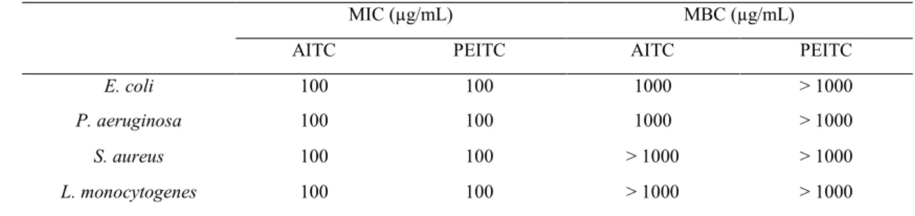
148
as the lowest concentration of ITCs at which no growth was detected (Borges et al. 2013).
149
All tests were performed in triplicate with three repeats.
152
Minimum bactericidal concentration
153
Bacterial cells were grown overnight in batch culture using MHB at 30 ºC and 150 rpm.
154
After the overnight growth, the bacterial suspension was centrifuged (3772 g, 6 min),
155
washed two times with saline solution (0.85% NaCl) and resuspended in saline solution
156
to obtain an OD640nm of 0.2 ± 0.02 (1 × 10⁸ cells/mL). Then, an aliquot of this suspension
157
was collected and maintained 30 min in contact with different concentrations of the ITCs
158
(100, 500 and 1000 µg/mL). Subsequently, bacterial suspensions were diluted to an
159
adequate cellular concentration (from 107 to 100) in saline solution. A volume of 100 µL
160
of each suspension (dilution 107 to 104) was transferred onto MHA plates and incubated
161
at 30 ºC. Colony enumeration was carried out after 24 h. Cell suspensions without
162
phytochemical were used as controls. The minimum bactericidal concentration (MBC)
163
was taken as the lowest concentration of phytochemicals at which no colony forming
164
units (CFU) were detected on solid medium (Borges et al. 2013). All experiments were
165
performed in triplicate with three repeats.
166 167
Physicochemical characterization of the bacterial surfaces
168
Bacterial suspensions were prepared in ultrapure water (Milli-Q®) (pH 6). No significant
169
osmotic pressure effects were found when comparing the planktonic bacterial viability in
170
water and in saline solution (0.85% NaCl), for a period of up to 150 min (P > 0.05).
171
Afterward, their physicochemical properties were determined by the sessile drop contact
172
angle measurement on bacterial lawns, prepared as described by Busscher et al. (1984).
173
Contact angles were determined automatically using an OCA 15 Plus (DATAPHYSICS,
174
Germany) video-based optical measuring instrument, allowing image acquisition and data
analysis. Contact angle measurements were carried out according to Simões et al. (2007).
176
Hydrophobicity was evaluated after contact angle measurement, following the van Oss
177
approach (van Oss et al. 1987; van Oss et al. 1988; van Oss et al. 1989), where the degree
178
of hydrophobicity of a given surface (s) is expressed as the free energy of interaction
179
between two entities of that surface, when immersed in water (w) – (ΔGsws mJ/m2). If the
180
interaction between the two entities is stronger than the interaction of each entity with
181
water, ΔGsws < 0, the material is considered hydrophobic. Conversely, if ΔGsws>0, the
182
material is hydrophilic. ΔGsws can be calculated through the surface tension components
183
of the interacting entities, according to:
184
LW 2 s w s w s s w w w LW s sws 2 4 G ; (1) 185where
LW accounts for the Lifshitz-van der Waals component of the surface free energy186
and γ+ and γ- are the electron acceptor and electron donor parameters, respectively, of the
187
Lewis acid-base component (
AB), with AB 2 . The surface tension components,188
of a solid material, can be obtained by measuring the contact angles of the three liquids
189
(l): the apolar α-bromonaphthalene; the polar formamide and water. The liquid surface
190
tension components reference values were obtained from the literature (Janczuk et al.
191
1993). Once the values are obtained, three equations of the type below can be solved:
192
w s w s LW w LW s Tot 2 θ cos 1
w
; (2) 193where θ is the contact angle and
Tot
LW
AB. At least three independent experiments194
were performed for each condition tested.
195 196 197
Bacterial surface charge - zeta potential
The zeta potential of bacterial suspensions, before and after the contact with different
199
AITC and PEITC concentrations (100, 500 and 1000 µg/mL), was determined using a
200
Nano Zetasizer (Malvern Instruments, UK). Cell suspensions in ultrapure water (pH 6),
201
without phytochemical, were used as controls. The zeta potential was measured by
202
applying an electric field across the bacterial suspensions. Bacteria in the aqueous
203
dispersion with non-zero zeta potential migrated towards the electrode of opposite charge,
204
with a velocity proportional to the magnitude of the zeta potential. The experiments were
205
repeated at least three times.
206 207
Assessment of membrane integrity due to propidium iodide uptake
208
The Live/Dead BacLightTM kit (Invitrogen/Molecular Probes, Leiden, Netherlands)
209
assesses membrane integrity by selective stain exclusion (Simões et al. 2005). This fast
210
method was applied to estimate both viable and total counts of bacteria. BacLight is
211
composed of two nucleic acid-binding stains: SYTO 9TM and propidium iodide (PI).
212
SYTO 9TM penetrates bacterial membranes, staining the cells green; PI only penetrates
213
cells with damaged membranes, binding to single and double-stranded nucleic acids. The
214
combination of these two stains generates red fluorescing cells. After overnight growth,
215
the cells were centrifuged (3772 g, 10 min) and washed one time with saline solution
216
(0.85%). Afterwards, bacteria were resuspended in saline solution to obtain an OD640nm
217
of 0.2 ± 0.02 (1 × 10⁸ cells/mL). Then, an aliquot of 1 mL of this suspension was collected
218
and different concentrations of the ITCs were tested (100, 500 and 1000 µg/mL) for 30
219
min in contact with the bacteria. Cell suspensions without phytochemicals were used as
220
controls. Afterwards, bacteria were transferred to saline solution and diluted 1:10. Three
221
hundred microliters of each diluted suspension were filtered through a Nucleopore®
222
(Whatman, Middlesex, UK) black polycarbonate membrane (pore size 0.22 µm) and
stained with 250 mL of diluted SYTO 9TM and 250 mL of diluted component PI. The dyes
224
were left to react for 15 min in the dark, at 27 ± 3 ºC. The membrane was then mounted
225
on BacLight mounting oil, as described in the manufacturer’s instructions. The
226
microscope used for the observation of stained bacteria was a LEICA DMLB2 with a
227
mercury lamp HBO/100W/3, incorporating a CCD camera to acquire images using IM50
228
software (LEICA) and a 100× oil immersion fluorescence objective. The optical filter
229
combination for optimal viewing of stained mounts consisted of a 480–500 nm excitation
230
filter in combination with a 485 nm emission filter (Chroma 61000-V2 DAPI/
231
FITC/TRITC). A program path (Scan Pro 5) involving object measurement and data
232
output was used to obtain the total number of cells (both stains) and the number of
PI-233
stained cells (damaged cells). Both the total number of cells and the number of PI-stained
234
cells on each membrane was estimated from counts of ≥ 20 fields of view. The total
235
number of cells counted per field of view ranged from 50 to 200 cells. Three independent
236
experiments were performed for each condition tested.
237 238
Potassium (K+) leakage
239
Flame emission and atomic absorption spectroscopy were used for K+ titration in bacteria 240
suspensions treated with 1000 µg/mL of each ITC. The samples were filtrated after
241
contact with the phytochemicals, using a sterile cellulose nitrate membrane filter (pore
242
size 0.22 μm) (Whatman, Maidstone-England), and then the filtrates were analyzed in a
243
GBC AAS 932plus device using GBC Avante 1.33 software. The experiments were
244
repeated three times.
245 246
Statistical analysis
The data were analysed using the statistical program SPSS (Statistical Package for the
248
Social Sciences) version 20.0 (IBM® SPSS® Statistics Corporation). The mean and
249
standard deviation within samples were calculated for all cases. One-way Anova with
250
Bonferroni test was used to assess the statistical significance value (confidence level ≥
251 95%). 252 253 Results 254
Inhibitory and bactericidal concentration of isothiocyanates
255
The MIC is the lowest concentration that inhibits visible microbial growth, while the
256
MBC is the lowest concentration at which no CFU were detected on solid medium. In
257
this study, the MIC of both ITCs against the four bacterial strains was 100 µg/mL (Table
258
1). The MBC for S. aureus and L. monocytogenes was > 1000 µg/mL for AITC and
259
PEITC (Table 1). E. coli and P. aeruginosa had MBC of 1000 µg/mL for AITC and >
260
1000 µg/mL for PEITC.
261 262
Effects of isothiocyanates on bacterial physicochemical surface properties
263
The physicochemical cell surface properties were determined using the van Oss approach,
264
which allows the assessment of the total degree of hydrophobicity of any surface in
265
comparison with their interaction with water (Table 2). All the bacteria used in this study
266
had hydrophilic properties (ΔGTOT > 0 mJ/m2), before exposure to the ITCs. It is possible
267
to observe changes in the bacterial membrane physicochemical character with the
268
application of ITCs, particularly with PEITC (P < 0.05). E. coli cell surface (31.3 mJ/m2)
269
became less hydrophilic in the presence of AITC (at 500 µg/mL - 30.9 mJ/m2 and 1000
270
µg/mL - 28.3 mJ/m2) and PEITC (at 100 µg/mL - 31.0 mJ/m2 and 1000 µg/mL - 21.9
271
mJ/m2) (P < 0.05). The application of both ITCs promoted the increase of hydrophilic
character of P. aeruginosa (particularly with PEITC) and S. aureus (P < 0.05). However,
273
for P. aeruginosa with AITC a decrease of hydrophilic character was verified with the
274
increase of phytochemical concentration (P < 0.05). The same behavior was observed for
275
S. aureus with PEITC (P < 0.05). The opposite effect was observed for L. monocytogenes,
276
i.e. AITC and PEITC induced a cell surface hydrophobic character (P < 0.05), except
277
with AITC at 100 µg/mL. The values of the surface tension components demonstrated
278
that the E. coli and L. monocytogenes acquired polar character after treatment with ITCs
279
(except for E. coli with PEITC at 500 and 1000 µg/mL), as reflected by an increase in γAB
280
(P < 0.05). However, P. aeruginosa and S. aureus acquired apolar properties after
281
exposure to AITC and PEITC (P < 0.05). The apolar and polar components (γLW and γAB)
282
of L. monocytogenes was almost unaffected by the exposure to AITC at 100 µg/mL (P >
283
0.05). The electron acceptor component (γ+), increased with ITCs application for E. coli
284
(except with PEITC at 500 and 1000 µg/mL) and L. monocytogenes (P < 0.05) and
285
decreased for P. aeruginosa and S. aureus (P < 0.05).
286 287
Effects of isothiocyanates on bacterial surface charge
288
The assessment of zeta potential is based on the mobility of cells in the presence of an
289
electrical field under defined pH and salt concentrations and allows the determination of
290
the surface charge of cells. The results obtained from the zeta potential measurements
291
(Fig. 2) allowed a better understanding on the cellular changes induced by AITC and
292
PEITC. The bacteria tested had a negative surface charge: -14.4 mV for E. coli, -12.5 mV
293
for P. aeruginosa, -20.2 mV for S. aureus and -34.9 mV for L. monocytogenes. The
294
exposure of S. aureus and L. monocytogenes to ITCs changed the surface charge of cells
295
to less negative values (P < 0.05). In contrast, for the Gram-negative bacteria, no
296
significant changes were caused by AITC and PEITC on the surface charge (P > 0.05).
298
Effects of isothiocyanates on bacterial membrane integrity
299
The PI uptake results suggest that AITC and PEITC compromise the integrity of the
300
cytoplasmatic membrane (Fig. 3). It is possible to observe that the percentage of cells
301
with damaged membrane increased considerably with ITCs concentration. For AITC and
302
PEITC at 100 µg/mL the percentages of PI stained cells of E. coli (AITC – 11%; PEITC
303
– 12%), P. aeruginosa (AITC – 32%; PEITC – 34%), S. aureus (AITC – 26%; PEITC –
304
7%) and L. monocytogenes (AITC – 12%; PEITC – 3%) were low. A concentration of
305
500 µg/mL increased significantly the membrane damage of E. coli for PEITC (P < 0.05),
306
and P. aeruginosa for both ITCs (P < 0.05). For AITC at 1000 µg/mL, the percentage of
307
cells of E. coli and S. aureus stained with PI was higher than 90%. However with PEITC,
308
this percentage was 68% and 67%, respectively. For P. aeruginosa exposed to AITC and
309
PETIC at 1000 µg/mL the damage in cytoplasmatic membrane was about 64% and 58%,
310
respectively, of the total cells. Although the MBC for this bacterial strain is 1000 µg/mL,
311
the results obtained for PI uptake at this concentration can be due to the presence of viable
312
but not cultivable cells.
313
L. monocytogenes was the microorganism less sensitive to both ITCs with 44% and 18%
314
of the cells with cytoplasmatic membrane damaged for ATIC and PEITC, respectively.
315 316 317 318
Effects of isothiocyanates in intracellular potassium release
319
The results of intracellular release of K+ by E. coli, P. aeruginosa, S. aureus and L.
320
monocytogenes after exposure to 1000 µg/mL of AITC and PEITC during 30 min are
321
presented in Table 3. It is possible to observe that, when compared to the control
experiments, the K+ leakage occurred due to the action of phytochemicals (P < 0.05).
323
However, no K+ release was found for P. aeruginosa due to phytochemicals exposure (P
324
> 0.05). Moreover, the release of K+ by Gram-positive bacteria was considerably higher
325
than for the Gram-negative (P < 0.05).
326 327
Discussion 328
Foodborne infections resulting from consumption of food contaminated with pathogenic
329
bacteria has been widely reported and constitutes an enormous public health problem.
330
Moreover, some foodborne bacteria that cause human diseases are less susceptible to the
331
existing treatments, rising the need of using different disinfection methods, with new
332
products, in order to successfully eliminate these contaminants (Oussalah et al. 2007). To
333
reduce health hazard due to foodborne microorganisms, natural products from plants have
334
gained importance as antibacterial compounds (Burt 2004; Luciano and Holley 2009;
335
Tiwari et al. 2009). The antimicrobial activity of some dietary phytochemicals produced
336
by cruciferous vegetables such as ITCs has been demonstrated against diverse bacteria
337
(Chen et al. 2012; Jang et al. 2010; Lin et al. 2000a; Masuda et al. 2001; Saavedra et al.
338
2010). However, their antimicrobial mode of action is still unknown.
339
In the present study, the antimicrobial activity and mode of action of AITC and
340
PEITC against E. coli, P. aeruginosa, S. aureus, and L. monocytogenes were
341
characterized. With this aim, the MIC and MBC were assessed followed by the
342
characterization of physiological changes induced by ITCs on the bacterial cells. The
343
analysis of antimicrobial activity showed that AITC and PEITC display a MIC of 100
344
µg/mL against all bacteria tested. The MICs obtained are in the range of those described
345
in other studies. Kyung and Fleming (1997) tested the antimicrobial activity of various
346
sulfur compounds including AITC, against 15 species of bacteria, namely L.
monocytogenes (F 5069 and ATCC 19115), S. aureus (B 31) and E. coli (ATCC 33625)
348
and found a MIC of 200 µg/mL, 100 µg/mL and 50 µg/mL, respectively. Other study
349
demonstrated that MIC values of AITC against E. coli O157:H7 ranged between 25.5
350
µg/mL to 510 µg/mL with the raising of pH (Luciano and Holley 2009). In a study
351
performed by Pang et al. (2013), AITC demonstrated to be an effective antimicrobial
352
agent against a cocktail of P. aeruginosa (ATCC 15442, 10145 and 27853), extending
353
the shelf life of fresh catfish fillets. A mixture of ITCs (AITC, benzylisothiocyanate and
354
PEITC) was tested by Conrad et al. (2013) against clinical important bacterial
355
(Haemophilus influenzae, Moraxella catarrhalis, Serratia marcescens, Proteus vulgaris,
356
S. aureus, S. pyogenes, Streptococcus pneumoniae, Klebsiella pneumoniae, E. coli and P.
357
aeruginosa) and fungal (Candida spp.) pathogens including antimicrobial resistant
358
isolates. The results obtained showed positive inhibitory activity.
359
The MBC of both ITCs was > 1000 µg/mL for the Gram-positive bacteria. The same
360
result was obtained for E. coli and P. aeruginosa with PEITC. These bacteria were the
361
most susceptible to AITC, with a MBC of 1000 µg/mL. The bactericidal effect was found
362
at a concentration ten times higher than that needed for the bacteriostatic effect (10 ×
363
MIC). The result of MIC and MBC determinations proposes that AITC and PEITC exert
364
non-specific antimicrobial effects on both Gram-negative and –positive bacteria. In fact,
365
the presence of an outer membrane, in addition to the cytoplasmic membrane, in
Gram-366
negative bacteria, did not increase antimicrobial resistance of E. coli and P. aeruginosa.
367
In a study performed by Lin et al. (2000b), AITC demonstrated bactericidal activity
368
against strains of E. coli and L. monocytogenes at a concentration of 500 µg/mL and 2500
369
µg/mL, respectively. Moreover, strong activity was obtained by Shin et al. (2004) with
370
AITC from roots of Korean and Japanese wasabi against six foodborne pathogenic
371
bacteria, including E. coli O157:H7 ATCC 43889 (MBC of 660 µg/mL) and S. aureus
ATCC 25923 (MBC of 5210 µg/mL). Others reports showed that AITC had high
373
bactericidal activity against many foodborne pathogens, including L. monocytogenes, S.
374
aureus, Salmonella enterica serovar Typhimurium, and enterohemorrhagic E. coli
375
O157:H7 (Lin et al. 2000a; Park et al. 2000; Rhee et al. 2003).
376
It is known that phytochemicals may inhibit the bacterial growth using different
377
mechanisms than those of the presently used antibiotics, providing an interesting
378
approach to drug-resistant microorganisms (Cowan 1999). Although there are numerous
379
studies reporting the antimicrobial properties of ITCs, the specific mechanisms of their
380
action are not completely understood. Hence, more studies are needed in order to know
381
the exact target of these phytochemicals in the bacterial cells. Zsolnai (1966)
382
hypothesized that the antimicrobial activity of ITCs may be linked to intracellular
383
inactivation of sulphydryl-enzymes through oxidative cleavage of disulfide bonds. Other
384
researchers found that ITCs can react with amino acids and microbial proteins forming
385
reactive thiocyanate radicals (Cejpek et al. 2000; Delaquis and Mazza 1995; Luciano et
386
al. 2008; Verma 2003).
387
The tested ITCs, in particular PEITC, had the ability to change bacterial
388
hydrophobicity of the bacteria used in this study. The differences verified relative to the
389
chemical properties and biological activity among ITCs are generally dependent on the
390
chemical structure and on the bacteria tested (Aires et al. 2009b; Borges et al. 2014a; Kim
391
and Lee 2009). The smallest effect detected for AITC can be explained by its less
392
chemical reactivity comparatively to PEITC, which have electron donating benzene rings
393
that increase the reactivity of their –N=C=S groups. Also, AITC has a higher water
394
solubility and higher volatility (Saavedra et al. 2010). It was also verified that ITCs
395
changed the polar, apolar and the electron acceptor (γ+) components of the bacterial cells.
396
The electron acceptor ability, after exposure to AITC and PEITC, increased for E. coli
and L. monocytogenes and decreased for P. aeruginosa and S. aureus. This result
398
demonstrates that AITC and PEITC are products with electrophilic potential that appears
399
to interact significantly with the bacterial surface components, modifying its
400
physicochemical properties. So, it is possible to hypothesize that the alteration of
401
hydrophobicity of bacterial membranes, after exposure to ITCs, can lead to perturbation
402
of the amphiphilic nature of lipid bilayer and eventually affect the integrity of
403
cytoplasmatic membrane of Gram-positive bacteria. Given that the hydrophobicity of
404
Gram-negative bacteria was also changed, these compounds may also have affected the
405
hydrophobic character of lipopolysaccharides (LPS) of their outer membrane in addition
406
to cytoplasmatic membrane. Consequently, this can lead to inactivation and/or dead of
407
both Gram-negative and -positive bacteria. Moreover, ITCs are well known to bind to
408
the external proteins of cell membranes, and penetrate to the cell cytoplasm (Gómez De
409
Saravia and Gaylarde 1998; Troncoso et al. 2005). Some researchers have shown the
410
ability of AITC to cross the membrane and achieve the cytoplasm of prokaryotic (Ahn et
411
al. 2001) and eukaryotic cells (Tang and Zhang 2005). Therefore, this interaction can
412
cause growth inhibition and, consequently, the cell death.
413
The charge properties of the cell surfaces can play a vital role in the microbial
414
homeostasis and resistance to antimicrobial agents (Ferreira et al. 2011). Under
415
physiological conditions, bacterial cells have normally negative surface charge, due to
416
the presence of anionic groups (e.g. carboxyl and phosphate) in their membranes (Gilbert
417
et al. 1991; Lerebour et al. 2004; Palmer et al. 2007). However, the magnitude of the
418
charge varies from species to species and can be influenced by various conditions, namely
419
age of the culture, ionic strength and pH (Ahimou et al. 2002; Palmer et al. 2007). Zeta
420
potential measurements demonstrated that after ITCs exposure, the cells become less
421
negatively charged. This surface charge alteration was particularly verified for the
positive bacteria. The results of the alteration of electrostatic potential of membrane
423
corroborate previous studies, where the Gram-negative bacteria were less sensitive than
424
Gram-positive to various ITCs (Aires et al. 2009b; Jang et al. 2010; Saavedra et al. 2010).
425
This can be attributed to the presence of an outer membrane, in addition to the
426
cytoplasmic membrane in Gram-negative bacteria (Simões et al. 2008). In Gram-negative
427
bacteria, the passage through the outer membrane is regulated by the presence of
428
hydrophilic channels (porins) that usually exclude the entry of hydrophobic compounds
429
such as ITCs. Moreover, the outer membrane of these bacteria lacks phosphoglycerides
430
and, hence, lacks the effective channels for hydrophobic diffusion (Bos et al. 2007; Cohen
431
2011; Liu and Yang 2010). However, the results obtained with the zeta potential
432
measurements are not correlated with the antimicrobial susceptibility tests. Both
Gram-433
negative and Gram-positive bacteria had similar susceptibilities to AITC (aliphatic
434
molecule) and PEITC (aromatic molecule). This result proposes once more that the
435
presence of an outer membrane for the Gram-negative E. coli and P. aeruginosa was not
436
relevant for antimicrobial resistance.
437
Cytoplasmic membrane permeabilization was observed based in the uptake of PI, a
438
nucleic acid stain to which cell membrane is usually impermeable. The results obtained
439
demonstrate that ITCs compromise the integrity of the cytoplasmatic membrane. The
440
percentage of cells with damaged membranes can be correlated with ITCs concentration.
441
It was also possible to verify that L. monocytogenes was the bacterium less susceptible to
442
both ITCs, with the minor percentage of cells with damaged membrane. The exact
443
mechanism of bacterial resistance to ITCs is not completely understood (Dufour et al.
444
2012; Tajima et al. 1998). Dufour et al. (2012) have proposed that the efficacy of the ITCs
445
may depend on both the rate of spontaneous degradation of ITC-thiol conjugates and of
the detoxification mechanisms of the bacterial isolate. The addition of exogenous thiol
447
groups can also suppress the antimicrobial effect of ITC.
448
The cytoplasmatic membrane of bacteria acts as a barrier between cytoplasm and
449
extracellular medium. The internal ionic environment of prokaryotic and eukaryotic cells
450
is generally rich in potassium and, therefore, leakage of this ion has been used to monitor
451
the membranolytic events in bacteria. On the other hand, K+ leakage is usually the
452
primary indicator of membrane damage in microorganisms (Lambert and Hammond
453
1973). According to Carson et al. (2002), the marked leakage of cytoplasmatic material
454
is considered indicative of gross and irreversible cytoplasmatic membrane damage. In
455
this work, significant release of K+ was verified particularly for S. aureus and L.
456
monocytogenes. So, the antimicrobial effects promoted by ITCs can be related with their
457
ability to react with cytoplasmatic membrane. This result together with those related from
458
PI uptake, zeta potential and contact angles assessment demonstrate that AITC and
459
PEITC interacted with the surface of Gram-negative and -positive bacteria, promoting
460
membrane damage, release of intracellular content and the consequent cell death. This
461
effect was dependent on the bacterial species.
462
Considering the results obtained in this study, it seems that ITCs have antimicrobial
463
activity, targeting mainly the bacterial membranes. It is possible to hypothesize that the
464
antimicrobial activity of AITC and PEITC is associated with their interaction with cell
465
surface constitutes, especially proteins and other critical biological macromolecules
466
necessary for microbial growth and survival, forming a monolayer around the cell that
467
changes the electrostatic potential, hydrophobicity and so disturbs the membrane
468
integrity.
469
It has been estimated that as many as 30% of people in industrialized countries suffer
470
from a foodborne disease each year (Burt 2004). Hence, it is also important to refer that
ITCs are frequently used as safe natural preservatives in food industry due to their
472
recognized antimicrobial activity against foodborne pathogens (Aires et al. 2009a;
473
Delaquis and Mazza 1995; EFSA 2010). In addition, these products are promising food
474
preservative candidates because they do not influence the organoleptic properties of
475
processed food (Al-Gendy et al. 2010). This is in part due to their higher volatility
476
(Saavedra et al. 2010; Sun et al. 2011). In a previously report, AITC was proposed as a
477
potential industrial disinfectant, due to its relatively simple and economical synthesis, and
478
also due to its rapid degradation in the environment (Gómez De Saravia and Gaylarde
479
1998). AITC is easily decomposed due to its electrophilic character. This relatively
480
immediate aqueous degradation of AITC is an advantage when considering it as a
481
disinfectant because it will not persist in the environment (Liu and Yang 2010; Mushantaf
482
et al. 2012). Moreover, in a study about the safety of AITC for the use as a food additive,
483
the European Food Safety Authority (EFSA) Panel on Food Additives and Nutrient
484
Sources added to Food (ANS) concluded that no significant safety concerns are expected
485
with its use as anti-spoilage agent (EFSA 2010).
486
For the design and development of effective antimicrobial strategies, it is crucial to
487
understand the mechanisms of action of antimicrobial agents as well as the mechanisms
488
of bacterial resistance. Phytochemical products can be a new attractive source of
489
environmentally friendly antimicrobials. The present work showed that ITCs may have
490
capacity to control the growth and proliferation of common foodborne microorganisms,
491
with pathogenic potential. It is also important to conclude that the electrophilic nature of
492
ITCs disrupt bacterial cell membranes and cause breakdown of the transmembrane
493
potential with leakage of important cytoplasmatic constituents. AITC and PEITC are not
494
promising molecules for clinical antimicrobial therapy due to their high cytotoxicity
495
(Borges et al. 2014b). However, these products can be promising alternatives or
synergists/complements to synthetic antimicrobials for disinfection in the food industry.
497
Their green status can contribute to the reduction of the environmental and health risks
498
associated with the intensified use of synthetic antimicrobial chemicals (Heidler et al.
499
2006; Wu et al. 2010). At this moment, additional studies are required to validate their
500
disinfectant potential, particularly the tests with adhered cells using standard protocols
501
(EN 13697, 2001). In fact, AITC and PEITC already demonstrated a significant potential
502
to prevent and control biofilm formation on polystyrene surfaces (Borges et al. 2014a).
503 504
Acknowledgements
505
This work was supported by Operational Programme for Competitiveness Factors –
506
COMPETE, FCT/MEC (PIDDAC) and FEDER through Projects Bioresist -
PTDC/EBB-507
EBI/105085/2008; Phytodisinfectants - PTDC/DTP-SAP/1078/2012 (COMPETE:
508
FCOMP-01-0124-FEDER-028765) and the PhD grants awarded to Ana Abreu
509
(SFRH/BD/84393/2012), Anabela Borges (SFRH/BD/63398/2009) and the post-doctoral
510
awarded to Lúcia C. Simões (SFRH/BPD/81982/2011).
511 512
References
513
Abreu AC, McBain AJ, Simões M. 2012. Plants as sources of new antimicrobials and resistance-modifying 514
agents. Nat Prod Rep 29(9):1007-1021. 515
Abreu AC, Borges A, Simões LC, Saavedra MJ, Simões M (2013) Antibacterial activity of phenyl 516
isothiocyanate on Escherichia coli and Staphylococcus aureus. Med Chem 9(5):756-761. 517
Ahimou F, Denis FA, Touhami A, Dufrêne YF (2002) Probing microbial cell surface charges by atomic 518
force microscopy. Langmuir 18(25):9937-9941. 519
Ahn ES, Kim YS, Shin DH (2001) Observation of bactericidal effect of allyl isothiocyanate on Listeria 520
monocytogenes. Food Sci Biotechnol 10:31-35.
Aires A, Mota VR, Saavedra MJ, Monteiro AA, Simões M, Rosa EAS, Bennett RN (2009a) Initial in vitro 522
evaluations of the antibacterial activities of glucosinolate enzymatic hydrolysis products against plant 523
pathogenic bacteria. J Appl Microbiol 106(6):2096-2105. 524
Aires A, Mota VR, Saavedra MJ, Rosa EAS, Bennett RN (2009b) The antimicrobial effects of 525
glucosinolates and their respective enzymatic hydrolysis products on bacteria isolated from the human 526
intestinal tract. J Appl Microbiol 106(6):2086-2095. 527
Al-Gendy AA, El-gindi OD, Hafez AS, Ateya AM (2010) Glucosinolates, volatile constituents and 528
biological activities of Erysimum corinthium Boiss. (Brassicaceae). Food Chem 118(3):519-524. 529
Ananou S, Valdivia E, Martínez Bueno M, Gálvez A, Maqueda M. 2004. Effect of combined physico-530
chemical preservatives on enterocin AS-48 activity against the enterotoxigenic Staphylococcus aurus 531
CECT 976 strain. J Appl Microbiol 97(1):48-56. 532
Barbieri G, Pernice R, Maggio A, De Pascale S, Fogliano V (2008) Glucosinolates profile of Brassica rapa 533
L. subsp. Sylvestris L. Janch. var. esculenta Hort. Food Chem 107(4):1687-1691. 534
Black MT, Hodgson J. 2005. Novel target sites in bacteria for overcoming antibiotic resistance. Adv Drug 535
Deliv Rev 57(10):1528-38. 536
Borges A, Ferreira C, Saavedra MJ, Simões M (2013) Antibacterial activity and mode of action of ferulic 537
and gallic acids against pathogenic bacteria. Microb Drug Resist 19(4):256-265. 538
Borges A, Saavedra MJ, Simões M (2012) The activity of ferulic and gallic acids in biofilm prevention and 539
control of pathogenic bacteria. Biofouling 28(7):755-767. 540
Borges A, Simões LC, Saavedra MJ, Simões M (2014a) The action of selected isothiocyanates on bacterial 541
biofilm prevention and control. Int Biodeter Biodegr 86, Part A(0):25-33. 542
Borges A, Serra S, Abreu AC, Saavedra MJ, Salgado A, Simões M (2014b). Evaluation of the effects of 543
selected phytochemicals on quorum sensing inhibition and in vitro cytotoxicity.Biofouling 30(2):183-95. 544
Bos MP, Robert V, Tommassen J (2007) Biogenesis of the Gram-negative bacterial outer membrane. Ann 545
Rev Microbiol 61(1):191-214. 546
Burt S (2004) Essential oils: their antibacterial properties and potential applications in foods - a review. Int 547
J Food Microbiol 94(3):223-253. 548
Busscher HJ, Weerkamp AH, Van Der Mei HC (1984) Measurement of the surface free energy of bacterial 549
cell surfaces and its relevance for adhesion. Appl Environ Microbiol 48(5):980-983. 550
Carson CF, Mee BJ, Riley TV (2002) Mechanism of action of Melaleuca alternifolia (tea tree) oil on 551
Staphylococcus aureus determined by time-kill, lysis, leakage, and salt tolerance assays and electron
552
microscopy. Antimicrob Agents Chemother 46(6):1914-1920. 553
Cartea M, Velasco P (2008) Glucosinolates in Brassica foods: bioavailability in food and significance for 554
human health. Phytochem Rev 7(2):213-229. 555
Cejpek K, Valusek J, Velisek J (2000) Reactions of allyl isothiocyanate with alanine, glycine, and several 556
peptides in model systems. J Agric Food Chem 48(8):3560-3565. 557
Chen H, Wang C, Ye J, Zhou H, Chen X (2012) Antimicrobial activities of phenethyl isothiocyanate 558
isolated from horseradish. Nat Prod Res 26(11):1016-1021. 559
Chorianopoulos NG, Tsoukleris DS, Panagou EZ, Falaras P, Nychas GJE (2011) Use of titanium dioxide 560
(TiO2) photocatalysts as alternative means for Listeria monocytogenes biofilm disinfection in food 561
processing. Food Microbiol 28(1):164-170. 562
Cohen GN (2011) The outer membrane of Gram-negative bacteria and the cytoplasmic membrane. In: 563
Microbial biochemistry. Springer Netherlands, pp 11-16. doi:10.1007/978-90-481-9437-7_2 564
Conrad A, Biehler D, Nobis T, Richter H, Engels I, Biehler K, Frank U (2013) Broad spectrum antibacterial 565
activity of a mixture of isothiocyanates from nasturtium (Tropaeoli majoris herba) and horseradish 566
(Armoraciae rusticanae radix). Drug Res 63(1):65-68. 567
Cowan MM (1999) Plant products as antimicrobial agents. Clin Microbiol Rev 12(4):564-582. 568
D'Antuono LF, Elementi S, Neri R (2009) Exploring new potential health-promoting vegetables: 569
Glucosinolates and sensory attributes of rocket salads and related Diplotaxis and Eruca species. J Sci Food 570
Agric 89(4):713-722. 571
Dangl JL, Jones JDG (2001) Plant pathogens and integrated defence responses to infection. Nature 572
411(6839):826-833. 573
Delaquis PJ, Mazza G (1995) Antimicrobial properties of isothiocyanates in food preservation. Food 574
Technol 49(11):73-84. 575
Diab Y, Atalla K, Elbanna K. 2012. Antimicrobial screening of some Egyptian plants and active flavones 576
from Lagerstroemi indica leaves. Drug Discov Ther 64 (4):212-217. 577
Dixon RA (2001) Natural products and plant disease resistance. Nature 411(6839):843-847. 578
Dufour V, Alazzam B, Thepaut M, Ermel G, Baysse C (2012) Antimicrobial activities of isothiocyanates 579
against Campylobacter jejuni isolates. Front Cell Infect Microbiol 2:1-13. 580
EFSA (2010) Panel on Food Additives and Nutrient Sources added to Food (ANS). Scientific opinion on 581
the safety of allyl isothiocyanate for the proposed uses as a food additive. EFSA Journal 8(12):1943-1983. 582
European Standard EN-1276. 1997. Chemical disinfectants and antiseptics-Quantitative suspension test for 583
the evaluation of bactericidal activity of chemical disinfectants and antiseptics used in food, industrial, 584
domestic, and institutional areas-Test method and requirements (phase 2, step 1). 585
European standard EN 13697. 2001. Chemical disinfectants and antiseptics-Quantitative non-porous 586
surface test for evaluation of bactericidal and/or fungicidal activity of chemical disinfectants used in food, 587
industrial, domestic and institutional areas - Test method and requirements without mechanical action 588
(phase 2, step 1). 589
Fahey JW, Zalcmann AT, Talalay P (2001) The chemical diversity and distribution of glucosinolates and 590
isothiocyanates among plants. Phytochemistry 56(1):5-51. 591
Ferreira C, Pereira AM, Pereira MC, Melo LF, Simões M (2011) Physiological changes induced by the 592
quaternary ammonium compound benzyldimethyldodecylammonium chloride on Pseudomonas 593
fluorescens. J Antimicrob Chemother 66(5):1036-1043.
594
Gilbert P, Evans DJ, Evans E, Duguid IG, Brown MRW (1991) Surface characteristics and adhesion of 595
Escherichia coli and Staphylococcus epidermidis. J Appl Bacteriol 71(1):72-77.
596
Gómez De Saravia SG, Gaylarde CC (1998) The antimicrobial activity of an aqueous extract of Brassica 597
negra. Int Biodeterior Biodegradation 41(2):145-148.
598
Grubb CD, Abel S (2006) Glucosinolate metabolism and its control. Trends Plant Sci 11(2):89-100. 599
Halkier BA, Du L (1997) The biosynthesis of glucosinolates. Trends Plant Sci 2(11):425-431. 600
Halkier BA, Gershenzon J (2006) Biology and biochemistry of glucosinolates. Annu Rev Plant Biol 57:303-601
333. 602
Heidler J, Sapkota A, Halden RU. 2006. Partitioning, persistence, and accumulation in digested sludge of 603
the topical antiseptic triclocarban during wastewater treatment. Environ Sci Technol 40(11):3634-3639. 604
Holst B, Williamson G (2004) A critical review of the bioavailability of glucosinolates and related 605
compounds. Nat Prod Rep 21(3):425-447. 606
Hong E, Kim GH (2008) Anticancer and antimicrobial activities of β-phenylethyl isothiocyanate in 607
Brassica rapa L. Food Sci Technol Res 14(4):377-382.
608
Jacob C, Anwar A (2008) The chemistry behind redox regulation with a focus on sulphur redox systems. 609
Physiol Plant 133(3):469-480. 610
Janczuk B, Chibowski E, Bruque JM, Kerkeb ML, Caballero FG (1993) On the consistency of surface free 611
energy components as calculated from contact angles of different liquids: An application to the Cholesterol 612
Surface. J Colloid Interface Sci 159(2):421-428. 613
Jang M, Hong E, Kim GH (2010) Evaluation of antibacterial activity of 3-butenyl, 4-pentenyl, 2-614
phenylethyl, and benzyl isothiocyanate in Brassica vegetables. J Food Sci 75(7):M412-M416. 615
Jones RN, Stilwell MG. 2013. Comprehensive update of dalbavancin activity when tested against 616
uncommonly isolated streptococci, Corynebacterium spp., Listeria monocytogenes, and Micrococcus spp. 617
(1357 strains). Diagn Microbiol Infect Dis 76(2):239-240. 618
Kim MG, Lee HS (2009) Growth-inhibiting activities of phenethyl isothiocyanate and its derivatives 619
against intestinal bacteria. J Food Sci 74(8):M467-M471. 620
Kolm RH, Danielson UH, Zhang Y, Talalay P, Mannervik B (1995) Isothiocyanates as substrates for human 621
glutathione transferases: Structure-activity studies. Biochem J 311(2):453-459. 622
Kyung KH, Fleming HP (1997) Antimicrobial activity of sulfur compounds derived from cabbage. J Food 623
Prot 60(1):67-71. 624
Lambert PA, Hammond SM (1973) Potassium fluxes, first indications of membrane damage in micro 625
organisms. Biochem Biophys Res Commun 54(2):796-799. 626
Langsrud S, Sidhu MS, Heir E, Holck AL (2003) Bacterial disinfectant resistance - a challenge for the food 627
industry. Int Biodeterior Biodegradation 51(4):283-290. 628
Lerebour G, Cupferman S, Bellon-Fontaine MN (2004) Adhesion of Staphylococcus aureus and 629
Staphylococcus epidermidis to the Episkin® reconstructed epidermis model and to an inert 304 stainless
630
steel substrate. J Appl Microbiol 97(1):7-16. 631
Lin CM, Kim J, Du WX, Wei CI (2000a) Bactericidal activity of isothiocyanate against pathogens on fresh 632
producer. J Food Prot 63(1):25-30. 633
Lin CM, Preston Iii JF, Wei CI (2000b) Antibacterial mechanism of allyl isothiocyanate. J Food Prot 634
63(6):727-734. 635
Liu T-T, Yang T-S (2010) Stability and antimicrobial activity of allyl isothiocyanate during long-term 636
storage in an oil-in-water emulsion. J Food Sci 75(5):C445-C451. 637
Luciano FB, Holley RA (2009) Enzymatic inhibition by allyl isothiocyanate and factors affecting its 638
antimicrobial action against Escherichia coli O157:H7. Int J Food Microbiol 131(2-3):240-245. 639
Luciano FB, Hosseinian FS, Beta T, Holley RA (2008) Effect of free-SH containing compounds on allyl 640
isothiocyanate antimicrobial activity against Escherichia coli O157:H7. J Food Sci 73(5):M214-M220. 641
Masuda H, Harada Y, Kishimoto N, Tano T (2001) Antimicrobial activities of isothiocyanates. vol 794. 642
McCabe-Sellers BJ, Beattie SE (2004) Food safety: Emerging trends in foodborne illness surveillance and 643
prevention. J Acad Nutr Diet 104(11):1708-1717. 644
Munday R, Mhawech-Fauceglia P, Munday CM, Paonessa JD, Tang L, Munday JS, Lister C, Wilson P, 645
Fahey JW, Davis W, Zhang Y (2008) Inhibition of urinary bladder carcinogenesis by broccoli sprouts. 646
Cancer Res 68(5):1593-1600. 647
Mushantaf F, Blyth J, Templeton MR (2012) The bactericidal effects of allyl isothiocyanate in water. 648
Environ Technol 33(21):2461-2465. 649
Negi PS (2012) Plant extracts for the control of bacterial growth: Efficacy, stability and safety issues for 650
food application. Int J Food Microbiol 156(1):7-17. 651
Oussalah M, Caillet S, Saucier L, Lacroix M (2007) Inhibitory effects of selected plant essential oils on the 652
growth of four pathogenic bacteria: E. coli O157:H7, Salmonella Typhimurium, Staphylococcus aureus 653
and Listeria monocytogenes. Food Control 18(5):414-420. 654
Palmer J, Flint S, Brooks J (2007) Bacterial cell attachment, the beginning of a biofilm. J Ind Microbiol 655
Biotechnol 34(9):577-588. 656
Pang Y-H, Sheen S, Zhou S, Liu L, Yam KL (2013) Antimicrobial effects of allyl isothiocyanate and 657
modified atmosphere on Pseduomonas aeruginosa in fresh catfish fillet under abuse temperatures. J Food 658
Sci 78(4):M555-M559. 659
Park CM, Taormina PJ, Beuchat LR (2000) Efficacy of allyl isothiocyanate in killing enterohemorrhagic 660
Escherichia coli O157:H7 on alfalfa seeds. Int J Food Microbiol 56(1):13-20.
661
Rahman A, Kang SC (2009) Inhibition of foodborne pathogens and spoiling bacteria by essential oil and 662
extracts of Erigeron ramosus (Walt.) B.S.P. J Food Safety 29(2):176-189. 663
Rhee MS, Lee SY, Dougherty RH, Kang DH (2003) Antimicrobial effects of mustard flour and acetic acid 664
against Escherichia coli O157:H7, Listeria monocytogenes, and Salmonella enterica serovar Typhimurium. 665
Appl Environ Microbiol 69(5):2959-2963. 666
Russell AD (2000) Do biocides select for antibiotic resistance? J Pharm Pharmacol 52(2):227-233. 667
Russell AD (2003) Biocide use and antibiotic resistance: the relevance of laboratory findings to clinical 668
and environmental situations. Lancet Infect Dis 3(12):794-803. 669
Saavedra MJ, Borges A, Dias C, Aires A, Bennett RN, Rosa ES, Simões M (2010) Antimicrobial activity 670
of phenolics and glucosinolate hydrolysis products and their synergy with streptomycin against pathogenic 671
bacteria. Med Chem 6(3):174-183. 672
Saleem M, Nazir M, Ali MS, Hussain H, Lee YS, Riaz N, Jabbar A (2010) Antimicrobial natural products: 673
An update on future antibiotic drug candidates. Nat Prod Rep 27(2):238-254. 674
Sarker SD, Nahar L, Kumarasamy Y (2007) Microtitre plate-based antibacterial assay incorporating 675
resazurin as an indicator of cell growth, and its application in the in vitro antibacterial screening of 676
phytochemicals. Methods 42(4):321-324. 677
Shin IS, Masuda H, Naohide K (2004) Bactericidal activity of wasabi (Wasabia japonica) against 678
Helicobacter pylori. Int J Food Microbiol 94(3):255-261.
679
Simões M, Bennett RN, Rosa EA (2009) Understanding antimicrobial activities of phytochemicals against 680
multidrug resistant bacteria and biofilms. Nat Prod Rep 26(6):746-757. 681
Simões M, Pereira MO, Vieira MJ (2005) Validation of respirometry as a short-term method to assess the 682
efficacy of biocides. Biofouling 21(1):9-17. 683
Simões M, Rocha S, Coimbra MA, Vieira MJ (2008) Enhancement of Escherichia coli and Staphylococcus 684
aureus antibiotic susceptibility using sesquiterpenoids. Med Chem 4(6):616-623.
685
Simões M, Simões LC, Cleto S, Machado I, Pereira MO, Vieira MJ (2007) Antimicrobial mechanisms of 686
ortho-phthalaldehyde action. J Basic Microbiol 47(3):230-242. 687
Sofrata A, Santangelo EM, Azeem M, Borg-Karlson AK, Gustafsson A, Pütsep K (2011) Benzyl 688
isothiocyanate, a major component from the roots of Salvadora persica is highly active against Gram-689
negative bacteria. PLoS ONE 6(8):1-10. 690
Sun B, Liu N, Zhao Y, Yan H, Wang Q (2011) Variation of glucosinolates in three edible parts of Chinese 691
kale (Brassica alboglabra Bailey) varieties. Food Chem 124(3):941-947. 692
Tabata A, Magamune H, Maeda T, Murakami K, Miyake Y, Kourai H. 2003. Correlation between 693
resistance of Pseudomonas aeruginosa to quaternary ammonium compounds and expression of outer 694
membrane protein OprR. Antimicrob Agents Chemother 47(7): 2093–2099. 695
Tajima H, Kimoto H, Taketo Y, Taketo A (1998) Effects of synthetic hydroxy isothiocyanates on microbial 696
systems. Biosci Biotechnol Biochem 62(3):491-495. 697
Tang L, Zhang Y (2005) Mitochondria are the primary target in isothiocyanate-induced apoptosis in human 698
bladder cancer cells. Mol Cancer Ther 4(8):1250-1259. 699
Tegos G, Stermitz FR, Lomovskaya O, Lewis K (2002) Multidrug pump inhibitors uncover remarkable 700
activity of plant antimicrobials. Antimicrob Agents Chemother 46(10):3133-3141. 701
Tiwari BK, Valdramidis VP, O’ Donnell CP, Muthukumarappan K, Bourke P, Cullen PJ (2009) Application 702
of natural antimicrobials for food preservation. J Agric Food Chem 57(14):5987-6000. 703
Troncoso R, Espinoza C, Sánchez-Estrada A, Tiznado ME, García HS (2005) Analysis of the 704
isothiocyanates present in cabbage leaves extract and their potential application to control Alternaria rot in 705
bell peppers. Food Res Int 38(6):701-708. 706
UNE-CEN ISO/TS 11133-2 2006. Microbiology of food and animal feeding stuffs - Guidelines on preparation 707
and production of culture media - Part 2: Practical guidelines on performance testing of culture media. 708
van Oss CJ, Chaudhury MK, Good RJ (1987) Monopolar surfaces. Adv Colloid Interface Sci 28(C):35-64. 709
van Oss CJ, Good RJ, Chaudhury MK (1988) Additive and nonadditive surface tension components and 710
the interpretation of contact angles. Langmuir 4(4):884-891. 711
van Oss CJ, Ju L, Chaudhury MK, Good RJ (1989) Estimation of the polar parameters of the surface tension 712
of liquids by contact angle measurements on gels. J Colloid Interface Sci 128(2):313-319. 713
Verma RP (2003) Synthesis and reactions of 3-oxobutyl isothiocyanate (OB ITC). European J Org Chem 714
2003(3):415-420. 715
Wang SY, Chen CT, Yin JJ (2010) Effect of allyl isothiocyanate on antioxidants and fruit decay of 716
blueberries. Food Chem 120(1):199-204. 717
Wu C, Spongberg AL, Witter JD, Fang M, Czajkowski KP. 2010. Uptake of pharmaceutical and personal 718
care products by soybean plants from soils applied with biosolids and irrigated with contaminated water. 719
Environ Sci Technol 44(16):6157-6161. 720
Zhang Y (2012) The molecular basis that unifies the metabolism, cellular uptake and chemopreventive 721
activities of dietary isothiocyanates. Carcinogenesis 33(1):2-9. 722
Zsolnai T (1966) The antimicrobial activity of thiocyanates and isothiocyantes. Drug Res (Stuttg) 723
16(7):870-876. 724
Figures and tables 726
727
a b
728
Fig. 1 Chemical structures of allylisothiocyanate (a) and 2-phenylethylisothiocyanate (b) 729
730 731 732 733 734 735 736 737 738 739 740 741 742 743 744 745 746
Fig. 2 Zeta potential values (mV) of suspensions of E. coli (♦), P. aeruginosa (■), S. 747
aureus (▲) and L. monocytogenes (●) when exposed to different concentrations (0, 100,
748
500 and 1000 µg/mL) of AITC (a) and PEITC (b) for 30 min. The means ± SD for at
749
least three replicates are illustrated
750
a
751 752 753 754 755 756 757 758 759 760 761 762 763 764 765 766 767 768 769 770 771
Fig. 3 Permeability of E. coli, P. aeruginosa, S. aureus and L. monocytogenes to PI after 772
treatment with AITC (a) and PEITC (b) at different concentrations, 0 ( ), 100 ( ) , 500 (
773
) and 1000 (■) µg/mL for 30 min. The percentage of cells non-stained with PI
774
corresponds to the fraction of viable cells. The means ± SD for at least three replicates
775
are illustrated
776
a
Table 1 MIC and MBC of AITC and PEITC for E. coli, P. aeruginosa, S. aureus and L. 777
monocytogenes
778
MIC (µg/mL) MBC (µg/mL)
AITC PEITC AITC PEITC
E. coli 100 100 1000 > 1000
P. aeruginosa 100 100 1000 > 1000
S. aureus 100 100 > 1000 > 1000
L. monocytogenes 100 100 > 1000 > 1000