Universidade de Lisboa
Faculdade de Ciências
Departamento de Biologia Vegetal
A comparison of genomic and viral expressed sequences of
a virus gene manipulating TLR responses
Maria Solange Gonçalves Barbas Baptista Martins
Dissertação
Mestrado em Biologia Molecular e Genética
Universidade de Lisboa
Faculdade de Ciências
Departamento de Biologia Vegetal
A comparison of genomic and viral expressed sequences of
a virus gene manipulating TLR responses
Maria Solange Gonçalves Barbas Baptista Martins
Dissertação orientada pela Doutora Sílvia Maurício Correia (IGC) e
Prof/ª Doutora Maria Filomena Ribeiro Alcobia da Silva
Trabucho Caeiro (FCUL)
Mestrado em Biologia Molecular e Genética
i
Acknowledgments
I would like to thank my supervisor Sílvia Correia, for her patience and support throughout the project. She helped me to evolve professionally and I appreciate how she tried for me not to be too pessimistic. She is a great advisor and coworker, and I wish her the best in her life. To my other supervisor and head of the group, Michael Parkhouse, I could not be more grateful for all the help he gave me during the project and while writing this thesis. Even when not present in the lab or even in the same country, he never ceased to be concerned about all of the I329L work.
I also appreciate all the help from my supervisor in FCUL, Filomena Caeiro, who gave me the final reviews of the thesis and guided me in its corrections.
Many thanks to my partners in the lab, Rute Nascimento and Diogo Dias, who were always a nice company to be with and who also offered help when needed. I am also grateful to Susana Ferreira, our lab technician who recently had to leave the group, but her help and assistance and of course, good disposition, will never be forgotten. I can say the same for Tomás Cruz, an intern who stayed only for a few months with us. Although his skills in the bench were not exactly the best, he was a nice person to be with and who could make us all have a good laugh.
And last but not least, I thank my love and best friend, César Santos, for his patience and for listening to all of my whining when things went wrong. Of course, I also thank my mother, for her unconditional love and support, even when she understood nothing of my thesis problems.
ii
Abbreviations
ASF ASFV CLR DC dsRNA EBV ECD eIF ER GFP HA ICD IFN IFNAR IFNGR Ig IkB IKKε IL IRF Jak LPS LRRs MAL MGF MHC MyD88 NFAT NF-kB NK NLRAfrican Swine Fever African Swine Fever Virus C-type Lectin Receptors Dendritic Cell
Double stranded RNA Epstein-Barr virus Extracellular Domain eukaryotic Initiation Factor Endoplasmatic Reticulum Green Fluorescent Protein Haemaglutinin Intracellular Domain Interferon IFN-α receptor IFN-γ receptor Immunoglobulin Inhibitor of kappa-B Inhibitory protein kB-ε InterLeukin
Interferon Regulatory Factor Janus activated kinase LipoPolySaccharide Leucine Rich Repeats MyD88-adaptor-like Multi Gene Family
Major Histocompatibility Complex
Myeloid Differentiation primary response gene 88 Nuclear Factor of Activated T cells
Nuclear Factor kB Natural Killer
iii NLS NOD ORF PAMP PBS PKR Poly (I:C) PRR RIG-I RLR STAT TANK TBK-1 TIR TLR TRAM TRIF Nuclear-Localization Signal
Nucleotide binding and Oligomerization Domain Open Reading Frame
Pathogen Associated Molecular Pattern Phosphate Buffered Saline
dsRNA-dependent Protein Kinase R Polyinosine-Polycytidylic Acid Pattern Recognition Receptor Retinoic Acid Inducible Gene I Rig-I Like Receptor
Signal Transducer and Activator of Transcription TRAF family member-associated NF-kB activator TANK-Binding Kinase 1
Intracellular Toll-IL-1 Receptor Toll Like Receptor
TRIF-related adaptor molecule
iv
Summary
The I329L ORF of the African swine fever virus is a host evasion gene coding for a transmembrane protein inhibiting the induction of interferon through activation of Toll Like Receptor 3 (TLR3). Additionally, by sharing sequence similarities with TLR3, and also inhibiting TRIF mediated NF-kb activation, the protein has been characterized as a viral TLR3 antagonist.
The key observation formed backing the project was that the I329L protein expressed in transfected cells not only yielded the predicted 50 kDa component, but also another one of 35 kDa. Thus the objective of this work was to explain the origin of the 35 kDa component. Two hypotheses were proposed to explain the origin of the 35 kDa molecule: (a) Translation of the I329L from a second AUG, predicted to yield a molecule of the expected size. (b) Proteolytic degradation of the 50 kDa full length protein, as indeed has been observed for other TLRs. Clear cut evidence for the first hypothesis was obtained as an I329L mutated at the second AUG only yielded a 50 kDa component in transfected cells. Importantly, both full length and second AUG 35 kDa constructs induced a similar inhibition of TLR3 signalling in luciferase reporter assays. Moreover, the full length and 35 kDa components were found to similarly localize to the endosome after activation of the TLR3 with double stranded RNA.
v
Resumo
A peste suína africana (PSA) é uma doença hemorrágica letal, muito contagiosa que afecta os porcos domésticos, com implicações socio-ecónomicas, tendo em conta que sem uma vacina o único controlo da doença implica o sacrifício dos porcos afectados e quarentena dos animais da mesma exploração. O vírus causa uma infecção persistente e assintomática nos seus hospedeiros naturais, o porco-bravo, javalis e carraças. Já existe sinais de contaminação na Rússia e o risco da doença se espalhar pela europa depende da população animal selvagem e das quintas com pouca segurança biológica, mas também do comércio entre os países africanos e a Europa.
Quando uma célula é infectada por um vírus, ela é capaz de reconhecer o microorganismo invasor ao reconhecer os padrões moleculares associados ao patogéneo (PMAP) graças aos seus receptores de reconhecimento de patogéneos (RRP), e por vários caminhos diferentes é capaz de induzir a expressão do interferão (IFN), uma citocina, de tipo I. A célula desenvolve um estado anti-viral e pelo impacto do IFN secretado, estabelece um estado anti-viral semelhante nas células vizinhas. Os receptores TLR fazem parte dos RRP e têm um papel importante na imunidade inata contra infecções, ao se ligarem a moléculas microbianas. As células infectadas por vírus dependem principalmente dos TLR3, TLR7/TLR8 e TLR9 para induzir a expressão do IFN do tipo I e assim detectar os ácidos nucleicos. Foi relatado que estes TLR, localizados em endossomas, são clivados por catepsinas sendo essencial para uma sinalização mais eficiente e maior estabilidade.
Os vírus de ADN possuem vários mecanismos estratégicos/genes que modulam de forma positiva ou negativa a biologia celular do hospedeiro, bem como a resposta imunitária. Existem várias estratégias contra o sistema do IFN, que incluem a inibição da sua produção, a inibição das suas vias de sinalização, e bloqueio de enzimas induzidas pelo IFN com actividade antiviral. Neste caso, o Vírus da Peste suína africana (VPSA), desenvolveu vários genes de evasão às respostas imunes do hospedeiro, entre os quais a ORF I329L, que codifica uma proteína transmembranar do tipo I, contendo quatro zonas de repetição de leucinas no seu domínio extracelular e com homologia no domínio intracelular com as Box1 e Box2 do domínio TIR intracelular do TLR3.
Recentemente, análises à proteína revelaram que inibe um componente da resposta inata, a indução do IFN de tipo I pela activação da via do TLR, isto é, a indução e secreção do IFN-β, bem como a activação do NF-kB. Essa inibição é adquirida por dois mecanismos distintos,
vi
nomeadamente pelo domínio extracelular que inibe a interacção do TLR com o ligando de RNA de dupla cadeia (dsRNA), e pelo domínio intracelular que causa um impacto na transdução do sinal pela associação com o intermediário TRIF.
O I329L foi então caracterizado como um antagonista viral do TLR3, afectando de forma negativa a resposta antiviral do hospedeiro pela via do IFN tipo I. Por este motivo, seria uma abordagem racional eliminar o I329L do vírus para a construção de uma vacina viral atenuada e por isso é essencial compreender os mecanismos de acção deste gene.
Como o TLR3, assim como o TLR7 e TLR9, precisam de ser proteoliticamente processados para uma melhor capacidade de sinalização, existe a possibilidade de que o I329L pode ser igualmente processado por um mecanismo semelhante. Ao estudar o I329L em células transfectadas, foi possível observar dois componentes proteicos, um com a massa molecular prevista de ~50 kDa, e outro de ~35 kDa. A origem do componente de menor massa molecular pode ter origem em um processamento da proteína de 50 kDa ou, alternativamente, ser o produto de uma tradução a partir do segundo codão AUG que codifica uma metionina, no dominio extracellular do I329L, o que levaria à produção de uma proteína do tamanho esperadode 35 kDa. Deste modo, os principais objectivos da tese foram determinar se o I329L sintetizado a partir do segundo AUG inibe a activação do TLR3, e se existe processamento proteolítico após activação com RNA de dupla cadeia, isto é poly (I:C).
Para responder a estas questões, foi primeiro necessário clonar o I329L com uma “immunotag” Myc no N terminal e uma “immunotag” HA no C terminal. A seguir, de modo a responder à questão colocada pelo primeiro objectivo, o I329L foi mutado no segundo codão ATG, usando mutagénese dirigida. Células transfectadas com o I329L mutante e TLR3 foram estimuladas com poly (I:C) durante 30 minutos. Após as amostras das células previamente lisadas após o estímulo terem sido transferidas para uma membrana por Western blot e reveladas com anticorpo anti-HA, foi possível ver que a banda de 35 kDa tornou-se ausente no mutante. Tal indicou que o I329L pode ser traduzido por ambos os codões AUG, originando dois produtos diferentes.
De modo a verificar que o produto originado a partir do segundo AUG é funcional, foram feitos ensaios de luciferase, medindo a activação do IFN-β após a activação de células a expressar o TLR3, estimuladas com poly (I:C) e sobre expressão do TRIF. Os resultados demonstraram que ambos os constructs (o I329L de tamanho completo e o “curto”) eram funcionais, podendo inibir a activação do TLR3 pela via do IFN-β após estimulação com dsRNA. Foi igualmente demonstrado que a inibição deriva do domínio extracelular ao
vii
interferir com a estimulação por poly (I:C) e deriva do domínio intracelular ao interagir com a sinalização mediada por TRIF.
Como o TLR3 se localiza nos endossomas, foi igualmente importante verificar a localização intracelular do I329L após estimulação com poly (I:C). Para tal, células foram cultivadas em lamelas e transfectadas com plasmídeos contendo a sequência completa ou “curta” do I329L marcado com as “tags” Myc e HA. Foi possível visualizar que ambas as formas se encontravam no endossoma, provando que seguem vias semelhantes de localização intracelular. Infelizmente as experiências realizadas com o anticorpo para a “tag” do Myc não deu um bom sinal para poder analisar a localização com mais afinco.
Por último, de forma à responder à questão do segundo objectivo, foi necessário transfectar células com o plasmídeo contendo o I329L completo, e após estimulo com poly (I:C), revelar as amostras transferidas por Western blot com um anticorpo para Myc. De modo desapontante os resultados foram inconclusivos devido ao alto ruído de fundo, que impediu verificar correctamente as bandas apresentadas. Em todo o caso, foi possível observar que após 30 minutos de estímulo com poly (I:C), a banda de 50 kDa desapareceu, o que não descartou a hipótese de um processamento proteolítico do I329L completo, e é necessário mais investigação sobre o assunto.
Resumindo e concluindo, I329L é uma proteína antagonista de TLR3 com duas formas funcionais sintetizadas a partir do primeiro e do segundo codão AUG, capazes de inibir a activação de IFN-β extracelularmente por dsRNA e intracelularmente pelo TRIF; ambas co-localizam nos endossomas, e existe ainda a possibilidade do I329L ser processado como um TLR3.
Para trabalho futuro é necessário confirmar as conclusões obtidas pelos ensaios funcionais de luciferase, fazendo análises de expressão por microarrays, de modo a comparar os domínios extracelulares dos I329L completo e “curto”, o que pode ser importante para futura exploração da sua capacidade de manipulação celular tal como da resposta imune. Adicionalmente, também é importante determinar se o I329L traduzido a partir do segundo AUG é igualmente produzido em células infectadas com o VPSA de modo a perceber melhor o mecanismo de acção do I329L após infecção viral.
viii Acknowledgments i Abbreviations ii Summary iv Resumo v
Table of Contents
1. Introduction ... 1 1.1. Innate Immunity ... 1 1.1.1. Toll-like-receptor Pathway ... 2 1.1.2. Interferon System ... 41.2. Viral Mechanisms of Immune Evasion ... 7
1.2.1. Viral Evasion of IFN-Responses ... 7
1.3. African Swine Fever Virus ... 8
1.3.1. Virus Structure and Genome Organization ... 9
1.3.2. Pathogenesis and Modulation of Host Immune Responses ... 10
1.3.3. The I329L protein ... 11
1.4. Aims of the Project ... 13
2. Materials and Methods ... 14
2.1. Cell Lines ... 14
2.2. Cloning “Short” Length, Full Length I329L and Respective Extracellular Domains ... 14
2.3. Luciferase Reporter Gene Assays ... 15
2.4. Immunofluorescence ... 16
2.5. Western Blot ... 16
2.6. Site-Directed Mutagenesis ... 17
2.7. Statistical Analysis ... 17
3. Results ... 18
3.1. The I329L protein is transcribed from both the first and second AUG sequence ... 18
3.2. I329L transcribed from both the first and second AUG sequence inhibit TLR3 activation... 20
3.3. Both full length and “short” length I329L colocalize to the early endosome ... 23
3.4. Possible proteolytic processing of I329L ... 25
4. Discussion ... 26
1
1. Introduction
The Immune system is an astoundingly resourceful defence system which has evolved to protect animals from invading pathogens. It is able to generate a number of different cells and molecules that act together and can specifically recognize and eliminate a wide variety of invader microorganisms pursing different life styles.
The Mammalian immune system has evolved two kinds of immune responses that allow the elimination of infective pathogens: innate and acquired. The innate immune system, being initiated within minutes of pathogen entry, is the first and immediate line of defence [1] while acquired immunity is involved in the late phase of infection and includes the generation of an immunological memory. The last is very specific and it is characterized by clonal selection from lymphocytes that have antigen-specific receptors produced by gene rearrangement [2]. In contrast, innate immunity involves defence mechanisms that are encoded in their mature functional forms by the germline genes of the host. Importantly, cytokines and chemokines released from infected and specialised antigen presenting cells, such as macrophages and dendritic cells control two major functions: the inflammating response and the functional activation of the later acquired immune response [1].
1.1. Innate Immunity
Innate immunity is the most immediate line of response against pathogens, particularly bacteria and viruses. The rapid and fast activation of the innate immune response is thus distinct from the later activation of adaptive immunity. The major objective of the innate response is to eliminate and prevent the spread of the pathogen and control the subsequent direction of the adaptive immune response. The innate immune system is mediated by cytokines, local sentinel cells such as dendritic cells and macrophages, the complement system and natural (NK) cells [1]. For control of viral infections, the interferon family of cytokines is particularly important.
The cytokines and chemokines released by the innate response activated cells, regulate the function of other cells and attract inflammatory leukocytes, lipid mediators of inflammation, bioactive amines and enzymes that contribute to tissue inflammation. This kind of immunity is then mediated by macrophages and dendritic cells (DCs) and although it is nonspecific, it is
2
able to distinguish between self and other pathogens. This system recognizes microorganisms via a limited number of germline-encoded pattern-recognition receptors (PRRs) [3].
PRRs are expressed constitutively in the host and recognize pathogen associated molecular patterns (PAMPs) which are essential for the survival of microorganisms. PRRs are expressed on all cells of a given type and independent of immunologic memory. Different PRRs react with specific PAMPs and lead to distinct responses, activating specific signalling pathways. These signalling PRRs include the families of cytoplasmic nucleotide-binding oligomerization domain (NOD)-like receptors (NLR) that recognize cytoplasmic PAMPs [4], RIG-I-like receptors (RLR) [5] and dsDNA cytoplasmic sensors [6] which are critical for host antiviral responses, C-type lectin receptors (CLR) involved in fungal recognition [7], and membrane-bound Toll-like receptors (TLR) [8] important for the early innate immune response [3] [9].
1.1.1. Toll-like-receptor Pathway
Toll-like receptors (TLRs) are a family of PRRs that play central roles in innate immune defence against infection by binding to microbial molecules.
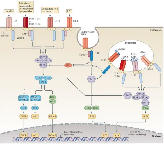
TLRs are type I integral membrane glycoproteins characterized by the extracellular domains containing several leucine-rich-repeats (LRR) motifs and a cytoplasmic signalling domain that is homologous to the interleukin 1 receptor (IL-1R), named the Toll/IL-1R homology (TIR) domain. Signalling pathways activated via the TIR domain lead to the activation of downstream kinases and transcriptions factors such as nuclear factor kappa B (NF-kB) and involve the TIR-domain containing Myeloid Differentiation Factor 88 (MyD88) adaptor protein [10]. TLRs dimerize and undergo conformational changes required for the recruitment of a downstream signalling molecule, either MyD88 upon interaction with the relevant PAMP, or TIR-domain-containing adaptor protein-inducing IFN-β (TRIF), leading to the activation of signalling pathways controlling the production of proinflammatory cytokines and type I IFNs, respectively (Fig. 1.1) [11] [9].
Thirteen members of the TLR family have been identified in mammals until now. Based on their primary sequences, TLRs have been divided into various subfamilies, each recognizing distinct PAMPs. While the subfamily of TLR1, TLR2 and TLR6 recognizes lipids, TLR7, TLR8 and TLR9 recognize nucleic acids [12]. However there are cases where some are able to recognize more than one structure, which is the case of TLR4 that recognizes
3
lipopolyssacharides (LPS), fibronectin and heat-shock proteins for example [13] [14] [15]. They can be expressed extra- or intracellularly and are present on various immune cells, including macrophages, B cells, DCs and can also be present in epithelial cells. The cellular localization of TLRs has important consequences for ligand accessibility and can also affect downstream signalling pathways. TLR1, 2, 4, 5 and 6, involved in the recognition of microbial membrane components, are located on the cell surface, and cellular TLRs recognizing microbial nucleic acids are mainly located within endolysosomal compartments (TLR3, 7, 8 and 9). TLR3 is the only one capable of recognizing viral dsRNA and poly (I:C), and it is the only TLR that does not use the MyD88 pathway [12] [16].
Figure 1.1: TLR Signalling Pathways (from O’Neill et al., 2013)
Endosomes contain pH-activated proteases such as cathepsins that have been documented to cleave TLR7 and TLR9 in their ectodomain [17]. It was also found that TLR3 was proteolytically processed into two fragments in endosomes, requiring the Loop 1 within the LRR12 of its ectodomain. Unc93b1, an endoplasmic reticulum-resident molecular chaperone,
4
is responsible for the translocation of TLRs 3, 7 and 9 to the endolysosomes where they are processed. The proteolytic processing of TLR3, done by cathepsins B and H, is essential for signalling and it is important for higher stability and modulation of the degree of response toward viral dsRNA [18] [19] [20]. Virus infected cells mainly depend on TLR3, TLR7/TLR8 and TLR9 to induce the expression of type I IFN, following detection of nucleic acids. However, TLR4 is also capable of inducing IFN type I by the recognition of non-nucleic acid ligands, such as LPS. TLR4 initiates signalling transduction pathways through both MyD88 and TRIF adaptors and induces both a late pro-inflammatory response (NF-kB dependent) and type I IFN expression (Fig. 1.1) [21] [22] [23].
1.1.2. Interferon System
1.1.2.1. Heterogeneity
The interferons (IFNs), a family of secreted cytokines that produce specific antiviral effects, are grouped into three classes called type I, II and III IFNS, according to their aminoacid sequence. Type I IFNs may be induced by essentially all nucleated cells and consist of a large family of molecules: mammals have several distinct IFN-α genes and up to three IFN-β genes. In man, there are thirteen IFNα and one IFNβ gene. There are other genes, such as IFNω, -ε, -τ, -δ and -κ [24]. IFN-α and -β genes are the principle cytokines induced in response to a viral infection, while the others possess a different kind of role. There is only one member that belongs to the type II IFN group, called IFN-γ, also known as “immune IFN”, and it is secreted by activated T cells and NK cells. Type III IFNs, recently described, comprise IFN-λ1, -λ2 and -λ3, also referred as IL-29, IL-28A and IL-28B respectively, and are also induced in response to viral infections but with a limited tissue distribution [25] [26] [27]. It is convenient to consider the IFN system in term of its induction, its subsequent impact on other cells through interaction of secreted IFN and cell surface expressed IFN receptors.
When a cell is infected by a virus, it is able to recognize the invading microorganism through PAMP recognition by PRRs and through multiple distinct routes induces the expression of type I IFN. The infected cell develops an antiviral state and, through the impact of secreted IFN, establishes a similar antiviral state in nearby cells.
5
1.1.2.2. Induction of IFN
Type I IFN expression can be induced by various stimuli. However, the downstream kinases and transcription factors are common to all. Sequences present in the 5’ flanking region of the IFN-α/β genes control the virus- or dsRNA-induced expression of type I IFN, requiring two families of transcriptional factors: NF-kB and interferon regulatory factors (IRF) [28]. Type I IFN transcription is regulated by IRF-1, IRF-3, IRF-5, IRF-7 and IRF-9, in concert with NF-kB (composed of p65 and p50 proteins) [29], but IRF-3 and IRF-7 are critical for the transcriptional activation of IFN-α/β genes and the IFN-β enhanceosome contains both IRF-3 and IRF-7 [30] [31] [32].
Prior to induction, IRF-3 and NF-kB are both cytoplasmic, NF-kB being associated with the inhibitor molecule IkB. Upon virus infection, the C-terminus of IRF-3 is phosphorylated by the IKK-related kinases, TANK-binding kinase (TBK)-1 and IKKε, which causes a conformational change leading to dimerization and translocation to the nucleus due to the unveiling of a nuclear-localization signal (NLS). Similarly, the signal generated upon viral infection causes phosphorylation of IkB and subsequent ubiquitination and degradation by proteasomes, thus revealing the NLS of the p65 subunit of NF-kB and its translocation to the nucleus. Both IRF-3 and p65 bind to the type I IFN promoter, allowing initiation of transcription and subsequent secretion of type I IFN. Like IRF-3, IRF-7 transcription factor is also expressed as an inactive monomer in the cytoplasm of cells, and after virus induction it is phosphorylated on C-terminal serine residues by the same kinases, TBK-1 and IKKε. Activated IRF-7 forms either homodimers with itself or heterodimers with IRF-3 and then translocates to the nucleus [25] [26].
When a host is invaded by pathogens, an early pro-inflammatory response is initiated and type II IFN is produced by activated CD4+ and CD8+ T cells after secretion of type I IFN by macrophages and NK cells. The majority of IFN-γ produced is therefore part of a secondary response and the main cytokines responsible for its induction are IL-12 and IL-18 [33]. Type III IFN, however, has functional similarities with type I IFN but instead of exerting antiviral activity on all cell types, it targets primarily epithelial cells, playing an important role in innate antiviral defence at epithelial surfaces. Type III IFNs are induced through independent actions of IRFs and NF-kB [34].
6
1.1.2.3. Signalling Responses to Interferon
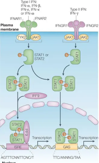
Type I IFNs (IFN-α/β), type II IFN (IFN-γ) and type III IFNs (IFN-λ) bind to distinct cell receptors but they can all activate a common intracellular signalling pathway, regulating many of the same biological activities, including the antiviral immune responses. The JAK-STAT pathway was the first signalling pathway shown to be activated by IFNs and extensive research revealed its relevance in the interferon system (Fig. 1.2) [25] [26] [27].
Figure 1.2: Interferon receptors and activation of classical JAK–STAT pathways by type I IFN and
7
1.2. Viral Mechanisms of Immune Evasion
As viruses are intracellular pathogens, they must enter the cell and take control of its machinery in order to replicate and then disseminate. In turn, the vertebrate host has evolved an elaborate system of innate and adaptive immune mechanisms in order to recognize and destroy pathogen-infected cells. Viruses, in response to the pressure of the immune system, have evolved multiple strategies for host evasion.
To avoid recognition, for example, the influenza virus, escapes humoral immunity through antigenic variation of the principal target of neutralising antibodies, the haemagglutinin protein [35]. Other viruses, like the Epstein-Barr virus (EBV), target the MHC class I and II biogenesis and transportation pathways, by blocking antigen presentation, in this way interfering with the activation of CD8+ and CD4+ T cells [36]. Lastly, some have also evolved strategies to subvert phagocytic activity, where they evade nitric oxide and reactive oxygen radicals generated by macrophages. Herpesviruses and poxvirus, for example, express surface proteins that mimic CD200, a host regulator that delivers inhibitory signals to macrophages [37].
1.2.1. Viral Evasion of IFN-Responses
The important role of IFN was dramatically demonstrated by the observation that mice deficient in IFN receptors were unable to control infection with a normally non-lethal virus [26]. Therefore, viral anti-IFN strategies are predictable.
Viral strategies against the IFN system are numerous and include the inhibition of IFN production, the inhibition of IFN-mediated signalling pathways, and blocking the action of IFN-induced enzymes with antiviral activity [38].
The strategy exploited by the virus depends on the biology of the infection and will be the main factor that will influence the pathogenesis of that virus infection [26].
In order to avoid recognition by PRRs, viruses usually manipulate production of PAMPs to achieve a minimum level of expression, hence minimizing the production of IFN in response to the infection. Minimizing the production of dsRNA through regulation of virus transcription and replication, or even capping viral RNA, making it indistinguishable from cellular mRNA, are some of the strategies used to achieve less IFN production [26].
8
IFN-antagonists have been identified in nearly 50% of the viruses, and are known to interfere with multiple steps of the IFN response. Approximately one third of the viruses encode an antagonist that interferes with more than one step of the IFN response system, underpinning the pleiotropic nature of viral proteins. Most of the IFN antagonists exert their action by (1) signalling cascade inhibiting cellular gene expression, (2) inhibiting post-translational modifications such as phosphorylation or ubiquitinylation, (3) sequestering molecules in the IFN pathway, (4) targeting these components for proteasomal degradation, (5) binding directly to components of the TLR and RLR signalling pathways [39] [40]. Thus blocking IFN production and suppressing host antiviral signal propagation [41].
Interferon-mediated signalling also stimulates antigen presentation through increased MHC expression and that is why viruses have also evolved strategies to inhibit the signal transduction pathways triggered upon binding of IFN to its specific receptor. Type I and type II IFN signal through distinct receptors, activating downstream components that can be unique or common to both signalling pathways. Thus, viruses can block the impact of IFN at several levels, inhibiting only one of these two pathways or both. Modulation of STAT activity is a very common viral strategy. According to the strain of the virus, IFN antagonists known as C and V encoded by members of the paramyxovirus, act by binding to STAT proteins inducing their degradation, or by inhibiting the Jak kinases [38].
Viruses can also inhibit or prevent the activation of the IFN-inducible antiviral effector protein PKR. The active PKR dimers phosphorylate eIF-2α, preventing the formation of the ternary translation complex, therefore repressing translation of RNAs [42]. Some viruses express RNA-binding proteins that sequester viral dsRNA, thus preventing PKR activation [43]. Another strategy is to encode small RNAs that compete with dsRNA for binding to PKR, thus inhibiting its activation [38].
1.3. African Swine Fever Virus
The African swine fever (ASF) is a lethal, highly contagious haemorrhagic disease that affects domestic pigs. This has socio-economic implications in which the livelihoods of pig keepers are affected in endemic countries of Africa. Due to the absence of a vaccine, control of this disease implies slaughter and quarantine of the pigs.
In marked contrast, ASFV causes a persistent but asymptomatic infection in its natural wild life hosts, namely bushpigs (Potamochoerus porcus), warthogs (Phacochoerus aethiopicus)
9
and soft ticks of the Ornithodorous spp. The tick remains infected for long periods of time and is able to transmit it to both wild and domestic animals [44]. There is a sylvatic cycle in which the neonatal warthog is infected by the soft tick in burrows, followed by a short period of viraemia, allowing the transmission of the virus to naïve ticks during blood meals. The Ornithodorous spp ticks have been found to infest the pig pens at first in African countries, and thus allowed the establishment of a domestic cycle of viral circulation, in which the virus is transmitted by direct contact and by fomites [45] [46].
First described in Kenya in 1921 as a result of the contact between domestic pigs and infected warthogs, ASF has been reported in south of the Sahara countries [47]. It appeared in Lisbon, Portugal, in 1957 through imported infected pork products [48] and again in Lisbon in 1960, leading it to spread to several countries in Europe remaining endemic in Portugal and Spain until mid-1990s. The ASF was eradicated from all European countries except for the Italian island of Sardinia since its introduction in 1982. There was an expansion of ASF in West African countries and Madagascar in more recent years [45]. It was also detected in Georgia, Armenia, Azerbaijan, and Russia, reaching as far as St. Petersburg [49], thus constituting a threat to the rest of Europe.
For example, countries bordering the Russian Federation, such as Finland, Estonia and Latvia are at risk since the disease appeared to have established itself in domestic and wild boar populations, making it important to monitor the region and have contingency plans. The risk of the ASF to spread further into Europe is dependent on the existence of wild boar population and its density but also on the characteristics of pig farms with low biosecurity. The commercial trade with African countries also imposes a bigger risk for the disease to spread in Europe and should be taken into account [50] [51].
1.3.1. Virus Structure and Genome Organization
ASFV is a large (≈200 nm) icosahedral virus that contains a linear double stranded ~200 kb DNA genome and is the only member of the genus Asfivirus in the family Asfarviridae [52] [53].
The virus replicates predominantly in the cytoplasm of porcine macrophages [54], although there may be an initial nuclear stage where small fragments of viral DNA are synthesized and are the precursors of larger cytoplasmic sequences. In addition, it was also detected dimeric concatemers which are head-to-head linked [55] [56]. The main morphogenesis occurs in viral
10
factories, located close to the cell nucleus and the microtubule organizing center. The viral DNA is packaged in the assembled capsid and core shell, exiting the host cell by budding from the plasma membrane [53].
The viral genome varies in length from 170 to 190 kbps, encoding between 151 and 167 open reading frames, due to sequence deletions and additions at both genome ends, and varies between different virus isolates [57]. These ends are formed by inverted terminal repeats and hairpin loops, composed by incompletely paired A and T residues [58].
Around 110 open reading frames are present as a single copy in the genomes of all isolates. The other ORFs belong to six different multigene families (MGFs) located close to the DNA ends, namely MGF 100, 110, 300, 360 and 505/530, and PZZ families, named after the average number of codons present in each gene [59]. Studies have shown that the N-terminal regions of members of MGFs 300, 360 and 505/530 have many similarities between them and genes in MGF 360 and 505/530 determine the host range and virulence [60] [61]. The genes, on the other hand, were named based on EcoRI restriction enzyme fragmentation, gene orientation left or right (L or R) and number of amino acids encoded [59].
1.3.2. Pathogenesis and Modulation of Host Immune Responses
The pathogenesis of ASF ranges from rapidly lethal to very attenuated and chronic disease depending on the nature of the isolate [62]. Although macrophages are the main target of ASFV, endothelial cells may also be infected late in the infection thus contributing to the haemorrhagic pathogenesis [63]. While in the domestic pig there is an uncontrolled lymphocyte apoptosis along with an impaired immune response and death in a matter of a few days [64], in the natural wild life host, the bush pig, the pathological damage and apoptosis in lymphoid tissue are controlled in order to ensure the hosts survival.
The immune response to ASFV infection is highly complex and virus elimination probably requires both humoral and cellular immunity. Cell mediated immune response studies revealed a positive correlation between the stimulation of NK activity and the absence of clinical symptoms, suggesting that NK cells play an important role in this model of protective
immunity [65]. In addition, CD8+T cell mediated immunity also plays a role in protection, as
an established immune response of pigs was abrogated by blocking CD8+T cells in vivo with
anti-CD8 monoclonal antibody [66]. Both IFN-α and IFN-γ were also shown to reduce virus replication in swine monocytes and macrophages [67], and the cooperative action of both was
11
able to cure lytically and persistently infected cell, which suggested an IFN response for protection. Furthermore, the report of virus replication of ASFV in IFN-treated cells suggested the possibility of ASFV subverting IFN response [68].
Large DNA viruses have many proteins involved in the evasion of host immune responses, ASFV contains approximately 90 proteins predicted to be involved in the virus replication, and several others have been identified as host evasion genes [69], including A238L, an inhibitor of NF-kB [70] and nuclear factor of activated T cells (NFAT) [71], the apoptosis inhibitors A179L and A224L [72] [73], the protein phosphatase 1 activator DP71L [74], and genes inducing apoptosis [75] [76].
In addition and as previously mentioned, ASFV has also evolved multiple mechanisms for the inhibition of IFN responses. The MGF360 and MGF530 protein family are involved on the impact on IFNα/β production [61]. Namely, the MGF360-18R protein has been found to interfere with the Jak-STAT signalling pathway, inhibiting both type I and type II IFN signalling cascades, by inducing STAT1 ubiquitination and subsequent degradation by the 26S proteasome [77]. Another host evasion gene, the I329L ORF, which inhibits the induction of type I IFN by activation of the TLR3 pathway [78], is the focus of this project.
1.3.3. The I329L protein
The ASFV I329L protein recently described as an inhibitor of the TLR3 signalling pathway, evolved to diminish the corresponding IFN and proinflammatory response, thus enhancing virus proliferation and propagation [79]. This gene was identified through the combination of bioinformatic analysis and functional assays. Consistent with this, the functional assays revealed that this protein inhibits TLR3-mediated NF-kB activation and induction of IFN-β by subsequent activation of TLR3 with poly (I:C). Its deletion provided a rational approach for the development of an attenuated virus vaccine.
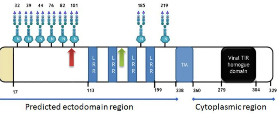
The ASFV I329L protein contains a signal peptide (positions 1-17 amino-acids) and the final chain covers a length of 312 residues (18-329 amino-acids). An extracellular N terminal domain is predicted to have 222 residues (18-239 amino-acids), while 69 residues (261-329 amino-acids) are predicted to localize in the carboxyterminal cytoplasmic domain. The 240-260 amino-acid region of 21 residues is a potential transmembrane region (Fig. 1.3).
Interestingly, the analysis of the sequence reveals that there are three potential AUG/methionine start sites in the extracellular domain (ECD) situated on amino acids 1, 99
12
and 149 (Figure ). The translation from the second AUG would yield a protein containing all four LRRs in the ECD and thus presumably capable of protein-protein interaction. The question that arises, and which is addressed in this work, is whether the translation from the second AUG would yield a protein still inhibiting TLR3 activation. Translation from the third AUG, however, would essentially yield a protein with a considerably shorter ECD containing only two LRRs.
In bioinformatics analysis, the I329L sequence shared some sequence similarities with TLR3. Both are type I transmembrane proteins with 20 % of intracellular sequence and 70 % highly glycosylated ECD composed by Leucine-Rich Repeats (LRR), and further analysis showed homology between I329L and the Box1 and Box2 of the TL3-TIR domain. I329L has been shown to inhibit TRIF-mediated NF-kB activation, suggesting that it acts at the level of TRIF in the signal transduction pathway [79].
As mentioned above (section 1.2.), TLR3, TLR7 and TLR9 are proteolytically cleaved after interaction with the relevant ligand, and this is essential for full biological activity. A pertinent question, and one that is addressed in this work, is whether the I329L TLR3 antagonist is proteolytically processed as part of its inhibitory action.
The ECD of TLR3 is involved in the recognition of the pathogen ligand. Modelling of the I329L protein showed a detectable, very low, homology between the ECD and ICD of the viral protein with TLR3 ECD and ICD. This supports the hypothesis that there might be formation of non-signalling TLR3-I329L heterodimers, leading to inhibition of the downstream signalling pathway [80].
I329L has been therefore characterized as a viral TLR3 antagonist, affecting negatively the type I IFN antivirus response of the host.
Figure 1.3: The I329L protein structure. The second AUG is located at position 99 (red arrow) and the
13
1.4. Aims of the Project
In order to survive, ASFV, as an acute and persistent virus of pigs, must have evolved genes for evasion and manipulation of interferon. The Toll-like receptors (TLRs) are particularly important in the induction of innate immunity, IFN in particular. The fact that the virus is adapted to survive in both vertebrate and invertebrate hosts, and only innate immunity, in particular TLR responses, is common to both, led us to search for a TLR agonist in the ASFV genome.
Although the homology search performed at the level of the entire genome to identify a possible ASFV TLR homologue was disappointing, the ASFV I329L ORF was predicted to be a type I transmembrane protein containing four leucine-rich repeats (LRR’s) in its extracellular domain (ECD), and with a detectable, but very low, homology in the intracellular domain (ICD) with Box1 and Box2 of the TLR3 intracellular TIR domain.
This structural homology correlated with luciferase reporter assays which clearly demonstrated that I329L inhibits TLR3-mediated induction of IFN-β and activation of NF-κB [79]. A more sophisticated modeling exercise on the I329L viral protein provided a molecular basis for subversion of TLR3 signaling by I329L, with both intracellular and extracellular domains potentially capable of contributing, as indeed is supported by our own investigation [80]. Thus the ECD inhibits the dsRNA stimulated activation via the ECD of the TLR, whereas the ICD interferes with the intracellular signalling cascade by targeting the downstream signalling molecule TRIF (Ventura S., unpublished work).
The fact that TLR3 needs to be proteolytically processed for optimal downstream signalling, raises the possibility that I329L is also processed by a similar mechanism as TLR3. Perhaps relevant for this, we observed two protein components in cells expressing I329L; one with the predicted molecular weight of ~50 kDa and the other ~35 kDa. An alternative possibility for this heterogeneity is suggested by the second methionine codon in the I329L ECD, which, if used by the virus to initiate transcription, would yield a protein fragment of 35 kDa.
The principal aims of this project, therefore, are to determine (1) whether an I329L protein synthesized from the second methionine encoding ATG sequence also inhibits TLR3 activation, and (2) whether I329L, like the TLR3, 7 and 9, is proteolytically processed following dsRNA activation.
14
2. Materials and Methods
2.1. Cell Lines
Vero cell line was cultured in Dulbecco’s modified Eagle’s medium (DMEM) supplemented with fetal bovine serum at 10 % (v/v) (FBS) (Gibco), 100 U/ml penicillin G sodium / 100 µg/mL streptomycin sulfate (Gibco) and 2 mM L-Glutamine (Gibco).
2.2. Cloning “Short” Length, Full Length I329L and Respective
Extracellular Domains
The I329L short length fragment (nucleotide 294 – 990) (Fig. 1.3) was obtained by PCR amplification of the pcDNA3-I329L-HA plasmid containing the I329L full length insert by
using the Pfu DNA polymerase (Fermentas). The primers used were
5’-GCGAATTCATGAAAAATATTTCTG-3’ and 5’-GCCTCGAGTTAAGCGTAATCTG-3’ which incorporated the restriction sites for EcoRI (upstream) and XhoI (downstream) respectively. The PCR conditions started with 5 minutes at 94 ºC for denaturation in the initial cycle, followed by a pause of 1 minute. Annealing required 44 ºC for 1 minute, and extension required 72 ºC for 1 minute and 30 seconds and 10 minutes on the last cycle. The total number of cycles between the phases was 30. The amplified product was then visualised on a 1.5 % agarose gel with ethidium bromide (EtBr) and the DNA extracted from the excised band. The fragment already containing the HA tag at the C’ terminal end was cloned in pcDNA3 plasmid using DH5α competent cells and both the HA peptide tag of the fragment and the EcoRI sequence previous to the second start codon of the gene were confirmed by automated sequencing. For the N-terminal Myc-tag (derived from the c-myc gene) insertion it was necessary to digest the pcDNA3-Myc plasmid and the pcDNA3 containing the I329L-HA fragment starting at the second ATG with EcoRI and XhoI enzymes. The fragment and pcDNA3-Myc were visualized with EtBr on 1 % agarose gel and DNA purified from the excised band. After purification, alkaline phosphatase was added to the constructed DNA fragment, incubated at 37 ºC and inactivated at 72 ºC. The fragment was ligated into pcDNA3-Myc using T4 ligase in a 1:1 and 1:3 ratio at RT and DH5α competent cells were then transformed.
Transformation of DH5α consisted in adding the construct and let stand for 30 minutes on ice, heat shock at 42 ºC for 45 seconds and 2 minutes again on ice. After adding 100 µl S.O.C
15
medium, cells were incubated for 1 hour at 37 ºC with agitation and then plated in plates with ampicillin.
The complete I329L fragment (nucleotide 1 – 990) (Fig. 1.3) (already containing the HA tag) was obtained by PCR amplification using the Pfu DNA polymerase (Fermentas) with the same conditions as the necessary to amplify the short I329L using the same restriction sites incorporated in the primers. They were GCGAATTCATGCTAAGGGTTTTC-3’ and 5’-GCCTCGAGTTAAGCGTAATCTG-3’. The amplified product was also visualized in 1 % agarose gel with EtBr and the DNA purified from the excised band. Both pcDNA3-Myc and PCR products were digested with EcoRI and XhoI and cloning proceeded as previously described for the short I329L construct.
The extracellular domains of the complete I329L fragment (nucleotide 1 – 780) and “short” I329L fragment (nucleotide 294 – 780) were obtained by PCR amplification using the Pfu DNA polymerase with the same conditions as the necessary to obtain the previously amplified products and using the EcoRI (upstream) and EcoRV (downstream) restriction sites incorporating primers. Primers were GCGAATTCATGCTAAGGGTTTTC-3’ and
5’-GCGATATCCAACAAGCATATGAA-3’ for the “long” ECD and
5’-GCGAATTCATGAAAAATATTTCTG-3’ together with
5’-GCGATATCCAACAAGCATATGAA-3’ for the “short” ECD. Amplified products were visualized in 1.5 % agarose gel with EtBr and DNA purified from the excised band. Both pcDNA3-HA and PCR products were digested with EcoRI and EcoRV and cloning proceeded as described above.
2.3. Luciferase Reporter Gene Assays
Vero cells (6x10⁴ cells/well, in a 24-well plate) were transfected with test plasmid pcDNA3
negative control (300 ng), or pcDNA3-full length I329L (300 ng), or pcDNA3-ECD full length I329L (300 ng), or pcDNA3-“short” I329L (300 ng) or pcDNA3-ECD “short” length I329L, IFN-β reporter luc (100 ng) or PRD2 reporter luc (100 ng), and β-galactosidase (25 ng) using Lipofectamine 2000 reagent (2 µl/50 µl Opti-MEM) (Invitrogen). After 72 hours cells were stimulated with Poly (I:C) (Amersham Biosciences) at a final concentration of 35 µg/mL for five hours or left untreated. Cells were then lysed with a lysis buffer (1 mM DTT, 2 mM EDTA, 1 % Triton, 0.1 M Potassium phosphate buffer pH 7.8). The luciferase activity was measured by using the luciferase assay system (Promega) and the internal control β-galactosidase activity was measured with the Galacton-Plus kit from Tropic (Bedford, MA),
16
both according to the manufacturer’s protocol. The luciferase activity was normalized relatively to the β-galactosidase activity of each sample in order to correct the transfection efficiency for variations between different cells.
2.4. Immunofluorescence
Vero cells were seeded onto coverslips previously prepared with polylysine (2x10⁵ cells/well,
in a 6-well plate) and incubated overnight at 37 ºC and 5 % CO2. Cells were transfected with
either pcDNA3 empty plasmid (4 µg) or Myc-I329L-HA expression plasmid (short or long) (4 µg), TLR3 expression plasmid (700 ng) and Xtreme 9 transfection reagent (15 µl) (Roche). After 48 hour transfection cells were either stimulated for 15 min and 30 min with 100 µg/ml Poly (I:C) (Amersham Biosciences) or left untreated. Medium was removed and cells were washed with PBS and fixed with Paraformaldehyde (PFA) at 4 % (w/v) for 20 min at RT. After the PBS wash, cells were permeabilized with PBS containing Triton X-100 at 0.1 % (v/v) for 20 min at RT then washed with PBS containing 0.05 % Tween-20 (v/v). Cells were blocked with the PBS-Tween containing 5 % normal goat serum for 30 min at RT. In order to detect the I329L-HA tagged, it was used a monoclonal mouse anti-HA-High Affinity (Roche) diluted in PBS-Tween-20 (blocking solution) at a previously determined optimal concentration 1:100 for 1h at RT, followed by a polyclonal goat anti-mouse Ig antibody Texas Red-conjugated (1:100) (Molecular Probes) 1 h at RT in the dark.
For detection of the early endosomes, cells were incubated 1 h RT with a polyclonal rabbit anti-EEA1 (Sigma), diluting the commercial product 1:50 in PBS-Tween-20 (blocking solution), followed by a polyclonal goat anti-rabbit 488 IgG at 1:500 dilution of the commercial product (Alexa) for 1 h at RT in the dark.
All washes after incubation with antibody were with PBS + 0.05 % Tween. Cells with no anti-rabbit 488 secondary antibody were stained with DAPI (200 ng/mL) for 1 min. All were mounted with Slow Fade (Molecular Probes) and observed under a fluorescence microscope. The fluorescent images were taken with a Leica DMRA2 microscope and pictures were analyzed with ImageJ 1.47u software.
2.5. Western Blot
Vero cells (2.5x10⁵ cells/well, in 6-well plates) were transfected with either pcDNA3 empty
plasmid or Myc-I329L-HA expression plasmid (short or long) (4 µg) and TLR3 expression plasmid (700 ng) using Xtreme Gene 9 (15 µl) (Roche), and TRIF expressing plasmid (250
17
ng) according to the Xtreme Gene 9 protocol. Forty-eight hours post-transfection cells were either stimulated with Poly (I:C) (100 µg/mL) (Amersham Biosciences) for 30 min, or left untreated. Cells were then harvested and lysed with lysis buffer (15 mM Tris-HCl, 120 mM NaCl, 25 mM KCl, 2 mM EGTA, 2 mM EDTA, 0.1 mM DTT and 1 % Triton X-100) containing a protease inhibitor cocktail (Roche). The lysate samples were resolved by sodium dodecyl sulphate-polyacrylamide gel electrophoresis (SDS-PAGE) and the proteins were transferred to a polyvinylidene difluoride (PVDF) membrane (Bio-Rad). Membranes were blocked with TBS + 0.05 % Tween-20 with 5 % non-fat milk for 1 h RT and probed with titrated dilutions of Rat anti-HA-horseradish peroxidase (HRP) conjugated (high affinity) or Rabbit-anti-Myc (Santa Cruz) which was followed by the titrated dilution of a HRP-conjugated goat anti-rabbit Ig antibody as secondary antibody, and a titrated dilution of Rat-anti-β-actin-HRP conjugated was included as loading control. Membranes were developed by chemiluminescence detection according to the manufacturer’s protocol with either Luminata Forte Western HRP Substrate (Millipore) or Pierce ECL western blotting substrate (Thermo Scientific).
2.6. Site-Directed Mutagenesis
With the purpose of obtaining a mutation in the second ATG, Phusion DNA polymerase (Fermentas) was used with the following forward and reverse primers at 10 µM: 5’-CGCTGCGGGGCGAAAAATATTTCTG-3’, 5’-CAGAAATATTTTTCGCCCCGCAGCG-3’. The PCR conditions were 30 sec at 98 ºC for the initial denaturation cycle, followed by 30 cycle repetition consisted of denaturating the samples at 98 ºC for 10 sec, annealing at 57 ºC for 30 seconds, and extension at 72 ºC for 15 sec. The last cycle finished with 10 min at 72 ºC. Dpn1 enzyme was then added to the reaction and left for a 1 hour incubation at 37 ºC. Inactivation required the sample to be at 80 ºC for 10 min. DH5α competent cells were transformed as previously described, DNA was extracted and mutation was confirmed by automated sequencing.
2.7. Statistical Analysis
Differences between experimental groups were determined by a two-tailed Student t test, using the Microsoft Excel software.
18
3. Results
3.1. The I329L protein is translated from both the first and second AUG
codons
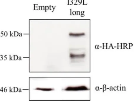
The full length I329L protein when expressed in transfected cells has two distinct molecular components (Fig. 3.1). These two components could be explained by transcription of two mRNA’s, one, the full length ~50 kDa component resulting from translation of the first AUG, and the second, corresponding to the ~35 kDa entity, resulting from translation from the second AUG (Fig. 1.3). Alternatively, since I329L has been described as a TLR3 analogue [79] it may be proteolytically processed, as is TLR3.
To address the first possibility, the I329L was mutated using directed mutagenesis of the second AUG codon, turning into a CUG (section 2.6). Vero cells were transfected with the
I329L-2nd AUG mutant together with TLR3 and stimulated with poly (I:C) for 30 minutes. As
positive and negative controls, Vero cells were transfected with the full length I329L and pcDNA3 respectively (section 2.5).
As it can be seen, the 35 kDa band, present in the cells transfected with the full length I329L, is absent in the mutant (Fig. 3.2). Thus it appears that I329L may be translated from the first and second AUG codons, to give two different products.
Figure 3.1: Expression of full length I329L protein. Western blot analysis of pcDNA3 (Empty) and
Myc-I329L-Ha transfected Vero cell lysates. Cells were transfected with 4 µg of the respective plasmid and 700 ng TLR3 in 6 well plates. Cells were lysed and total cell extracts were immunoblotted with anti-HA-HRP conjugated antibody. Anti-β-actin was used as loading control.
19
Figure 3.2: Mutant I329L expression. Western blot analysis of pcDNA3 (Empty), mutant
I329L-HA, and full length Myc-I329L-HA transfected Vero cell lysates. Cells were transfected with 4 µg of the respective plasmid and 700 ng TLR3 in 6 well plates followed by 30 min 100 µg/ml poly (I:C) stimulation after 48 hours. Cells were lysed and total cell extracts were immunoblotted with anti-HA-HRP conjugated antibody. Anti-β-actin was used as loading control.
20
3.2. I329L translated from both the first and second AUG codons inhibit
TLR3
activation
The full length I329L protein has been previously demonstrated to inhibit poly (I:C) and TRIF-mediated activation of NF-kB and IFN-β induction through a Myd88 independent pathway [79].
In order to test for a similar function of an I329L molecule translated by the second AUG sequence, this was cloned (section 2.2) and compared with full length I329L for inhibition of IFN-β expression in a luciferase assay (section 2.3). Thus Vero cells were transfected with an empty plasmid or the I329L expressing plasmid together with either an IFN-β or PRD2 (domain containing only the NF-kB binding site of the IFN-β promoter) luciferase reporter plasmid and stimulated with poly (I:C). Activation of the IFN-β promoter reporter plasmid was also induced via ectopic expression of TRIF, by transfecting the TRIF plasmid vector. As it can be seen, the “short” length (second AUG) I329L was able to inhibit both IFN-β and NF-kB pathways upon poly (I:C) stimulation (Fig. 3.3 A, B) and TRIF over expression (Fig. 3.3 C), as does the full length protein (Fig. 3.3).
In cells expressing “short” I329L and stimulated with poly (I:C), the inhibition of the luciferase activity might be due to the intracellular domain of the protein and without any contribution from the smaller extracellular domain (ECD). Therefore, the ECD of the “short” length protein, the “short” length ECD was cloned and tested in IFN-β luciferase reporter assays, stimulating with poly (I:C). As it can be observed, the extracellular domain of the “short” length I329L also inhibits the IFN-β induction pathway upon poly (I:C) stimulation (Fig. 3.4).
It may therefore be concluded that I329L translated from the second AUG sequence is fully functional, being capable of inhibiting TLR3 activation, the ECD through interference with poly (I:C) stimulation and the ICD through its negative impact on TRIF mediated signalling. Although these results were reproducible, it was necessary to confirm that the inhibition of the luciferase assays was indeed correlated with the expression of the recombinant proteins, and this was indeed confirmed by identification of the “short” length I329L by fluorescence antibody staining of transfected cells (section 3.3).
21
-Figure 3.3: “short” length I329L inhibits the activation of IFN-β (A) and PRD2 (B) promoter.
Vero cells were transfected with 300 ng pcDNA3-I329L plasmids (long or short), 100 ng of IFN-β (A) or PRD2 (B) reporter plasmid, and 25 ng of β-gal reporter plasmid (black bars) and incubated for 72 hours prior to reporter assays. The negative control was the plasmid vector without insert (pcDNA3) (white bars). Cells were stimulated with poly (I:C) at a concentration of 35 µg/µl. C) “short length
I329L inhibits TRIF mediated activation of IFN-β. In an alternative protocol, Vero cells were
transfected such as (A), but with 15 ng TRIF plasmid vector and without poly (I:C) (black bars). Luciferase activity was normalized to β-galactosidase activity as a control for transfection efficiency. Data are expressed as means of Relative Luciferase Units (RLU) and standard deviations are shown by error bars. Statistical significance is represented as p≤0.05 (*)
* * 0 5 10 15 20 25 30 35 40 45
empty vector I329L long I329L short
RLU (IF N -β ac tivat ion ) Medium Poly (I:C) * * 0 10 20 30 40 50
empty vector I329L long I329L short
RLU (P RD 2 ac vivation ) Medium Poly (I:C) * * 0 2 4 6 8 10 12
empty vector I329L long I329L short
RLU (IF N -β ac tivat ion ) Medium TRIF A B C
22
Figure 3.4: Full length (A) and “short” length (B) I329L extracellular domains inhibit the activation of IFN-β promoter. Vero cells were transfected with 300 ng pcDNA3-I329L plasmids
(long or short or respective ECDs), 100 ng of IFN-β reporter plasmid, and 25 ng of β-gal reporter plasmid (black bars) and incubated for 72 hours prior to reporter assays. The negative control was the plasmid vector without insert (pcDNA3) (white bars). Cells were stimulated with poly (I:C) at a concentration of 35 µg/µl. Luciferase activity was normalized to β-galactosidase activity as a control for transfection efficiency. Data are expressed as means of Relative Luciferase Units (RLU) and standard deviations are shown by error bars. Statistical significance is represented as p≤0.05 (*)
0 5 10 15 20 25 30 35 40 45 50
empty vector I329L short ECD short
RLU (IF N -β ac tivat ion ) Medium Poly (I:C) 0 10 20 30 40 50 60 70 80 90 100
empty vector I329L long ECD long
RLU (IF N -β ac tivat ion ) Medium Poly (I:C) * A B
23
3.3. Both full length and “short” length I329L colocalize to the early
endosome
Since TLR3 localizes to endosomes, it is relevant to determine the intracellular localization of I329L after stimulation with poly (I:C). For this study, we took advantage of the N’ terminal Myc - entire I329L - C’ terminal HA construct. In addition, we similarly investigated an N’
terminal Myc – 2nd AUG I329L – C’ terminal HA construct (section 2.3).
Vero cells were seeded onto coverslips and transfected with empty pcDNA3 vector plasmid or containing I329L (long or short) and TLR3. They were stimulated with poly (I:C) for 30 minutes or left untreated. For simultaneous detection of the I329L protein and early endosomes, cells were stained for I329L with HA and for early endosomes with anti-EEA1 antibodies (Fig. 3.5) (section 2.6).
It can be observed using the HA-tag antibody that both the full length and “short” I329L species colocalize to the early endosome, and that the expression of both increase following poly (I:C) stimulation.
Unfortunately the Myc antibody failed to give a signal due to high background as might have been predicted from the Western Blot results (see 3.4. below).
Thus both the full length and “short” I329L species follow similar pathways of intracellular localization. These results also demonstrate that both full length and “short” length I329L sequences are expressed as protein in transfected cells and therefore further justify the conclusions from the luciferase assays.
24
Figure 3.5: I329L colocalizes to the early endosome. Immunofluorescence analysis of pcDNA3
(Empty), full and short length Myc-I329L-HA transfected Vero cells. Vero cells were seeded onto coverslips and transfected with 4 µg empty pcDNA3 vector plasmid or containing Myc-I329L-HA (long or short) and 700 ng TLR3. After 48 hours, they were stimulated with 100 µg/ml poly (I:C) for 30 min. Coverslips were incubated with anti-EEA1 (green) and anti-HA antibodies (red). Third lane corresponds to DAPI staining and the last are EEA1-HA composite images, revealing the targets localization.
25
3.4. Possible proteolytic processing of I329L
To investigate the possible proteolysis of the I329L molecule after stimulation with poly (I:C), a recombinant I329L was constructed with both an N-terminal Myc “immunotag” and a C-terminal HA “immunotag” (section 2.3).
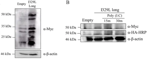
To address the question of the existence of proteolytic processing in the full length I329L, Vero cells were transfected with either an empty vector plasmid or plasmid containing Myc-I329L-HA (full length and “short”) and TLR3, and later stimulated with poly (I:C). The membrane in which samples were transferred was either developed with α-HA-HRP or α-Myc antibodies (section 2.5).
Processing, similar to that occurring with TLR3, would yield a smaller protein revealed by anti-HA, but not anti-Myc. Unfortunately development of the Western blot with the anti-Myc resulted in unacceptable levels of background (Fig. 3.6 A) and so the experiment was disappointingly inconclusive. However, when lysate of poly (I:C) stimulated cells from a time course experiment were Western blotted and developed with the anti-HA antibody, the 50 kDa I329L component had almost disappeared 30 minutes after poly (I:C) stimulation. Thus the possibility of intracellular proteolytic processing of I329L in poly (I:C) stimulated cells cannot be excluded, but has not been conclusively demonstrated (Fig. 3.6 B).
Figure 3.6: Full length I329L expression without (A) and upon poly (I:C) stimulation (B).
Western blot analysis of pcDNA3 (Empty) and Myc-I329L-Ha transfected Vero cell lysates. Cells were transfected with 4 µg of the respective plasmid and 700 ng TLR3 in 6 well plates followed by 15 min and 30 min 100 µg/ml poly (I:C) stimulation after 48 hours. Cells were lysed and total cell extracts were immunoblotted with anti-HA-HRP conjugated antibody or anti-Myc antibody to detect expression of Myc-I329L-HA tagged protein. Anti-β-actin was used as loading control. (A) I329L expression without stimulus. (B) I329L expression with poly (I:C) stimulation at different time points.
26
4. Discussion
The I329L ORF of the African swine fever virus is a host evasion gene coding for a transmembrane protein inhibiting the induction of interferon through activation of TLR3. This inhibition is achieved by two entirely distinct mechanisms; the extracellular domain inhibiting interaction of TLR3 with its corresponding dsRNA ligand and the intracellular domain impacting intracellular signal transduction through its association with the downstream signalling intermediate TRIF. Deletion of I329L from the virus is a rational approach for the construction of an attenuated virus vaccine; as such a recombinant virus might be expected to have a reduced advantage over the host. For licensing of such a vaccine it is essential to understand the mechanism of the deleted gene, which is the focus of this work.
Equally important, there is an enormous intellectual and industrial interest in inhibition of TLR activation as possible novel drugs for the modulation of cell biology and immune responses in health and in disease [81] [82] [83]. Thus understanding the mechanism used by I329L in the modulation of TLR signalling may have relevance to its further exploitation. This project focused on the observation that I329L was expressed in transfected cells as two distinct molecular entities, one of ~50 kDa, corresponding to the predicted molecular weight, and another unexpected component of ~35 kDa. Two hypotheses were proposed to explain expression of the 35 kDa component: (a) expression of I329L results from translation from the first AUG, to yield the 50 kDa component, whereas the ~35 kDa molecule is the product of translation initiating at the second AUG (see Fig. 1.3) and (b) the ~35 kDa component Is due to proteolytic processing of the ~50 kDa full length I329L protein.
To address the first hypothesis, we used the direct approach of changing the second AUG to CUG by site directed mutagenesis. We then compared the expression, function and intracellular localization of the wild type and mutated forms of I329L in transfected cells. The results of the expression were clear, whereas the wild type I329L yielded both ~50 kDa and ~35 kDa molecules, the construct mutated in the second AUG only yielded the ~50 kDa component. Thus we may conclude that the ~35 kDa protein results from translation of the I329L on its second AUG. The next step was to determine whether the ~35 kDa component also inhibited TLR3 activation.
Functional analysis was done by luciferase reporter assays directed on activation of IFN-β and NF-kB transcription factor after activation of TLR3 expressing cells transfected with dsRNA poly (I:C). The result was once again very clear, with both constructs exhibiting a similar