Abstract
Submitted: March 2, 2018 Modification: June 13, 2018 Accepted: July 13, 2018
Comparison of
in vitro
erosion
protocols in bovine teeth to simulate
natural erosion lesion: analysis of
mechanical properties and surface
gloss
Objective: The aim of this study was to compare two in vitro erosion
protocols, in which one simulates in vivo conditions experienced by patients with gastroesophageal disorders or bulimia (HCl-pepsin protocol), and the other simulates the diet of an individual who consumes a high volume of erosive beverages (citric acid protocol). In addition, the mechanical properties and surface gloss of eroded human dentin were compared with those of sound human dentin. Materials and Methods: Blocks of cervical dentin were used: sound human dentin (n=10), human dentin with erosive lesions (n=10), and bovine dentin (n=30). Twenty bovine blocks were subjected to either of
two erosion protocols (n=10/protocol). In the first protocol, samples were
demineralized using HCl-pepsin solution, then treated with trypsin solution. In the second protocol, samples were demineralized with 2% citric acid. Toothbrushing was performed in both protocols using a toothbrushing machine (15 s with a 150 g load). Ten bovine dentin blocks were not subjected to any erosive treatment. All samples of bovine and human dentin were analyzed to obtain Martens hardness values (MH), elastic modulus (Eit*) and surface gloss. One-way ANOVA and Tukey’s test were performed to analyze the
data (α=0.05). Results: Sound human and eroded human dentin groups
showed similar MH and Eit* values (p>0.05); however, sound human dentin showed a higher surface gloss value when compared to eroded human dentin (p<0.05). Sound bovine dentin and HCl-pepsin-treated bovine dentin treatments resulted in similar values for both MH and Eit* (p>0.05), but HCl-pepsin-treated bovine dentin and citric acid-treated bovine dentin resulted in lower surface gloss than sound bovine dentin (p<0.05). Conclusions: The
HCl-pepsin protocol modified bovine dentin properties that could be similar
to those that occur on human dentin surfaces with erosive lesions.
Keywords: Citric acid. Dentin. Tooth erosion. Pepsin A.
Mariana Dias MODA1
Ticiane Cestari FAGUNDES1
Eduardo BRESCIANI2
André Luiz Fraga BRISO1
Paulo Henrique DOS SANTOS3
1Univ. Estadual Paulista, Faculdade de Odontologia de Araçatuba, Departamento de Odontologia
Restauradora, Araçatuba, São Paulo, Brasil.
2Univ. Estadual Paulista, Instituto de Ciência e Tecnologia, Departamento de Odontologia
Restauradora, São José dos Campos, São Paulo, Brasil.
3Univ. Estadual Paulista, Faculdade de Odontologia de Araçatuba, Departamento de Materiais Odontológicos e Prótese, Araçatuba, São Paulo, Brasil.
Introduction
The occurrence of non-carious cervical lesions (NCCL) is a very common clinical situation, resulting
from multifactorial etiology.1,2,3 Establishing
differentiation among NCCL types is difficult because
most cases involve the association of abfraction, attrition, erosion, and abrasion.4,5 Acids from
gastroesophageal disorders, foods and drinks play key
roles in the development of erosive lesions, which may
cause irreversible loss of dental tissue.1,2,6,7
Given this context, dentin is a complex tissue which
undergoes constant change due to the network of
tubules that extend from the pulp to the dentinoenamel
junction; thus, being closely related to the pulp.8,9 Dentin hypersensitivity is common in cases of erosion.1
The erosion process starts in the peritubular dentin,
which shows a greater degree of mineralization and
involves dentinal tubules.10 Following, demineralization of hydroxyapatite crystals occurs in the intertubular
dentin, thus exposing the collagen fibrils of the organic
matrix.10 In addition, the erosive process can involve
metalloproteinases, natural constituents of dentin that
function in the degradation of collagen fibers, which
may accentuate the erosion process by degrading
the collagen matrix.11 Some modifications occur in
eroded dentin areas, such as changes in mechanical properties12 and brightness.13
Due to the high incidence of in vivo dental erosion,
there is a great need for protocols and studies that
accurately reproduce the erosive process in vitro, to facilitate the better understanding of dentin tissue.4,14
However, simulating all complex conditions that occur
in the oral environment is very difficult4. In addition, the use of enzymes or acids to cause in vitro erosion requires optimization to ensure that the outcomes
have similar characteristics to in vivo lesions4. Some
prior studies have used gastrointestinal tract enzymes
to simulate gastroesophageal disorders.6,7 Another
in vitro protocol involves the use of citric acid, a key
ingredient in some beverages, which are associated
with tooth erosion.15,16
However, the literature lacks in vitro protocols that accurately simulate clinical conditions of erosive tooth
wear. Therefore, the objective of this study was to
compare two in vitro erosion protocols, in which one
simulates in vivo conditions experienced by bulimia patients (HCl-pepsin protocol), and the other simulates
the diet of an individual who consumes high volumes of
erosive beverages (citric acid protocol); these protocols
reproduce alterations of human dentin subjected to natural erosive lesions. The second objective was to
compare the mechanical properties and surface gloss
of eroded human dentin and sound human dentin. Two
null hypotheses were tested: (1) there would be no differences in mechanical properties and surface gloss
between sound and eroded human dentin; (2) there
would be no differences in mechanical properties and
surface gloss of bovine dentin after exposure to two
in vitro erosion protocols when compared to sound
bovine dentin.
Materials and methods
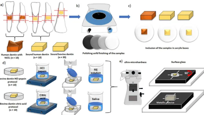
Sample preparation
Twenty human pre-molars and lower incisors
[sound human dentin (n=10) and human dentin with
erosive lesions (n=10)] were collected from patients at a private clinic; the clinician who performed the
surgical procedures provided information regarding
the type of lesion in each case. Moreover, the clinician
was able to detect these lesions through anamnesis, in which the patients reported their eating habits
(ingestion of acidic beverages) or gastroesophageal
disorders. All patients provided written informed
consent to donate their teeth for the study. The research project was approved by the local Human
Research Ethics Committee, #32545114100005420.
Additionally, 30 bovine teeth were used in this study.
All teeth were cleaned and stored in 0.1% thymol solution. The animal portion of this research project
was approved by the local Animal Research Ethics
Committee, #2016-00150.
Blocks of cervical dentin were obtained by using a slow-speed diamond saw (Isomet 2000, Buehler,
Aurora, OH, USA). The blocks (4x4 mm) of sound
human and bovine dentin were obtained by cutting into
the dentin-enamel junction and extending up to 4 mm of the root dentin. Blocks from the erosive lesion group
were restricted to the lesion area. All dentin blocks
were mounted on acrylic bases and polished (Arotec
APL 4, Cotia, SP, Brazil) with aluminum oxide abrasive papers (600-, 800- and 1200-grit). Final polishing
was performed with felt discs embedded in a 1-μm diamond polishing suspension (Extec Corp., Enfield,
Campo Mourão, PR, Brazil) in distilled water for 10
min. For eroded human dentin, after being mounted on the acrylic bases, only the edges were removed
during the flattening process. The central features of
these lesions remained unchanged, preserving the
tissue characteristics for the analysis.
All blocks of human dentin (sound or eroded)
and 10 blocks of bovine dentin were stored at 100%
relative humidity for 2 weeks in an incubator (ECB-2.
Adamo Products for Laboratory Ltda., Piracicaba, SP, Brazil) at 37°C. The other 20 bovine dentin blocks were
treated with the erosion protocols, as described below.
All samples had their Martens hardness (MH) values,
elastic modulus (Eit*), and surface gloss determined.
Erosion protocols
Twenty bovine blocks were treated with either of
two erosion protocols (n=10/protocol):
HCl-pepsin protocol:7 10 bovine specimens were
cyclically demineralized over 9 days. HCl-pepsin solution was used to perform six demineralization
cycles (2 min each) per day, under smooth stirring
(30/min) in a water bath at 37ºC. The demineralizing
solution was prepared by dissolving 5 mg/ml NaCl in distilled water and adjusting the pH to 1.6 with HCl.
Finally, 1.5 mg/ml pepsin (4800 U/ml; P-6887, pepsin
from porcine gastric mucosa, Sigma-Aldrich, Seelze,
Germany) was added to the HCl-pepsin solution. After each erosive process, all specimens were treated with
a trypsin solution that was prepared by dissolving
2000 BAEE units/ml trypsin (T-9201, trypsin from
bovine pancreas, Sigma-Aldrich, Seelze, Germany) in a mineral salt solution for 10 min. The trypsin solution
contained 4.08 mM H3PO4, 20.10 mM KCl, 11.90 mM
Na2CO3, and 1.98 mM CaCl2; maintaining a 6.7 pH.
This solution was also used for sample storage (up to 18 h overnight) and for the composition of the slurry.
After the first and last trypsin treatments, specimens were mechanically brushed for 15 s with a
fluoride-free toothpaste (slurry with remineralization solution,
1:3 w/w). A brushing machine (150 g axial load, five
strokes/s; Elquip, São Carlos, SP, Brazil) was used for
this process. After each intervention, specimens were
thoroughly rinsed for 1 min with distilled water. Citric acid protocol:15 10 bovine dentin blocks were
immersed in 2% citric acid (pH 2.8) for 5 min, under
smooth stirring (30/min) in a water bath at 37°C, over 5
days. After each erosion cycle, abrasion was performed on the blocks by using a mechanical brushing machine
(150 g axial load, five strokes/s; Elquip, São Carlos,
SP, Brazil). Brushing was performed for 15 s each time, by using a dentifrice slurry (diluted 1:3 w/w in distilled
water) containing carboxymethylcellulose, sodium
saccharin, glycerol, peppermint oil, and water, in which
fluoride was incorporated. The fluoride concentration was 4500 μgF/g. This process was repeated every 2
h, for a total of four cycles each day.
All samples had their mechanical properties and
surface gloss evaluated. The complete experimental design is illustrated in Figure 1. Sample size was based
on previous studies for both mechanical properties17
and gloss surface18 analysis.
Evaluation of mechanical properties
For each sample, MH and Eit* measurements were performed using a digital dynamic ultra-microhardness
tester (DUH-211S, Shimadzu, Kyoto, Japan) with a
Vickers indenter tip under a 500 mN load, at 70.0670
mN/s loading speed for 5 s of holding time. Five indentations were made in the central region of each
sample with 100 μm between each one. Mean MH and
Eit* measurements were obtained for each sample.
The MH value (N/mm2)is defined as the maximum force (F max) divided by the surface area of the
indenter, multiplied by the squared penetration depth
(h):
The Eit* value was calculated according to the
following equation:
Here v and vi are Poisson’s coefficient (defined
as the property between the specific transverse and
longitudinal deformations) of the sample and indenter,
respectively, and Ei is the elastic modulus of the indenter.
Surface gloss
Surface gloss analysis was performed by using
the Micro-Gloss 60 device, Novo-Curve Glossmeter
(Novo-Curve, Rhopoint TM, England), on a 2x2 mm area with 60° geometry (light incidence). Surface
gloss measures were expressed in gloss units (GU)
and ranged from 0 to 100. The measuring principle
of this device is based on light beam incidence at Fmax
MH=
26.43 h2
1 (1-v2) (1-vi2)
60° to the object surface, where the glossmeter
measures the intensity of the reflected light at 60°
and compares it to a reference value (polished black
standard -1.567 refractive index). The equipment was
calibrated prior to each analysis using the reference
standard. Three readings were made at the center of each specimen, which was then turned 90°, calculating
a mean to obtain a single value for each specimen. The
positioning of specimens at the center of the device
was guided by the intersection of the white lines marked on its sole plate. For each reading, specimens
were covered by a metallic device to avoid interference
from external light.
Specimens were analyzed in dry conditions because
of the formation of water film, to avoid changes in the
refractive index of the samples.
Statistical analysis
All statistical analyses were performed with the
StatView statistical software version 5.0.1 (SAS Institute, Cary, NC, USA). Normal distribution of data
was confirmed with the Shapiro-Wilk test. Homogeneity
of variances was checked by using Bartlett’s test.
One-way ANOVA was used for data analysis and Tukey’s test was used for multiple comparisons (α=0.05). The
power analysis was done for all non-significant results
using the OpenEpi webpage.
Results
Sound and eroded human dentin showed similar MH and Eit* values (Table 1), with no statistically
significant differences (p>0.05). The power analysis
was 100% for MH and 29% for Eit*. However, surface
gloss showed higher values for sound human dentin than for eroded human dentin (p<0.05). Table 1
summarizes all data regarding mechanical properties
and surface gloss.
Sound bovine dentin was not statistically different from bovine dentin that were subjected to the
HCl-pepsin protocol considering MH and Eit* values (p>0.05
for both). Bovine dentin that underwent the citric acid
protocol showed the lowest MH and Eit* values when compared to both sound and HCl-pepsin-eroded bovine
dentin (p<0.05). These values are summarized in Table
Groups MH Eit* Surface
gloss
Human dentin with
NCCL 1.5±0.09
A 38.7±2.2A 54.4±2.9B
Sound human dentin 1.7±0.07A 37.9±1.3A 65.7±2.5A
*Different letters in columns indicate that values are significantly different from each other (p<0.05). NCCL=non-carious cervical lesions
2. Sound bovine dentin showed higher surface gloss
values than other groups (p<0.05, Table 2). Regarding
surface gloss, no statistical differences were found
between bovine dentin that were subjected to either of the erosion protocols (p>0.05).
Discussion
According to the results obtained in this study,
MH and Eit* values for both sound and eroded
human dentin showed no statistical differences (Table 1). This may be a result of reparative or sclerotic
processes that occur in eroded human dentin.10,19 After
experiencing erosion, odontoblasts within dentinal
tubules can start the formation of tubular dentin with a new morphology,10,20 create minerals that occlude
the dentinal tubules10,19 and form atubular dentin.10
These processes may contribute to the maintenance
of the mechanical properties of eroded human dentin, in relation to sound human dentin. The demineralized
surface layer was softened after the erosive challenge21
and could be removed by mechanical abrasions, such
as toothbrushing,21,22 increasing the hardness values of eroded human dentin.
In general, sound human dentin showed higher
MH and Eit* values than sound bovine dentin. This
result is consistent with a previous study23 that found greater hardness values in human dentin because
bovine dentin has a higher density of dentinal
tubules.23,24 Despite this finding, some authors
consider bovine dentin to be suitable for in vitro experiments, especially when combined erosion and
abrasion are applied.25 Furthermore, some studies
found no significant differences between human dentin
and demineralized and mineralized bovine dentin in conventional hardness tests.26
When comparing the citric acid and HCl-pepsin
protocols, the citric acid protocol was found to be more
aggressive in terms of dentin mineral dissolution (Table 2). Citric acid chelates with calcium ions and may
dissolve the smear layer of dentinal tubules,15 thus
contributing to the reductions in mechanical properties
found in this study (Table 2). Moreover, the exposure of a demineralized organic surface layer27 could also
result in lower mechanical properties, especially
with low loads. According to Wiegand, et al.4 (2011), there are considerable differences among the various studies that use erosion protocols, such as pH values,
durations of erosive-abrasive cycles, toothpaste
composition, and abrasive cycles.4 Other factors may
contribute to mild or severe erosion, including the degree of stirring of the samples in erosive solutions,
as well as the presence or absence of a smear layer.4,14
The protocols used in this study were chosen because
the first simulated patients with gastroesophageal
disorders or bulimia, and the second reproduced the
diet of an individual who consumes high volumes of
erosive beverages.
Pepsin can degrade the organic matrix by up to
25% without influencing mineral loss.6 Moreover, the remineralizing agent, consisting of a trypsin solution
(also used for sample storage and slurry formation),
is supersaturated when considering hydroxyapatite.7 These facts may explain the modified reduction
in mechanical properties of specimens that were
subjected to the HCl-pepsin protocol when compared
to the citric acid protocol, and may also explain the lack of difference from sound bovine dentin (Table 2).
Another important factor is that proteoglycans (PG)
and glycosaminoglycan (GAG) form supramolecular
aggregates that are interconnected with the collagen network; these can regulate the behavior of the
extracellular matrix.28 Proteoglycans may act in
the regulation of osmotic and hydrostatic pressure,
as well as in the elastic behavior of the adjacent dentinal tubules.28 Similarly, PGs and GAGs bind
primarily to the extracellular matrix and can regulate
extracellular fluid.28 The dentino-cemental complex were degraded by digestive enzymes to investigate
the influence of PGs and GAGs. Trypsin was used
to remove PGs, and their absence decreased the
resilience of the dentin by approximately 75% under
stress conditions.28 The absence of PGs decreased the strength and ductility of the extracellular matrix,
while the absence of GAGs decreased hardness and
the elastic modulus.28 However, in our study, a minimal
Groups MH Eit* Surface
gloss Bovine dentin with
citric acid protocol 0.7±0.04
B 20.5±1.2B 60.7±2.8B
Bovine dentin with
HCl-pepsin protocol 1.1±0.04
A 28.3±0.9A 60.3±1.7B
Sound bovine dentin 1.2±0.05A 29.2±1.0A 76.8±2.3A
*Different letters in columns indicate that values are significantly different from each other (p<0.05)
decrease in mechanical properties was observed for
dentins that were subjected to the HCl-pepsin protocol, with no difference from sound bovine teeth (Table
2). In addition, the abrasive load used for brushing
(150 g) may not be able to remove the demineralized
organic matrix. A study of different abrasive loads found that a toothbrushing abrasion load of up to 4
N (approximately 400 g) was unable to remove the
remaining organic matrix and protect the underlying
dentin; therefore, it could not decrease the progression of the erosion process.29
In vitro protocols do not expose the samples
to salivary dynamics that occur in the oral cavity;4
importantly, oral buffering dynamics that could minimize the mineral loss process.10,23 Although
saliva contains enzymes that can degrade the
organic matrix and accentuate the erosion process,
it contains neutralization mechanisms and promotes remineralization of the substrate.11,30 Furthermore, in
vivo erosion, which is strongly linked to the individual’s
lifestyle, is not continuous and standardized, as
occurs in in vitro protocols.10,23 To simulate erosive lesions more accurately the erosion should be similar
to the native pattern found in human dentin. The
bovine dentin that was subjected to the HCl-pepsin
protocol was exposed to enzymes that are natively in contact with the oral cavity when vomiting, or even in
cases of reflux. This erosion protocol resulted in the modification of dentin properties that could be similar
to those that occur in eroded human surfaces. The methods used in this study aimed to quantify
the mechanical behavior of dentin in different
situations. Using conventional micro-hardness tests
would not have been adequate, since the substrate presents elasticity due to possible contraction of
the exposed organic matrix, and other conditions
could alter the micro-hardness values.31 Indentations
made with very low loads are the most appropriate method to evaluate dentin.30 When lower loads are
applied under the indenter tip, dentin surfaces can
be measured more adequately during early stages
of erosion.31 Ultra-microindentation with the Vickers diamond was used in this study. A low load (500 mN)
was used to allow greater sensitivity to small variations
of the eroded substrate. Another advantage of the
ultra-microhardness test is that mechanical properties can be evaluated considering both elastic and plastic
deformations.17
Eroded human dentin showed lower surface gloss
than sound human dentin (Table 1), rejecting the
first null hypothesis of this study. Sound bovine and
human dentin groups were polished and not subjected
to any erosive protocol; therefore, their surface gloss
values represent the initial unaltered values. Both
erosive protocols decreased the surface gloss of bovine dentin when compared to sound bovine teeth (Table
2). Thus rejecting the second null hypothesis of this
study. Ganss, et al.10 (2014) showed that after an in
vitro erosion protocol the specimens were softened
and dull. This corroborates the results obtained in this
study, which showed loss of surface gloss regardless
of the erosive protocol employed. Both in vitro erosive
protocols resulted in similar decreases in surface gloss values. Nonetheless, when considering the mechanical
properties, the modifications in dentin caused by the HCl-pepsin protocol were closer to modifications that
occur in natural lesions than changes caused by the citric acid protocol. Namely, the comparison of the
mechanical properties (MH and Eit*) of sound human
dentin and eroded human dentin showed that they
were similar (Table 1), as is the case for sound and HCl-pepsin-treated bovine dentin (Table 2). Other
studies, such as profilometry and scanning electron microscopy, should be performed to confirm these
results.
Although baseline values are important to assure
that all samples were similar to mineral content,
the lack of baseline and ΔMH measurements can
be considered a limitation of this study, since it was not possible to conduct baseline measurements in
the human dentin with NCCL before the lesions had
occurred.
Conclusion
Bovine dentin that were subjected to the HCl-pepsin protocol, which uses enzymes present in the
gastrointestinal tract, showed similar mechanical
properties (MH and Eit*) when compared to sound
bovine teeth and a decrease in surface gloss values. Similarly, eroded human dentin showed similar
mechanical properties and less surface gloss when
compared to sound human dentin.
Conflict of Interest
The authors declare that they have no conflict of
Acknowledgments
The authors would like to thank Professor Juno
Gallego of the Department of Mechanical Engineering,
Ilha Solteira School of Engineering, Univ. Estadual
Paulista (UNESP) for his contribution in the analyses of the mechanical properties.
The authors also thank the São Paulo Research
Foundation (FAPESP) #2014/11734-8 for the financial
support.
References
1- Carvalho TS, Colon P, Ganss C, Huysmans MC, Lussi A, Schlueter
N, et al. Consensus report of the European Federation of Conservative Dentistry: erosive tooth wear - diagnosis and management. Clin Oral
Investig. 2015;19(7):1557-61.
2- Sawlani K, Lawson NC, Burgess JO, Lemons JE, Kinderknecht KE,
Givan DA, et al. Factors influencing the progression of noncarious
cervical lesions: a 5 year prospective clinical evaluation. J Prosthet
Dent. 2016;115(5):571-7.
3- Silva AG, Martins CC, Zina LG, Moreira AN, Paiva SM, Pordeus IA,
et al. The association between occlusal factors and noncarious cervical lesions: a systematic review. J Dent. 2013;41(1):9-16.
4- Wiegand A, Attin T. Design of erosion/abrasion studies - insights
and rational concepts. Caries Res. 2011;45 Suppl 1:53-9.
5- Nascimento MM, Dilbone DA, Pereira PN, Duarte WR, Geraldeli S,
Delgado AJ. Abfraction lesions: etiology, diagnosis, and treatment
options. Clin Cosmet Investig Dent. 2016;8:79-87.
6- Schlueter N, Hardt M, Klimek J, Ganss C. Influence of the digestive
enzymes trypsin and pepsin in vitro on the progression of erosion in dentine. Arch Oral Biol. 2010;55(4):294-9.
7- Schlueter N, Glatzki J, Klimek J, Ganss C. Erosive-abrasive tissue loss in dentine under simulated bulimic conditions. Arch Oral Biol.
2012;57(9):1176-82.
8- Dorvee JR, Deymier-Black A, Gerkowicz L, Veis A. Peritubular
dentin, a highly mineralized, non-collagenous, component of dentin: isolation and capture by laser microdissection. Connect Tissue Res.
2014;55:9-14.
9- Ryou H, Romberg E, Pashley DH, Tay FR, Arola D. Importance of age
on the dynamic mechanical behavior of intertubular and peritubular dentin. J Mech Behav Biomed Mater. 2015;42:229-42.
10- Ganss CA, Lussi A, Schlueter N. The histological features and physical properties of eroded dental hard tissues. Monogr Oral Sci.
2014;25:99-107.
11- Buzalaf MA, Kato MT, Hannas AR. The role of matrix metalloproteinases
in dental erosion. Adv Dent Res. 2012;24(2):72-6.
12- Mahoney E, Beattie J, Swain M, Kilpatrick N. Preliminary in vitro
assessment of erosive potential using the ultra-micro-indentation system. Caries Res. 2003;37(3):218-24.
13- Wada I, Shimada Y, Ikeda M, Sadr A, Nakashima S, Tagami J, et al.
Clinical assessment of non-carious cervical lesion using swept-source
optical coherence tomography. J Biophotonics. 2015;8(10):846-54.
14- Mistry M, Zhu S, Moazzez R, Donaldson N, Bartlett DW. Effect of
model variables on in vitro erosion. Caries Res. 2015;49(5):508-14.
15- Reis C, De-Deus G, Leal F, Azevedo E, Coutinho-Filho T, Paciornik S. Strong effect on dentin after the use of high concentrations of citric
acid: an assessment with co-site optical microscopy and ESEM. Dent Mater. 2008;24(12):1608-15.
16- Pancote LP, Manarelli MM, Danelon M, Delbem AC. Effect of fluoride
gels supplemented with sodium trimetaphosphate on enamel erosion
and abrasion: in vitro study. Arch Oral Biol. 2014;59(3):336-40.
17- Suzuki TY, Gomes-Filho JE, Gallego J, Pavan S, Dos Santos PH,
Briso AL. Mechanical properties of components of the bonding interface in different regions of radicular dentin surfaces. J Prosthet Dent.
2015;113(1):54-61.
18- Silva EM, Maia JN, Mitraud CG, Russo JD, Poskus LT, Guimarães
JG. Can whitening toothpastes maintain the optical stability of enamel over time? J Appl Oral Sci. 2018;26:e20160460.
19- Perdigão J. Dentin bonding - variables related to the clinical situation and the substrate treatment. Dent Mater. 2010;26(2):24-37.
20- Pashley DH, Carvalho RM. Dentine permeability and dentine adhesion. J Dent. 1997;25(5):355-72.
21- Assunção CM, Lussi A, Almeida Rodrigues J, Carvalho TS. Efficacy
of toothpastes in the prevention of erosive tooth wear in permanent
and deciduous teeth. Clin Oral Investig. 2018. doi:10.1007/s00784-018-2434-x. Epub ahead of print.
22- Carvalho TS, Lussi A. Combined effect of a fluoride-, stannous- and
chitosan-containing toothpaste and stannous-containing rinse on the
prevention of initial enamel erosion-abrasion. J Dent. 2014;42(4):450-9.
23- Turssi CP, Messias DF, Corona SM, Serra MC. Viability of using enamel and dentin from bovine origin as a substitute for human counterparts
in an intraoral erosion model. Braz Dent J. 2010;21(4):332-6. 24- Costa BM, Iwamoto AS, Puppin-Rontani RM, Pascon FM.
Comparative analysis of root dentin morphology and structure of human versus bovine primary teeth. Microsc Microanal. 2015;21(3):689-94.
25- Wegehaupt F, Gries D, Wiegand A, Attin T. Is bovine dentine an
appropriate substitute for human dentine in erosion/abrasion tests? J
Oral Rehab. 2008;35(5):390-4.
26- Yassen GH, Platt JA, Hara AT. Bovine teeth as substitute for
human teeth in dental research: a review of literature. J Oral Sci. 2011;53(3):273-82.
27- Ganss C, Lussi A, Scharmann I, Weigelt T, Hardt M, Klimek J, et
al. Comparison of calcium analysis, longitudinal microradiography and
profilometry for the quantitative assessment of erosion in dentine.
Caries Res. 2009;43(6):422-9.
28- Bertassoni LE, Swain MV. The contribution of proteoglycans to the mechanical behavior of mineralized tissues. J Mech Behav Biomed
Mater. 2014;38:91-104.
29- Ganss C, Hardt M, Blazek D, Klimek J, Schlueter N. Effects of
toothbrushing force on the mineral content and demineralized organic matrix of eroded dentine. Eur J Oral Sci. 2009;117(3):255-60.
30- Alghilan MA, Cook NB, Platt JA, Eckert GJ, Hara AT. Susceptibility of restorations and adjacent enamel/dentine to erosion under different
salivary flow conditions. J Dent. 2015;43(12):1476-82.
31- Schlueter N, Hara A, Shellis RP, Ganss C. Methods for the