1 INTRODUCTION
The current condition of many historical monuments clearly reveals that they are not immune to weather-ing factors such as biological decay or biodeteriora-tion. In addition to physical and chemical factors, microorganisms play a contributing role in deteriora-tion of stone monuments. Among the microorgan-isms dwelling on stone, the photosynthetic microor-ganisms possess the major ecological importance as pioneering microorganisms in the colonization of stone due to their photoautotrophic nature (Ortega-Calvo et al. 1995, Gómez-Alarcón et al. 1995, Al-bertano et al. 2000, Tomaselli et al. 2000b, Bellin-zoni et al. 2003, Lamenti et al. 2003, Herrera et al. 2004, Crispim et al. 2005). Since they require no more than light, water and mineral ions to grow, these microorganisms colonize stone surfaces of his-toric monuments, regardless of whether interior or exterior, and develop a visible biofilm of enriched organic biomass, which alters the appearance of the building and serves as a substrate for the growth of other biodeteriogens (Crispim et al. 2005). The exis-tence of such biofilms indicates the continuous pres-ence of water, and therefore a potential source of physical damage (Saiz-Jimenez 1999), and chemical deterioration due to the metabolic products they ex-crete (Zurita et al. 2005). The development of biofilms formed by phototrophic microorganisms is regulated by the environmental location and climatic conditions, together with the lithotype and its
sur-face features. The mineral composition, structure-texture, porosity and permeability of stone may in-fluence the distribution of such organisms in the monuments. Guillitte & Dreesen (1995) reported that the bioreceptivity of building materials was con-trolled mainly by surface roughness, initial porosity and the mineralogical nature of the substrate. The combination of environmental factors, such as high relative humidity, temperature and light availability usually enhance the growth of microbial communi-ties.
The purpose of this study was to identify the main phototrophic biodeteriogens and determine the envi-ronmental conditions presented inside of Vilar de Frades church, located in the North part of Portugal (city of Barcelos).
Vilar de Frades church was classified as a Na-tional Monument in 1910. It is integrated in the Vilar de Frades Monastery founded in 566 by the bishop S. Martinho de Dume. After a major destruc-tion of the building due to Moslem invasions, a complete reconstruction was ordered by D. Godinho Viegas. It was reconstructed in Romanesque style using granite as the main building material. During its history the monastery suffered other structural and architectural modifications as well as restoration works. The most relevant change was in the XVI century, modifying substantially the look of the old Romanesque monastery. Nowadays, the interior of the church has one nave and six bays, holding ten chapels. In its present state, the church is a good
ex-Mapping and characterization of a green biofilm inside of Vilar de
Frades Church (Portugal)
A. Miller & M.F. Macedo
Departamento de Conservação e Restauro, Faculdade de Ciências e Tecnologia, Universidade Nova de Lisboa, Monte da Caparica, 2829-516 Caparica, Portugal
ample of the biodeterioration problems. It can be seen at first glance that the chapels have a severe biological colonization characterised by an intense green biofilm.
2 MATERIALS AND METHODS
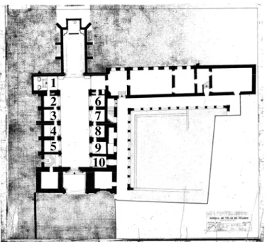
An accurate survey was carried out in April 2005 in-side Vilar de Frades church. During this survey de-termination of environmental conditions, collection of samples and also photographic record were made. Figure 1 presents the ground level plan of the church with the numbers attributed to each chapel in this work.
Figure 1. Ground level plan of Vilar de Frades church, present-ing the chapels numbered from 1 to 10.
2.1 Assessment of environmental conditions
Environmental parameters were recorded in 22nd April 2005 on the interior of the Vilar de Frades church. Relative humidity of the air (RH%), tem-perature (TºC), visible light and ultra-violet radia-tion (UV) were measured using a data logger Elsec 764 Environmental Monitor. The measurements were carried out with a periodicity of 10 minutes during two hours. The values of the exterior envi-ronmental parameters reported on that day were supplied by Institute of Meteorology.
Moisture content of the substrate (%) was meas-ured with a moisture meter Tramex Moisture En-counter Plus. The instrument measures the electrical impedance of the substrate by creating a low fre-quency alternating electric field between the elec-trodes. This field penetrates the material under test to a depth of approximately 30 mm.
2.2 Mapping of green biofilms
In order to map the green biological patina it was made a mapping of chapel 3 and 4 of the Vilar de Frades church, which represent the greater biologi-cal covering surface area. Mapping of biologibiologi-cal colonization was performed through the photo-graphic record with Photoshop CS image treatment software.
2.3 Biological sampling
With the permission from current authorities, bio-logical samples were collected from columns sur-faces in six chapels of the church (chapels 1 to 6). These were the chapels included in the conservation and restoration works. The biological material was collected on Rodac contact plates (∅ 5.5 cm) with BG-11 culture medium, at 1m above the ground. The plates were kept at 18ºC under a 12/12 light/dark cycle. The development of mixed cultures of phototrophs was followed using an inverted mi-croscope, and features of individual species were observed with an optical microscope equipped with a photographic camera. After 30 days, algae and cyanobacteria colonies growing on the agar medium were isolated and identified. Determination of the microorganisms’ genera and species was performed through direct observation by optical microscopy; taxonomic identification was carried out according to Bourrelly (1990) and Komárek & Anagnostidis (1999).
Biomass, calculated as the amount of chlorophyll
a per area, was determined for the two columns on
chapel 3. The samples were collected by scraping the biofilms covering the granite surface. This in-cluded scraping of fifteen areas (each of 9 cm2) from each column until 1.50 m above the ground. The samples were taken with the help of a grid (3 x 3 cm) and using sterile materials. The amount of chlo-rophyll a of the collected samples from chapel 3 was
determined spectrofluorometrically. Chlorophyll a
from the samples was extracted with 90% aqueous acetone (v/v). Chlorophyll a and phaeopigments
concentration was determined by the method of Yentsch & Menzel (1963) as modified by Holm-Hansen et al. (1965). The fluorescence measure-ments were performed in a SPEX Fluorolog F111 that was first calibrated with pure Chla (SIGMA).
3 RESULTS
The climatic conditions recorded outside of Vilar de Frades church in 22nd April 2005 are shown in Table 1. Air temperature did not vary during the day, pre-senting a mean value of 15ºC. Relative humidity (RH) was near 100% from 00 to 06 UTC (Universal 1
2 3 4 5
6 7 8
Time Coordinated). The minimum value of relative humidity was obtained for 12 UTC (78%). Between 06 UTC and 12 UTC it rained about 6 mm.
Table 1. Air temperature, relative humidity and rainfall for 00, 06, 12 and 18 hours corresponding to universal time coordi-nated (UTC) in 22nd April 2005.
Hour Temperature
(ºC) Relative hu-midity (%) Rainfall in the last six hours (mm)
00 UTC 14 96 0.0
06 UTC 14 97 0.0
12 UTC 17 78 6.0
18 UTC 15 84 0.0
The environmental conditions recorded inside the church in 22nd April 2005 are presented in Figures 2,3. Relative humidity ranged from 81.5% to a maximum of 88%. Between 15:10h and 17:10h the RH remained relatively constant at about 87.5%. Note that this value of air RH inside the church is higher than the one recorded outside for that after-noon. Moreover, a relative humidity above 80% is more than sufficient to promote microorganisms growth.
Air temperature was almost constant with a mean value of 13.4ºC. This temperature allows microor-ganisms to growth but is not an optimum value. With the recorded values of air temperature and rela-tive humidity the average dew point is 11.2 ºC. This implicates that if temperature decreases about 2ºC, the condensation of water in the interior walls of the church would occur. This situation is very liked to happen during the night.
Figure 2. Temperature and relative humidity recorded inside the Vilar de Frades church in 22nd April 2005.
Figure 3. Light and UV radiation recorded inside the Vilar de Frades church in 22nd April 2005.
The interior of the church is illuminated by natu-ral and artificial light source. During the day, the light irradiance is mainly from natural origin pro-vided by several windows located on the first level of the building. Light irradiance presented values be-low 50 Lux, with the exception of one obtained at 3:20h that reached 56.4 Lux (Fig. 3). The average value for light irradiance recorded was 45.5 Lux. The UV radiation ranged from 54.4 µW/lm to 77.8 µW/lm presenting an average value of 64.3 µW/lm.
Moisture content of the substrate (%) was meas-ured in the columns of the six chapels under study until 3 m high. The results showed that water con-tent of the substrate was always around 100% at all measured areas. This is in agreement with the tem-perature and relative humidity measured in the air.
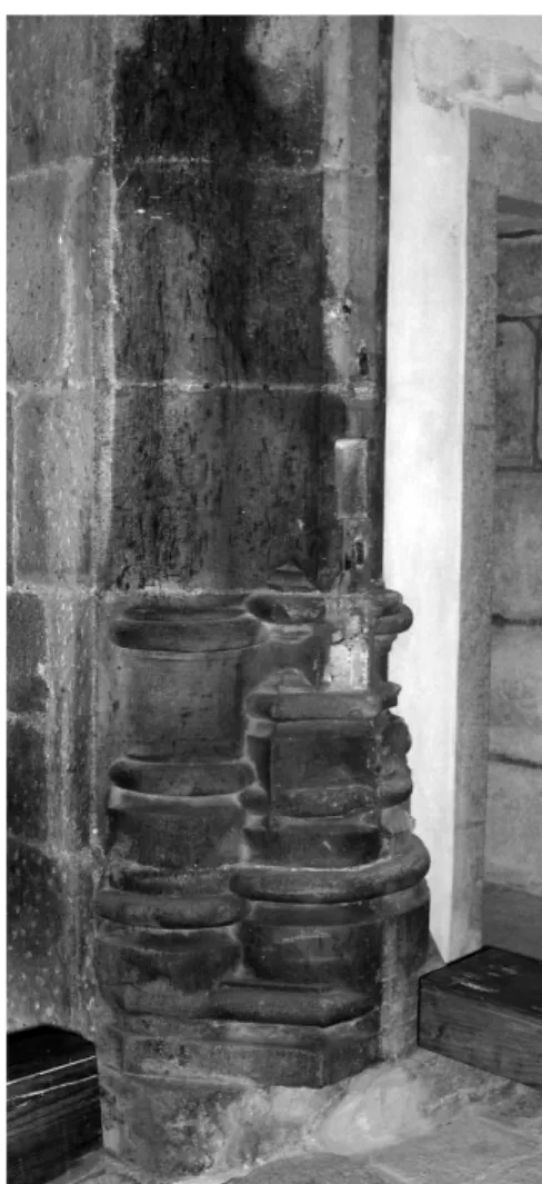
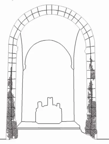
The granite surfaces of the chapels were abun-dantly covered with a green biological colonization. Biodeterioration phenomena were evident through these extensive and thick green biofilms. The biofilms from each chapel present different exten-sion and distribution, and were firmly bounded to the stone surfaces (Fig. 4). Mapping of green bio-logical patina, in chapels 3 and 4, is presented in Figures 5a, b, 6a, b.
Figure 5. a) Mapping of biological colonization from chapel 3.
Figure 6. a) Mapping of biological colonization from chapel 4.
b) Chapel 3.
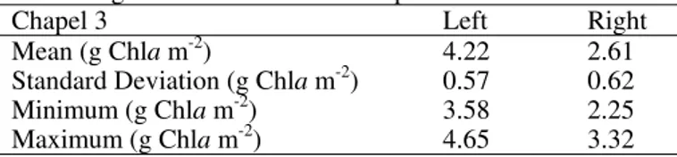
Concentration of chlorophyll a was determined
for the two columns of chapel 3 (Table 2). The left column of the chapel presented superior values of concentration of chlorophyll a in relation to the right
column.
Table 2. Chlorophyll a concentration (g Chla m-2) determined
from the right and left column of chapel 3.
Chapel 3 Left Right
Mean (g Chla m-2) 4.22 2.61
Standard Deviation (g Chla m-2) 0.57 0.62
Minimum (g Chla m-2) 3.58 2.25
Maximum (g Chla m-2) 4.65 3.32
Regarding species identification, one genus of cyanobacteria and seven genera of microalgae were found. Green biofilms consisted mainly of cyano-bacteria Gloeocapsa sp. that was present in all the
collected areas. The microalgae found on this green biofilms were essential Chlorophyta coccoid mor-photypes. The main microalgae identified were:
Chlamydocapsa sp., Chlorella ellipsoidea, Chloro-coccum sp., Desmococcus sp., Tetracystis sp., Tre-bouxia sp. and Trentepohlia sp..
4 DISCUSSION
The presence of green biofilms inside Vilar de Frades church is not a new problem, it remains for many years. The authorities in charge of that monu-ment have tried different approaches (cleaning, use of biocides, etc.) but the problem still persists. These green biofilms not only affect drastically the aes-thetic value of the church as they also promote the deterioration of stone due to their photosynthetic na-ture and retention of water.
The main biodeterioration agents identified were the cyanobacterium Gloeocapsa sp. and several
mi-croalgae. Gloeocapsa sp. was the genus that most
occurred in all samples. Cyanobacteria are generally adapted to resist adverse conditions because of their thick outer envelopes and the presence of protective pigments (Crispim et al. 2005). Gaylarde & Gay-larde (2005) found that, in laboratory experiments, the wall-inhabiting organisms tolerant to the highest salt levels were the cyanobacteria Gloeocapsa sp..
This may be an explanation for the large amount found on the granite surfaces of Vilar de Frades church.
The microalgae found on this green biofilms were essential Chlorophyta. Chlamydocapsa can be found
in very inhospitable places like the sub-Antarctic lakes (Hansson & Tranvik 1997). Chlorella ellip-soidea was also identified on a limestone monument
in the central part of Portugal, city of Coimbra (San-tos 2003). Chlorococcum has been reported by
sev-eral authors on granite, marble and limestone monu-ments (e.g. Leite Magalhães & Braga 2000, Tomaselli et al. 2000b, Santos 2003). Desmococcus
is able to habitat tree bark and desert crusts (Hu et al. 2003). Tetracystis is usually isolated from soils;
therefore is expected to be also found in stones sur-faces. The genus Trebouxia was identified on granite
from Cathedral of Lund by Ortega-Calvo et al. (1991), and Trentepohlia has also found on another
granite monument in Ourense, NW Spain (No-guerol-Seoane & Rifon-Lastra 1997). In summary, the microorganisms present on this biofilm are adaptable to a wide range of extreme environmental conditions.
The results of the present work show that envi-ronmental conditions inside the church promote the growth of microorganisms. These results also indi-cate that photoautotrophic microorganisms consti-tute the main portion of the biomass on these granite surfaces. Photosynthetic microorganisms are com-mon inhabitants of com-monuments and their growth represents a significant input of organic matter to the stone, as estimated through chlorophyll a
This study shows that stone-degrading cyanobac-teria and microalgae are present on the interior walls of Vilar de Frades church. The visible deterioration of the interior of Vilar de Frades church is mainly due to biological phenomena and it is strictly related to the environmental conditions. Therefore, in order to improve the conservation state of interior walls of the church the environmental conditions have to be altered. First, it would be recommended to avoid sunlight inside the church. In this way, there would not be enough light for the photosynthetic organisms growth, since artificial light are usually turned off. Secondly, the relative humidity and temperature of the church should be controlled to avoid the growing of heterotrophic microorganisms (fungi and bacte-ria). Values of relative humidity around 55 – 65% and temperature between 18ºC and 20ºC can be sug-gested. However, special care is necessary in order to avoid fast environmental changes that could dam-age the materials inside the church. The biofilms should be cleaned with a vacuum cleaner and a soft brush. Afterwards the moisture content of the walls should decrease. Meanwhile, different hydrophobic protective mixed with different biocide should be tested in situ, in order to select the mixture to be
ap-plied. After the application of the selected treatment monitoring plan should be implemented. This plan should include an hourly determination of air rela-tive humidity, temperature and light. And a survey with photographic records, samples collection and surface moisture content determination, on a monthly basis.
5 REFERENCES
Albertano, P., Bruno, L., Bellezza, S., Paradossi, G. 2000. Polysaccharides as a key step in stone bio-erosion. Proceed-ings of the 9th International Congress on Deterioration and Conservation of Stone, Elsevier: Venice, pp. 425-432.
Ariño, X., Saiz-Jimenez, C., Hernandez-Marine, M. 2002. Colonization of cryptoendolithic niches in mortars by photo-trophic microorganisms. Galán, E. & Zezza, F. (eds), Protec-tion and ConservaProtec-tion of the Cultural Heritage of the Medi-terranean Cities, Balkema: Lisse, pp. 127-131.
Bellinzoni, A.M., Caneva, G., Ricci, S. 2003. Ecological trends in travertine colonization by pioneer algae and plant com-munities. International Biodeterioration and Biodegradation
51: 203-210.
Bourrelly, P. 1990. Les algues d’eau douce – Initiation à la systématique. Les Algues Vertes. Paris, Société Nouvelle des
Éditions Boubée.
Crispim, C.A., Gaylarde, P.M., Gaylarde, C.C. 2003. Algal and cyanobacterial biofilms on calcareous historic buildings.
Current Microbiology 46: 79-82.
Gaylarde, C. & Gaylarde, P. (2005). A comparative study of the major microbial biomass of biofilms on exteriors of buildings in Europe and Latin América. International Biode-terioration and Biodegradation 55: 131-139.
Gómez-Alarcón, G., Muñoz, M., Ariño, X., Ortega-Calvo, J.J. 1995. Microbial communities in weathered sandstones: the
case of Carrascosa del Campo church, Spain. The Science of the Total Environment 167: 249-254.
Guillitte, O., Dreesen, R. 1995. Laboratory chamber studies and petrographical analysis as bioreceptivity assessement tool of building materials. The Science of the Total Environ-ment 167: 365-374.
Hansson, L.-A., Tranvik, L.J. 1997. Algal species composition and phosphorus recycling at contrasting grazing pressure: An experimental study in sub-Antartic lakes with two tro-phic levels. Freshwater Biology 37: 45-53.
Herrera, L.K., Arroyave, C., Guiamet, P., Saravia, S.G., Vi-dela, H. 2004. Biodeterioration of peridotite and other con-structional materials in a building of the Colombian cultural heritage. International Biodeterioration and Biodegradation
54: 135-141.
Holm-Hansen, O., Lorenzen, C.J., Holmes, R.W., Strickland, J.D.H. 1965. Fluorometric determinarion of chlorophyll.
Journal of Conseil International Exploration de la Mer 30:
3-15.
Hu, C., Zhang, D., Huang, Z., Liu, Y. 2003. The vertical mi-crodistribution of cyanobacteria and green algae within de-sert crusts and the development of the algal crusts. Plant and Soil 257: 97-111.
Komárek, J., Anagnostidis, K. 1999. Cyanoprocaryota I. In: Ettl, H., Gerloff, J., Heynig, H., Mollenhauer, D. (eds.),
Süßwasserflora von Mitteleuropa, Band 19/1, Gustav
Fischer Verlag, 548 p.
Leite Magalhães, S., Sequeira Braga 2000. Biological coloni-zation features on a granite monument from Braga. Proceed-ings of the 9th International Conference on Deterioration and Conservation of Stone, Elsevier: Venice, pp. 521- 529.
Lamenti, G., Tomaselli, L., Tiano, P. 2003. Cyanobacteria and biodeterioration of monumental stones. Saiz-Jimenez, C. (ed), Molecular Biology and Cultural Heritage, Balkema:
Lisse, pp. 73-77.
Noguerol-Seoane, A. & Rifon-Lastra, A. 1997. Epilithic phy-coflora on monuments. A survey of San Eastern de Ribas de Sil monastery (Ourense, NW Spain). Cryptogamie: Algologie 18: 351-361.
Ortega-Calvo, J.J., Hernandez-Marine, M., Saiz-Jimenez, C. 1991. Biodeterioration of building materials by cyanobacte-ria and algae. International Biodeterioration 28: 165-185.
Ortega-Calvo, J.J, Ariño X., Hernandez-Marine, M., Saiz-Jimenez, C. 1995. Factors affecting the weathering and colo-nisation of monuments by phototrophic microorganisms. The Science of the Total Environment 167: 329-341.
Saiz-Jimenez, C. 1999. Biogeochemistry of weathering proc-esses in monuments. Geomicrobiology Journal 16: 27-37.
Santos, M.F.A. 2003. Estudo em microscopia óptica das amostras recolhidas em três zonas distintas das paredes dos claustros da Igreja de Santa Cruz, Internal report from Universidade de Coimbra, Departamento de Botânica, Portugal.
Tomaselli, L., Tiano, P., Lamenti, G. 2000a. Occurrence and fluctuation in photosynthetic biocoenoses dwelling on stone monuments. Ciferri, O., Tiano, P., Mastromei, G., (Eds.), Of Microbes and Art – the role of microbial communities in the
degradation and protection of cultural heritage, Kluwer
Academic/Plenum Publishers: New York, pp. 63-76. Tomaselli, L., Lamenti, G., Bosco, M., Tiano, P. 2000b.
Biodi-versity of photosynthetic micro-organisms dwelling on stone monuments. International Biodeterioration and Biodegrada-tion 46: 251-258.
Yentsch, C.S., Menzel, D.W. 1963. A method for the determi-nation of phytoplankton chlorophyll and phaeophytin by fluorescence. Deep Sea Research 10: 221-231.