S
C
I
E
N
T
I
F
I
C
-T
E
C
H
N
I
C
A
L
The effect of andiroba oil and chitosan concentration
on the physical properties of chitosan emulsion film
Vanessa Tiemi Kimura
1, Cintia Satiyo Miyasato
1, Bianca Pereira Genesi
1, Patrícia Santos Lopes
1,
Cristiana Maria Pedroso Yoshida
1and Classius Ferreira da Silva
1*
1
Laboratório de Biotecnologia e Produtos Naturais – BIONAT, Instituto de Ciências Químicas,
Ambientais e Farmacêuticas, Universidade Federal de São Paulo – UNIFESP, Diadema, SP, Brazil
*[email protected]Abstract
Chitosan ilm is used as a dressing to heal burns. The physical and biological properties of the ilm can be modiied by the addition of phytotherapic compounds. This work used the casting -solvent evaporation technique to prepare chitosan ilm containing andiroba oil (Carapa guianensis) which has anti-inlammatory, antibiotic, and healing properties. The objective of this study was to determine the effect of the concentrations of chitosan and andiroba oil on the physical properties of chitosan ilms. The emulsion ilms were evaluated concerning the mechanical properties and luid handling capacity. Additionally, scanning electron microscopy and thermal analysis were performed. The results showed that the barrier and mechanical properties were affected by the addition of andiroba oil, and these may be modulated as a function of the concentration of oil added to the ilm. The thermal analysis showed no evidence of chemical interactions between the oil and chitosan.
Keywords: biopolymers, dressings, Carapa guianensis.
1. Introduction
Andiroba (Carapa guianensis) is a tree of the Meliaceae family found in Central and South America, especially in the Amazon basin region. Many studies have reported the pharmacological properties of products obtained from the andiroba flower[1,2], from the ethanolic extract of the andiroba
leaf[3,4] and especially products derived from andiroba seed
oil[5-7]. Andiroba oil is widely used in popular medicine in
the Amazon basin region.
According to Cabral et al.[8], andiroba oil is composed
mostly of triacylglycerols, with high levels of unsaturated and saturated fatty acids such as oleic (51.81%), palmitic (25.76%), stearic (9.08%), and linoleic (8.3%).The medicinal properties of andiroba oil have been attributed to the presence of limonoids, which are tetranortriterpenoids[6]. Andiroba
oil also contains triterpenes, tetraterpenes, alkaloids, and glycerides[9].
Andiroba seed oil is currently considered to be acaricides[10],
larvicidal against Aedes aegypti[11], as well as being an
antiplasmodial[5], anti-inflammatory[12], anti-allergic[7,13,14],
and also suitable for wound healing[3,4].
The ethanolic extract of Carapa guianensis leaves was evaluated for antibacterial and wound healing activity, using excision, incision, and dead space wound models in rats. The results showed an increased rate of wound contraction and hydroxyproline content (a biochemical marker for tissue collagen), which indicates the potential application of Carapa guianensis in wound healing[4].
Many wound dressings have been developed for the treatment of severe burn wounds or ulcers. Damaged tissue requires biocompatible materials like chitosan that has a high film-forming capacity. The most cited advantages of chitosan are its physico-chemical and biological
properties. Chitosan promotes activation and proliferation of inflammatory cells in granular tissues[15], stimulates cell
proliferation and histoarchitectural reorganization of the tissue[16], and affects the functioning of macrophages, thus
accelerating the healing process[17]. The use of chitosan
resulted in a substantial decrease in healing time and minimal scarring in several animals[18]. These and other properties
can be potentiated with the incorporation of andiroba oil. As previously described, andiroba oil has many interesting properties for use in dressings.
The preparation of emulsified films presents some challenges, for example, maintaining the stability of the emulsion during the process of drying the film. The stability of chitosan films can be achieved by adding surfactants but also by emulsification with rotor–stator homogenizer (> 20,000 rpm)[19-21], or even less vigorously (13,500 rpm)[22].
The small concentration of chitosan in oil emulsions also helps to maintain the stability of the emulsion. Usually, this concentration ranges from 0.1 to 1%[19-21], but it can reach
values up to 3%[22]. Therefore, in this work, we decided to
work with concentrations between 0.1 and 1% and agitation (24,000 rpm).
This work aims to study the effect that the concentrations of andiroba oil and chitosan have on the physical and chemical properties of chitosan film used for wound dressings.
2. Materials and Methods
2.1 Materials
Commercial chitosan (deacetylation of approximately 82% and molar mass of approximately 1.47 × 10⁵ g/mol)
purification. Acetic acid (Synth, Brazil) was used as an acidic medium. Andiroba oil was provided by MPR Indústria e Comércio de Óleos Vegetais Ltda (Brazil).
2.2 Chitosan suspension
Chitosan (1.0% or 2.0%, w/w) was dissolved in an aqueous acetic acid solution. The stoichiometric amount of acetic acid was calculated to achieve the protonation of all the NH2 sites and taking into account the sample weight
and the degree of acetylation. Using this base amount plus an extra 50% gave the total stoichiometric amount to be used. The suspension was homogenized for 2 h prior to the preparation of the chitosan film, in order to complete chitosan solubilization.
2.3 Preparation of chitosan film
The films were prepared by the casting technique. The chitosan suspension and the andiroba oil was emulsified beforehand (24000 rpm for 10 min). We tested three concentrations of andiroba oil (g of andiroba oil/100 g of solution, i.e. % w/w): 0.1%, 0.5%, and 1.0% w/w. The chitosan emulsion was poured into polyethylene Petri dishes. The films were dried in a forced air oven at 40 °C for 24 h. The mass of the suspension applied to the Petri dishes was kept constant (0.21 g/cm2).The films then underwent various analyses.
2.4 Scanning Electron Microscopy (SEM)
SEM analysis was performed on fractured cross-sections and the surfaces of gold-sputtered films using an LEO 440i scanning electron microscope (LEO Electron Microscopy Ltda.) with 10 kV and 100 pcA.
2.5 Fluid Handling Capacity (FHC)
The fluid handling capacity (FHC) of the film is defined as the sum of the Absorbency (ABS) and Moisture Vapor Transmission Rate (MVTR). The FHC was examined according to the BS EN 13726-1 method[23] for hydrocolloids
and dressings. In this test, samples of each film (or dressing) were applied to the modified Paddington cups (Figure 1), to which were added 20 mL of simulated exudate fluid (SEF).
The cups were weighed using a calibrated analytical balance, inverted so that the dressing came into contact with
the SEF – see Figure 1c – and the solution was placed in a temperature and humidity controlled incubator to maintain an environment of 37 °C ± 2 and a relative humidity below 20% for a period of 24 h. At the end of the test the cups were removed from the incubator and were allowed to equilibrate at room temperature for a period of 30 min prior to reweighing on the analytical balance. The FHC, ABS, and MVTR were calculated by the following equations:
x y
MVTR
time surface
− =
× (1)
b a ABS
time surface
− =
× (2)
FHC=MVTR+ABS (3)
where x is the complete system weight (film + SEF solution + cup) at the beginning of the test; y is the complete system weight (film + SEF solution + cup) after 24 h; b is the film weight at the beginning of the test; and a is the film weight after 24 h. Five repetitions were done per experiment.
2.6 Mechanical properties
Tensile testing was done in accordance with the ASTM D882 method[24]. Films were cut into 10.00 cm × 2.54 cm
strips. The tensile strength, elongation at breaking point, and Young’s modulus were measured using TexturePro CT V1.2 (Brookfield, CT3 50K Texturometer). The crosshead speed was set at 1 mm.s–1. Samples were pre-conditioned in a desiccator at 75% relative humidity for 48 hours. There were at least 10 repetitions per experiment.
2.7 Thermal analysis
Differential scanning calorimetry (DSC) and thermogravimetric analysis (TGA) studies were performed on chitosan film, pure chitosan, and andiroba oil. TGA was done with a TGA-60 (Shimadzu) analyzer. All analyses were performed with 5-10 mg samples in platinum pans in a dynamic nitrogen atmosphere (100 mL.min–1), between 30 °C and 700 °C. The experiments were done at a scanning rate of 10 °C.min–1. DSC analysis was performed with a DSC-60 (Shimadzu) analyzer. Samples (approx. 5-10 mg) were scanned in a sealed aluminum pan and heated to a
temperature of 450 °C at a rate of 10 °C.min–1, in a nitrogen atmosphere, and with a flow rate of 50 mL.min–1.
2.8 Statistical analysis
All the characterizations were done in replicate. The Tukey’s test was done for comparison of means, using BioEstat 5.3[25].
3. Results and Discussion
3.1 Scanning electron microscopy
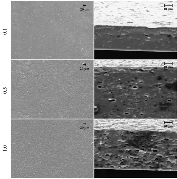
No macroscopic phase separation was observed after drying for any of the samples prepared from chitosan/andiroba oil, which indicates that oil droplets were stabilized in the chitosan suspension. SEM was used to evaluate the morphology and distribution of oil droplets in the films. This analysis allows us a better understanding of mechanical and barrier properties[26]. Figure 2 shows SEM micrographs of the chitosan
films containing andiroba oil for 2% chitosan. Micrographs of films for 1% (w/w) chitosan were quite similar, and they are not presented. The emulsified films showed structural discontinuities associated with the formation of two phases (lipid and polymer) in the matrix. The oil-free films had a smooth and homogeneous microstructure with no irregularities like air bubbles or oil droplets detected (micrograph not shown), as our group had previously published[27]. The number of
oil droplets increases as the concentration of oil increases. The cross-sections of the emulsified films show that droplets have an ellipsoidal shape, which was also verified by other authors[26,28,29]. This ellipsoidal shape can be attributed to the
weight of the chitosan over the droplets during the drying. It is important to mention that no surfactant was used in the preparation of our films.
Kokoszka et al.[30] studied whey protein/rapeseed oil
emulsion film.Unlike what was observed in this study, they verified that the oil is not well distributed throughout
Figure 2. SEM of the surface (left) of chitosan film viewed at a magnification of 500×, and cross-sections (right) viewed at a magnification
the film on both sides. According to Kokoszka et al.[30],
the oil droplets are more concentrated on the side exposed to the air since the film retraction during drying produces changes in its structure, which becomes denser, and the oil droplets migrate towards the side exposed to the air, thus favoring coalescence.
3.2 Fluid handling capacity
The moisture content of the wounds must be carefully controlled to achieve optimal rates of wound healing. The healing process can be influenced by changes in the moisture content of a wound and the skin around it. A wound that is too dry may delay or impair the healing while excess fluid can cause maceration or infection. Thus, the optimal healing environment is achieved by applying an appropriate dressing that should be removed in time to avoid maceration or adherence[31]. Donor sites, unspecified granulating wounds,
and third-degree burns generate between 3.4 and 5.1g of exudate per 10 cm2 over a 24 hour period[32].
The MVTR, ABS, and FHC are presented in Table 1. Thomas and Young[31] evaluated these properties for two
commercial dressings: ActivHeal (Advanced Medical Solutions) and Allevyn Adhesive (Smith & Nephew) – both are film-backed foam dressings. They verified the absorbencies to be 3.44 and 4.32 g/10 cm2/24 h, respectively, for ActivHeal
and Allevyn Adhesive. Our results for absorbency are very close to these values, especially when the concentration of chitosan is 2.0% w/w. Furthermore, the MVTR were 1.67 and 12.35 g/10 cm2/24 h, respectively, for ActivHeal
and Allevyn Adhesive[31]. Regarding the MVTR, our results
were close to those for ActivHeal. Also, it is important to mention that both these commercial dressings are foam dressings, so these properties are usually greater than those for film dressings.
The WVTR decreases with the oil concentration in both chitosan concentrations; this could be attributed to the hydrophobicity of the andiroba oil. Concerning the Absorbency, the incorporation of andiroba oil decreased this parameter especially for the 2% chitosan films.
3.3 Mechanical properties
To adequately protect a wound, the film must maintain its integrity against external stress during the manipulation and application, even when it is on the wound. Tensile strength indicates the maximum tensile stress that the film can sustain, elongation at breaking point is the maximum change in length of a test specimen before breaking, and the Young’s modulus is a measure of the stiffness of the film[21].
The mechanical properties of chitosan film are shown in Table 2. Both the concentrations significantly affected the mechanical properties of the emulsified films.
Young’s modulus and the tensile strength of the chitosan film increased when andiroba oil was incorporated into the chitosan matrix. These two properties are usually higher for samples with 2.0% of chitosan. The addition of low concentrations (0.1%) of andiroba oil causes an increase in these properties if compared to oil-free film. Whenever the concentration of oil increases, these two properties decrease
Table 1. Fluid handling properties of the film dressing for different concentrations of andiroba oil.
Concentration (% w/w)
MVTR (g/10cm2/24h) Absorbency
(g/10cm2/24h) FHC (g/10cm 2/24h) Chitosan Andiroba oil
1.0
0.0 1.73 ± 0.07ª 1.67 ± 0.12ª 3.39 ± 0.14
0.1 2.83 ± 0.28b 0.62 ± 0.06b 3.45 ± 0.29
0.5 1.53 ± 0.12c 1.59 ± 0.16ª 3.12 ± 0.20
1.0 1.31 ± 0.13d 1.49 ± 0.08ª 2.81 ± 0.15
2.0
0.0 3.24 ± 0.22e 3.43 ± 0.21c 6.67 ± 0.30
0.1 1.88 ± 0.12ª 2.32 ± 0.18d 4.20 ± 0.21
0.5 1.28 ± 0.13d 2.74 ± 0.26e 4.02 ± 0.29
1.0 1.36 ± 0.07c,d 2.63 ± 0.21d,e 3.98 ± 0.22
Different superscripts within the same column indicate significant differences among formulations (p<0.05).
Table 2. Effect of the concentrations of chitosan and andiroba oil on Young’s modulus, tensile strength, and elongation at breaking point,
for the emulsified films.
Concentration (% w/w)
Young’s modulus (MPa) Tensile strength (N/m2) Elongation at breaking point (%) Chitosan Andiroba oil
1.0
0.0 36.52 ± 1.49a 169.99 ± 14.06a 13.59 ± 1.20a
0.1 67.67 ± 5.32b 263.47 ± 22.62b 13.48 ±1.29a
0.5 55.39 ± 4.96c 204.16 ± 18.71c 13.07 ± 0.40a
1.0 34.63 ± 3.99a 172.60 ± 17.16a 13.69 ± 1.03a
2.0
0.0 62.02 ± 6.75b,c 171.21 ± 14.01a 22.56 ± 1.86b
0.1 98.50 ± 9.58d 272.55 ± 17.00b 9.26 ± 0.89c
0.5 81.98 ± 3.55e 250.20 ± 22.77b 9.23 ± 0.92c
1.0 57.32 ± 4.37c 182.67 ± 17.11a,c 12.43 ± 1.14a
and are closer to those of the control film (oil-free chitosan film). A decrease in these two properties with the increase of oil was also observed in emulsified films with chitosan/basil essential oil[19], chitosan/thyme essential oil[20], chitosan/tea
tree oil[22], cassava starch-chitosan/oregano essential oil[33],
and whey protein/olive oil[34].
The mechanical properties obtained by the addition of oil may be related to the structural arrangement of the lipid phase in the chitosan matrix. A number of discontinuities increases as the concentration of andiroba oil increases, which could explain the decrease in Young’s modulus and tensile strength.
No significant effect on elongation at breaking point was observed for 1% of chitosan and when the andiroba oil concentration increased. This effect was also reported by other authors when adding oil to a chitosan matrix[29,35,36] and
it could also be attributed to the structural discontinuities provoked by the incorporation of the oil. Moreover, the incorporation of oil promoted a substantial reduction in the elongation of the films containing 2% chitosan.
The effects of chitosan concentration on the mechanical and physical properties of the films seem to be greater than the effect of the oil. Most likely, the oil concentrations are not sufficiently high so that they could be more important than chitosan concentration. But it would be difficult to produce films with higher oil concentration because we would have exudation of the oil from the film, even if we had used one tensoative.
3.4 Thermogravimetric analysis
TGA was performed to evaluate the thermal stability of the chitosan powder, andiroba oil, and chitosan-andiroba oil films. Thermal degradation is displayed in Table 3 and Figures 3, 4.
Table 3 and Figure 3 show that chitosan powder mainly loses mass due to decomposition between 200 °C and 400 °C, and especially in the range of 200 °C to 300 °C. On the other hand, the weight loss of the pure andiroba oil is mainly between 300 °C and 400 °C, while above 500 °C andiroba oil is entirely decomposed. The TGA curves have different behavior above and below the temperature of the oil degradation for andiroba oil/chitosan films. Below the temperature of oil degradation, the oil seems to
Table 3. Thermal analysis of chitosan, andiroba oil, and andiroba oil/chitosan film in an N2 atmosphere.
Concentration (% w/w) % weight loss
Chitosan Andiroba oil 100 °C 200 °C 300 °C 400 °C 500 °C 600 °C 700 °C
1.0 0.0 12 17 43 59 63 66 69
0.1 14 22 44 59 66 71 75
0.5 10 15 34 54 74 76 78
1.0 9 13 30 54 77 80 82
2.0 0.0 12 19 44 59 63 66 67
0.1 14 21 44 59 65 69 74
0.5 10 17 38 52 68 72 75
1.0 10 15 32 48 73 76 79
Chitosan powder 4 6 32 50 56 60 62
Andiroba oil 0 1 2 21 100 100 100
Figure 3. TGA thermograms of andiroba oil and chitosan powder.
Figure 4. TGA thermograms of 1% and 2% (w/w) chitosan films
stabilize the film, but when the temperature is higher than the temperature of the oil degradation, the oil appears to promote the opposite. If we compare the weight loss up to 400 °C (Figure 4 and Table 3), the increase of andiroba oil concentration decreases the weight loss values (the TGA curves are in a superior position for 0.5 and 1.0% of andiroba oil). However, when the temperature is above 400 °C, there is one inversion and the TGA curves become inferior for these higher concentrations of andiroba oil. We also observed in Figure 4 that the incorporation of andiroba oil in chitosan film tends to shift the thermal degradation zone to higher temperatures. Such change is attributed to an increase in thermal stability by the incorporation of andiroba oil, and they were also supported by the differential thermogravimetric analysis - DTG (data not shown). Regardless, Figure 4 shows that both chitosan concentrations presented the same behaviour when the andiroba oil concentration increases.
Pelissari et al.[33] verified that the addition of oregano
essential oil to chitosan-starch films did not influence the thermal stability of these films; however, they observed an increase in residue percentage after the incorporation of the oregano essential oil.
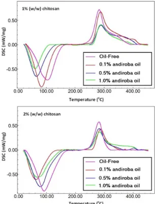
3.5 Differential scanning calorimetry
Figures 5, 6 shows the DSC results for the chitosan powder, andiroba oil, and andiroba oil/chitosan films. The results obtained from the DSC include the temperatures, and their respective ΔH values are presented in Table 4.
The DSC heating curve for pure andiroba oil showed one endothermic peak at 42.57 °C and some exothermic peaks between 180 °C and 360 °C. Oils are one complex mixture of triacylglycerols (TAGs) acting also as a solvent for minority components, such as vitamins, pigments, phenolic compounds, phospholipids, free fatty acids, and mono- and diacylglycerols[37]. The five top TAGs present in
Andiroba oil are, in descending order: Palmitic-Oleic-Oleic, Palmitic-Palmitic-Oleic, Palmitic-Oleic-Stearic, Oleic-Oleic-Oleic/Stearic-Oleic-Linoleic, and Palmitic-Linoleic-Oleic[8,37].
Thus, the first endothermic peak is probably associated with the fusion of free fatty acids. The exothermic peaks are probably associated with the decomposition of TAGs.
Matos[38] conducted a thermal study of pure fatty acids
by DSC. He observed endothermic peaks for melting temperatures at 64.55 °C ± 0.67 and 71.36 °C ± 0.30, respectively, for the palmitic acid and stearic acid. Exothermic peaks of decomposition were observed at 235.24 °C ± 7.57, 241.87 °C ± 5.61, 238.79 °C ± 12.11, and 268.26 °C ± 18.16, respectively, for the palmitic, stearic, oleic, and linoleic acids. These fatty acids also showed a sequence of many exothermic peaks above 350 °C[38].
The exothermic peaks between 200 °C and 300 °C are probably due to the decomposition of the TAGs and fatty acids into smaller chains while the sequence of peaks above 350 °C can be attributed to the decomposition of these compounds into even smaller chains. The chitosan-andiroba oil film presented two peaks (T1 and T2) while the andiroba
oil-free film showed also two peaks (T1 and T2) with a
shoulder on the descending side. These shoulders could be attributed to the decomposition described above (T > 350 °C). The increase in andiroba oil concentration promotes a decrease
in the temperature T1, which is attributed to the evaporation
of water associated with the chitosan and could also be attributed to the hydrophobic properties of the andiroba
Figure 5. DSC run curves for andiroba oil and chitosan powder.
Figure 6. DSC run curves for 1% and 2% (w/w) chitosan films
oil. Such decrease in this temperature was, even more, prominent for 1% chitosan films. This fact can be explained by the hydrophobic character of andiroba oil and also by the reduction in enthalpy related to this evaporation (ΔH1).
The temperature T2 for the decomposition of chitosan is
little affected by the addition of andiroba oil; however, the enthalpy (ΔH2) of the chitosan decomposition is reduced,
which shows that andiroba oil reduces the heat generated by the decomposition of chitosan, thus confirming the increased thermal stability of the film that was observed in the TGA results.
4. Conclusions
Chitosan films containing andiroba oil were obtained with satisfactory properties for use as a dressing to heal wounds. The addition of andiroba oil significantly modified the mechanical and barrier properties of the films and promoted greater thermal stability for the films. About the barrier properties, it was found that all of the films exhibited properties which were compatible with commercial dressings, and the films with the highest concentration of chitosan showed best results. Concerning the mechanical properties, the Young’s modulus and tensile strength increased with the addition of andiroba oil; however, as the concentration of andiroba oil increases, the values for these properties approach the values observed for the film without andiroba oil. While elongation remained practically unchanged for the films with lower concentrations of chitosan (1.0%), for the films with higher concentrations of chitosan, the addition of andiroba oil promoted a decrease in elongation.
5. Acknowledgements
The authors wish to thank São Paulo Research Foundation (FAPESP) for the financial support (2010/17.721-4).
6. References
1. Tanaka, Y., Sakamoto, A., Inoue, T., Yamada, T., Kikuchi,
T., Kajimoto, T., Muraoka, O., Sato, A., Wataya, Y., Kim, H.
S., & Tanaka, R. (2012). Andirolides H-P from the flower of
andiroba (Carapa guianensis, Meliaceae).Tetrahedron, 68(18), 3669-3677. http://dx.doi.org/10.1016/j.tet.2011.12.076. 2. Sakamoto, A., Tanaka, Y., Inoue, T., Kikuchi, T., Kajimoto, T.,
Muraoka, O., Yamada, T., & Tanaka, R. (2013). Andirolides
Q-V from the flower of andiroba (Carapa guianensis,
Meliaceae).Fitoterapia, 90, 20-29. http://dx.doi.org/10.1016/j.
fitote.2013.07.001. PMid:23850542.
3. Nayak, B. S., Kanhai, J., Milne, D. M., Swanston, W. H., Mayers,
S., Eversley, M., & Rao, A. V. C. (2010). Investigation of the
wound healing activity of Carapa guianensis L. (Meliaceae)
bark extract in rats using excision, incision, and dead space wound models.Journal of Medicinal Food, 13(5), 1141-1146.
http://dx.doi.org/10.1089/jmf.2009.0214. PMid:20828307.
4. Nayak, B. S., Kanhai, J., Milne, D. M., Pereira, L. P., & Swanston,
W. H. (2011). Experimental evaluation of ethanolic extract of
Carapa guianensis L. leaf for its wound healing activity using
three wound models. Evidence-Based Complementary and
Alternative Medicine, (419612), 6. http://dx.doi.org/10.1093/
ecam/nep160
5. Miranda, R. N. C., Jr., Dolabela, M. F., Silva, M. N., Póvoa,
M. M., & Maia, J. G. S. (2012). Antiplasmodial activity of
the andiroba (Carapa guianensis Aubl., Meliaceae) oil and
its limonoid-rich fraction.Journal of Ethnopharmacology,
142(3), 679-683. http://dx.doi.org/10.1016/j.jep.2012.05.037.
PMid:22659195.
6. Ferraris, F. K., Rodrigues, R., Silva, V. P., Figueiredo, R.,
Penido, C., & Henriques, M. G. M. O. (2011). Modulation
of T lymphocyte and eosinophil functions in vitro by natural tetranortriterpenoids isolated from Carapa guianensis Aublet. International Immunopharmacology, 11(1), 1-11. http://dx.doi.
org/10.1016/j.intimp.2010.09.010. PMid:20951667.
7. Penido, C., Costa, K. A., Pennaforte, R. J., Costa, M. F. S.,
Pereira, J. F. G., Siani, A. C., & Henriques, M. G. M. O. (2005). Anti-allergic effects of natural tetranortriterpenoids
isolated from Carapa guianensis Aublet on allergen-induced
vascular permeability and hyperalgesia.Inflammation Research, 54(7), 295-303. http://dx.doi.org/10.1007/s00011-005-1357-6.
PMid:16134059.
8. Cabral, E. C., Cruz, G. F., Simas, R. C., Sanvido, G. B.,
Gonçalves, L. V., Leal, R. V. P., Silva, R. C. F., Silva, J. C. T., Barata, L. E. S., Cunha, V. S., França, L. F., Daroda, R. J., Sá, G. F., & Eberlin, M. N. (2013). Typification and quality control
of the andiroba (Carapa guianensis) oil via mass spectrometry
fingerprinting.Analytical Methods, 5, 1385-1391. http://dx.doi.
org/10.1039/c3ay25743f.
9. Tappin, M. R. R., Nakamura, M. J., Siani, A. C., & Lucchetti,
L. (2008). Development of an HPLC method for the
determination of tetranortriterpenoids in Carapa guianensis
seed oil by experimental design.Journal of Pharmaceutical
and Biomedical Analysis, 48(4), 1090-1095. http://dx.doi.
org/10.1016/j.jpba.2008.08.027. PMid:18845411.
10. Vendramini, M. C. R., Mathias, M. I. C., Faria, A. U., Furquim,
K. C. S., Souza, L. P., Bechara, G. H., & Roma, G. C. (2012).
Action of andiroba oil (Carapa guianensis) on Rhipicephalus
sanguineus (Latreille, 1806) (Acari: Ixodidae) semi-engorged
females: morphophysiological evaluation of reproductive
Table 4. Temperatures and enthalpy measured by DSC.
Concentration (% w/w) Temperature (°C) Enthalpy (J/g)
Chitosan Andiroba oil T1 T2 ΔH1 ΔH2
1.0
0.0 103.98 283.02 –288.99 233.48
0.1 83.60 288.00 –297.25 268.72
0.5 66.51 288.88 –210.18 181.10
1.0 61.29 289.43 –112.70 155.37
2.0
0.0 91.07 282.37 –340.13 266.23
0.1 63.36 287.69 –203.67 102.58
0.5 77.92 282.47 –259.22 116.44
1.0 69.55 283.72 –228.50 138.21
system.Microscopy Research and Technique, 75(12), 1745-1754. http://dx.doi.org/10.1002/jemt.22126. PMid:22972770. 11. Silva, O. S., Prophiro, J. S., Nogared, J. C., Kanis, L., Emerick,
S., Blazius, R. D., & Roma, P. R. T. (2006). Larvicidal effect
of andiroba oil, Carapa guianensis (Meliaceae), against Aedes aegypti.Journal of the American Mosquito Control Association, 22(4), 699-701. http://dx.doi.org/10.2987/8756-971X(2006)2
2[699:LEOAOC]2.0.CO;2. PMid:17304939.
12. Penido, C., Conte, F. P., Chagas, M. S. S., Rodrigues, C.
A. B., Pereira, J. F. G., & Henriques, M. G. M. O. (2006).
Antiinflammatory effects of natural tetranortriterpenoids isolated
from Carapa guianensis Aublet on zymosan-induced arthritis
in mice.Inflammation Research, 55(11), 457-464. http://dx.doi. org/10.1007/s00011-006-5161-8. PMid:17122962.
13. Ferraris, F. K., Moret, K. H., Figueiredo, A. B. C., Penido,
C., & Henriques, M. G. M. O. (2012). Gedunin, a natural
tetranortriterpenoid, modulates T lymphocyte responses
and ameliorates allergic inflammation.International
Immunopharmacology, 14(1), 82-93. http://dx.doi.org/10.1016/j.
intimp.2012.06.002. PMid:22709475.
14. Penido, C., Costa, K. A., Costa, M. F. S., Pereira, J. F. G., Siani,
A. C., & Henriques, M. G. M. O. (2006). Inhibition of
allergen-induced eosinophil recruitment by natural tetranortriterpenoids is mediated by the suppression of IL-5, CCL11/eotaxin and
NFkappaB activation.International Immunopharmacology,
6(2), 109-121. http://dx.doi.org/10.1016/j.intimp.2005.07.011.
PMid:16399616.
15. Alemdaroğlu, C., Değim, Z., Çelebi, N., Zor, F., Öztürk, S., &
Erdoğan, D. (2006). An investigation on burn wound healing in
rats with chitosan gel formulation containing epidermal growth factor.Burns, 32(3), 319-327. http://dx.doi.org/10.1016/j.
burns.2005.10.015. PMid:16527411.
16. Muzzarelli, A. A. (1989). Amphoteric derivatives of chitosan and their biological significance. In G.Skjak-Braek, T.Anthonsen
& P.Sandford (Eds.), Chitin and Chitosan (pp. 87-99). London:
Elsevier.
17. Balassa, L. L., & Prudden, J. F. (1984). Applications of chitin and chitosan in wound healing acceleration. In J. P.Zikakis
(Ed.), Chitin, chitosan and related enzymes (pp. 296-305). San
Diego: Academic Press.
18. Paul, W., & Sharma, C. (2004). Chitosan and alginate wound
dressings: a short review.Trends in Biomaterials & Artificial Organs, 18, 18-23.
19. Bonilla, J., Vargas, M., Atarés, L., & Chiralt, A. (2011).
Physical properties of chitosan-basil essential oil edible films as affected by oil content and homogenization conditions.
Procedia Food Science, 1, 50-56. http://dx.doi.org/10.1016/j.
profoo.2011.09.009.
20. Bonilla, J., Atarés, L., Vargas, M., & Chiralt, A. (2012). Effect
of essential oils and homogenization conditions on properties of chitosan-based films.Food Hydrocolloids, 26(1), 9-16.
http://dx.doi.org/10.1016/j.foodhyd.2011.03.015.
21. Pereda, M., Amica, G., & Marcovich, N. E. (2012). Development
and characterization of edible chitosan/olive oil emulsion films.Carbohydrate Polymers, 87(2), 1318-1325. http://dx.doi.
org/10.1016/j.carbpol.2011.09.019.
22. Sánchez-González, L., González-Martínez, C., Chiralt, A.,
& Cháfer, M. (2010). Physical and antimicrobial properties
of chitosan–tea tree essential oil composite films.Journal of
Food Engineering, 98(4), 443-452. http://dx.doi.org/10.1016/j.
jfoodeng.2010.01.026.
23. British Standards Institute – BSI. (2002). BS EN 13726-1:2002: test methods for primary wound dressings. Part 1: Aspects of Absorbency, section 3.3 Fluid Handling Capacity. London:
BSI.
24. American Society for Testing and Materials – ASTM. (1995).
ASTM D882-95: standard test method for tensile properties of thin plastic sheeting. Philadelphia: ASTM. pp. 182-188.
25. Instituto de Desenvolvimento Sustentável Mamirauá. (2014).
BioEstat 5.3. Tefé. Retrieved in 25 Feb. 2014, de http://www.
mamiraua.org.br/pt-br/downloads/programas/
26. Ghasemlou, M., Khodaiyan, F., Oromiehie, A., & Yarmand, M.
S. (2011). Characterization of edible emulsified films with low
affinity to water based on kefiran and oleic acid.International Journal of Biological Macromolecules, 49(3), 378-384. http://
dx.doi.org/10.1016/j.ijbiomac.2011.05.013. PMid:21640752.
27. Estevam, L. S., Debone, H. S., Yoshida, C. M. P., & Silva, C.
F. (2012) Adsorption of bovine serum and bovine haemoglobin
onto chitosan film. Adsorption Science and Technology, 30( 8-9), 785-792.
28. Pereda, M., Aranguren, M. I., & Marcovich, N. E. (2010).
Caseinate films modified with tung oil.Food Hydrocolloids,
24(8), 800-808. http://dx.doi.org/10.1016/j.foodhyd.2010.04.007.
29. Sánchez-González, L., Vargas, M., González-Martínez, C.,
Chiralt, A., & Cháfer, M. (2009). Characterization of edible
films based on hydroxypropylmethylcellulose and tea tree essential oil.Food Hydrocolloids, 23(8), 2102-2109. http://
dx.doi.org/10.1016/j.foodhyd.2009.05.006.
30. Kokoszka, S., Debeaufort, F., Lenart, A., & Voilley, A. (2010).
Liquid and vapor water transfer through whey protein/lipid emulsion films.Journal of the Science of Food and Agriculture, 90(10), 1673-1680. http://dx.doi.org/10.1002/jsfa.4001.
PMid:20564446.
31. Thomas, S., & Young, S. (2008). Exudate-handling mechanisms
of two foam-film dressings.Journal of Wound Care, 17(7),
309-315. http://dx.doi.org/10.12968/jowc.2008.17.7.30524.
PMid:18705233.
32. Lamke, L. O., Nilsson, G. E., & Reithner, H. L. (1977). The
evaporative water loss from burns and water vapour permeability of grafts and artificial membranes used in the treatment of burns.Burns, 3(3), 159-165.
http://dx.doi.org/10.1016/0305-4179(77)90004-3.
33. Pelissari, F. M., Grossmann, M. V. E., Yamashita, F., & Pineda, E.
A. G. (2009). Antimicrobial, mechanical, and barrier properties
of cassava starch-chitosan films incorporated with oregano
essential oil.Journal of Agricultural and Food Chemistry,
57(16), 7499-7504. http://dx.doi.org/10.1021/jf9002363.
PMid:19627142.
34. Javanmard, M., & Golestan, L. (2008). Effect of olive oil and
glycerol on physical properties of whey protein concentrate films.Journal of Food Process Engineering, 31(5), 628-639.
http://dx.doi.org/10.1111/j.1745-4530.2007.00179.x.
35. Pranoto, Y., Rakshit, S. K., & Salokhe, V. M. (2005). Enhancing
antimicrobial activity of chitosan films by incorporating
garlic oil, potassium sorbate and nisin.LWT - Food Science
and Technology, 38, 859-865. http://dx.doi.org/10.1016/j.
lwt.2004.09.014.
36. Zivanovic, S., Chi, S., & Draughon, A. F. (2005). Antimicrobial
activity of chitosan films enriched with essential oils.
Journal of Food Science, 70(1), M45-M51. http://dx.doi.
org/10.1111/j.1365-2621.2005.tb09045.x.
37. Saraiva, S. A., Cabral, E. C., Eberlin, M. N., & Catharino, R.
R. (2009). Amazonian vegetable oils and fats: fast typification
and quality control via triacylglycerol (TAG) profiles from dry matrix-assisted laser desorption/ionization time-of-flight
(MALDI-TOF) mass spectrometry fingerprinting.Journal of
Agricultural and Food Chemistry, 57(10), 4030-4034. http://
dx.doi.org/10.1021/jf900043u. PMid:19358529.
38. Matos, F. C. (2012). Study of thermal decomposition of fatty acids through differential scanning calorimetry (Master’s
dissertation). Universidade Estadual de Campinas, Campinas.