Comet and cytogenetic tests as tools for evaluating genomic instability in
seeds of
Oryza sativa
L. and
Phaseolus vulgaris
L. from gene banks
Alisson F. Dantas
1, Renata M. Lopes
2, Maria L. Fascineli
1, Solange C.B.R. José
3, Juliano G. Pádua
3, Marcos
A. Gimenes
3and Cesar K. Grisolia
11
Laboratório de Genética Toxicológica, Departamento de Genética e Morfologia, Instituto de Ciências
Biológicas, Universidade de Brasília, Brasília, DF, Brazil.
2
Departmento de Botânica, Instituto de Ciências Biológicas, Universidade de Brasília, Brasília, DF, Brazil.
3Embrapa Recursos Genéticos e Biotecnologia, Brasília, DF, Brazil.
Abstract
This study aimed to assess the feasibility of comet and cytogenetic tests as tools for evaluating genomic instability in seeds ofOryza sativa L. (rice) and Phaseolus vulgaris (beans) L. from gene banks. Rice and beans were exposed to methyl methanesulfonate (MMS) as a reference DNA damaging agent. Seeds of two accessions of rice and beans were obtained from Embrapa Rice and Beans - Brazil. Seed groups were imbibed in three concentrations of MMS for three periods of time to carry out cytogenetic tests, and for one period for the comet test. At concentrations of 10 and 15 mg/L, MMS induced cytotoxic and/or mutagenic effects in the meristematic cells of roots from all the accessions of both species. In the comet test, MMS induced genotoxic effects at all the concentrations in the evaluated accessions of rice and beans, except in one accession of beans at the lowest concentration (5 mg/L). Both species showed sen-sitivity to MMS. The comet test can be proposed for the measurement of genomic instability in accessions of rice and beans in gene banks, as being more sensitive than the cytogenetic tests used.
Keywords:Oryza sativaL.,Phaseolus vulgarisL., methyl methanesulfonate, cytogenetic test, comet test. Received: January 23, 2017; Accepted: September 11, 2017.
Introduction
Common rice,Oryza sativaL. (Poaceae family), and common bean, Phaseolus vulgaris L. (Fabaceae family) play an important role in the nutrition of various countries, and in Brazil they are the main components of the staple diet (Barbosa, 2007). Common rice is an annual gramineous species of Asiatic origin, which can adapt to a wide range of environmental conditions (Sousa et al., 1995), and it is widely distributed in tropical and subtropi-cal regions and in some temperate regions (Vaughan, 1994). Common bean is a leguminous species that presents a large number of varieties and cultivars spread over differ-ent contindiffer-ents, due to the high value of its seeds for human nutrition (Peixotoet al., 2000). Consumption of both can benefit human health due to the presence of antioxidant compounds (Walter and Marchesan, 2011; Lópezet al., 2013).
Because of the importance of rice and beans as food items, large germplasm collections have been established, and there are studies reporting cytotoxic, mutagenic and/or
genotoxic evaluations in O. sativa (Mei et al., 1994; Todoriki and Toru, 1999; Mohantyet al., 2004; Weiet al., 2006; Shiet al., 2010; Endoet al., 2012; Kwonet al., 2013; Macovei and Tuteja, 2013; Macoveiet al., 2014) and inP.
vulgaris(Hallaket al., 1999; Khanet al., 2002, Cenkciet
al., 2010). The use of methodologies to detect damage to DNA based on chromosome breaks and mis-segregation would play an important role in understanding genomic sta-bility and the viasta-bility of seeds stored in gene banks, be-cause the loss of genomic stability is one of the first stages in the seed deterioration process (Peixotoet al., 2006). In this case, methyl methanesulfonate (MMS) was used as ref-erence compound for inducing such genomic damages in rice and beans. MMS is an alkylating agent capable of add-ing methyl groups to a series of nucleophilic sites in DNA bases (Wyatt and Pittman, 2006; Maliniet al., 2010), which can cause chromosome aberrations (Kaina, 2004). It is used for mutagenic and genotoxic evaluation in different organ-isms, from fungi (Myung and Kolodner, 2003) to rodents (Mutoet al., 2015; Plappert-Helbiget al., 2015).
The main goal of the present study was to test known cytogenetic methodologies and single cell gel electrophore-sis (comet test), as tools for the measurement of genomic instability in seeds stored in a gene bank.
DOI: http://dx.doi.org/10.1590/1678-4685-GMB-2017-0020
Send correspondence to Cesar Grisolia. Departamento de Genética e Morfologia, Instituto de Ciências Biológicas, Universidade de Brasília, Campus Darcy Ribeiro 7091-900 Brasília, DF, Brasil. E-mail: [email protected].
Plant material
Seeds from two accessions ofO. sativa(BGA012099 “Ferrinho” and BGA008070 “Primavera”) and from two accessions of P. vulgaris (GF004 and GF007) were ob-tained from the Active Germplasm Bank of Rice and Beans (Embrapa Rice and Beans, Santo Antônio de Goiás, GO, Brazil). They were stored for six months at 10 °C and 30% of humidity. Initial germination of the seeds was 97 and 94% for the accessions of O. sativa, BGA012099 “Ferrinho” and BGA008070 “Primavera”, respectively, and 56 and 99% for the accessions ofP. vulgaris, GF004 and GF007, respectively.
Chemicals
MMS (CAS N. 66-27-3), ethidium bromide, sodium dodecyl sulfate, tris (hydroxymethyl) methane, ethylene diamine tetraacetic acid (EDTA) and boric acid were ac-quired from Sigma-Aldrich (St. Louis, USA). Nor-mal-melting-point (NMP) agarose was acquired from Life Technologies (New York, USA) and low-melting-point (LMP) agarose from Laboratorios Conda (Madrid, Spain).
Cytogenetic tests
Twenty seeds from two accessions ofO. sativa(2n= 2x= 24) and 30 seeds from two accessions ofP. vulgaris
(2n= 2x= 22) were imbibed in different concentrations of MMS for three periods of time: MMS 5 mg/L for 4, 8 and 24 h; MMS 10 mg/L for 4, 8 and 24 h and MMS 15 mg/L for 4, 8 and 24 h. As well as these treatments, seeds from the different accessions were imbibed in only distilled water for 24 hours. As a control, seeds ofO. sativaandP. vulgaris
were used without exposure to MMS or distilled water. Next, the seeds from each treatment were sown in substrate of germitest paper, wetted with distilled water, at the pro-portion of 2.5 mL/g of dry paper. The germitest papers with seeds remained at room temperature (20-30 °C) until the roots reached between 1 and 2 cm forO. sativa, and be-tween 1 and 3 cm forP. vulgaris. The roots were fixed in Carnoy solution (ethanol/glacial acetic acid - 3:1 v/v) and stored in ethanol 70% until slides were prepared.
The slides were prepared according to the methodol-ogy proposed by Guerra and de Souza (2002), with modifi-cations. The roots were washed in distilled water for 5 min, hydrolyzed in hydrochloric acid (5N) for 25 min, washed again in distilled water for 2 min and arranged on slides to cut the meristematic region of the roots and then to color with acetic orcein 2% for 15 min. Next, the meristematic re-gion was covered with a coverslip and evaluated under an optical microscope (1,000 X).
Evaluation of 1,000 cells per root was carried out, with four roots per group, to a total of 4,000 cells analyzed per group tested. The parameters used to characterize the groups were: (a) Mitotic Index (MI) as an indication of
mutagenicity. For the analysis of chromosome alterations, we evaluated chromosome lagging and chromosome bridges in anaphase and/or telophase and chromosome fragments in metaphase, anaphase and/or telophase. The evaluation of the slides was carried out in a blind test.
Comet test
Ten seeds from the two accessions ofO. sativaand from the two accessions ofP. vulgariswere imbibed in dif-ferent concentrations of MMS for a single period of time: MMS 5 mg/L for 24 h; MMS 10 mg/L for 24 h and MMS 15 mg/L for 24 h. Similar to the cytogenetic tests, one group of seeds ofO. sativaandP. vulgariswas also submitted to im-bibition in distilled water for 24 h. The control consisted of seeds of O. sativa and P. vulgaris without exposure to MMS or distilled water.
Cell suspensions were obtained from seed embryos from the two accessions of rice and bean for consequent processing in the test. Cell suspensions were obtained in ac-cordance with Koppen and Cerda (1997), with some adap-tations. The embryos collected were transferred to 2 mL microtubes containing 1 mL of cold phosphate buffered sa-line (PBS), macerated and left for 1 h in the refrigerator. Next, the supernatant was used to carry out the comet test. The comet test was carried out in accordance with Cerdaet al.(1997), with modifications. Fifteen microliters of the cell suspension were mixed with 85 mL of LMP
agarose (0.8%) at 45 °C and arranged on slides pre-covered with NMP agarose (0.5%). Next, the slides were re-covered immediately with the coverslip, placed on a metal sheet, and put in the refrigerator for 5 min. After the agarose gel solidified, the coverslips were removed and the slides im-mersed in TBE buffer solution (45 mM Tris-borate, 1 mM EDTA, pH 8.4) containing 2.5% sodium dodecyl sulfate for 30 min. After the lysis phase, the slides were transferred to an electrophoresis tank containing TBE buffer solution and left to rest for 5 min before running electrophoresis at 0.5 V/cm for 2 min. To determine the most appropriate voltage for the observation of nucleoids ofO. sativaandP. vulgaris
embryos, SCGE tests were done with different voltages (0.5, 0.75, 1 and 2 V/cm for rice and 0.5, 0.75 and 1 V/cm for bean) for 2 min. After electrophoresis, the slides were plunged into ice-cold distilled water for 10 min, dried at room temperature and kept in the refrigerator until the mo-ment for staining. The slides were stained with 50mL of
ethidium bromide (20mg/mL) and analyzed under a
slide. The evaluation of nucleoids of O. sativa and P.
vulgarisembryos was done using theComet AssayIV
pro-gram, version 4.3.1, and the parameter of tail intensity was chosen to measure the damage to DNA as being indicative of genotoxicity. Slides were evaluated in a blind test.
Statistical analysis
The statistical analyses of the data from the cytogenetic and comet tests were done with GraphPad Prism software, version 5.00, using One-way ANOVA fol-lowed by the Tukey test with significance levels ofp<0.05.
Results and Discussion
Initially, seeds ofO. sativaandP. vulgariswere
ex-posed only to distilled water for 24 h to verify the influence of imbibition, as the excess of water could be damaging for the seeds and interfere indirectly in the oxygenation of the embryo (Blacket al., 2006). However, it was observed that
imbibition of the seeds in water for 24 h did not induce ob-servable damage (p ³0.05) in the cytogenetic and comet
tests in seeds from the two accessions ofO. sativaandP.
vulgaristhat were evaluated.
Cytogenetic tests
In theO. sativaaccessions evaluated, MMS signifi-cantly inhibited the MI of meristematic cells of the roots of accession BGA012099 “Ferrinho” at the concentration of 15 mg/L for 4 and 8 h, when compared with the control (Ta-ble 1). In addition, MMS significantly reduced the MI of
meristematic root cells of accession BGA008070 “Primavera” at the concentration of 15 mg/L for 4 h, when compared with the control (Table 2). In accession GF004 of
P. vulgaris, MMS at the concentration of 10 and 15 mg/L
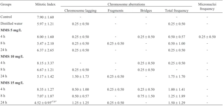
for 24 h reduced the MI of the meristematic root cells of ac-cession GF004, differing statistically from the other groups tested (Table 3). This indicated that exposure time may be a determining factor for the observation of these effects. In accession GF007, MMS significantly reduced the MI of the meristematic root cells at the concentration of 15 mg/L for 24 h, when compared to the control and to distilled water (Table 4).
In mammals, MMS induces a collapse in the replica-tion forks or a halt in replicareplica-tion due to the addireplica-tion of methyl groups to the DNA molecule, preventing the pro-gression of DNA replication in the cell cycle (Wyatt and Pittman, 2006). Although cytotoxic damages of MMS have not been previously reported in the literature with the plants evaluated in the present study, the cytotoxicity of MMS has been reported in meristematic cells of plant roots, such as
Allium cepaL. (Rank and Nielsen, 1993), and it is well
es-tablished as a positive control inA. cepatest (Limanet al., 2015).
A series of events occur in the seed up until its com-plete germination. Seed metabolism begins after hydration. Respiration and synthesis of proteins begin minutes after hydration, followed by RNA synthesis, repair mechanisms and DNA synthesis. The last event in germination is the ex-pansion of the cells in the radicle, preceding cell division. Most seeds do not manage to germinate when immersed in
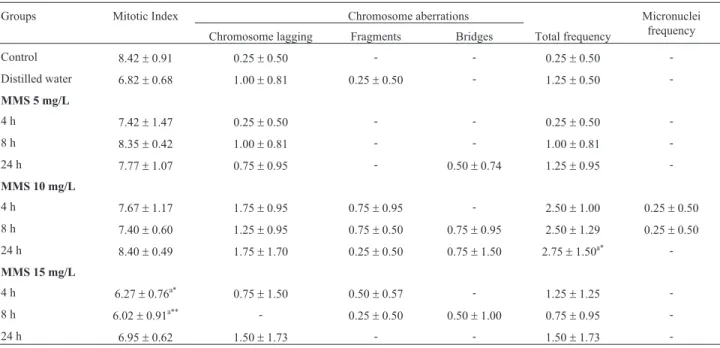
Table 1- Mitotic index, frequency of chromosome aberrations and of micronuclei obtained in the cytogenetic tests on cells from the meristematic region (n = 4000) in roots ofOryza sativaL. accession BGA012099 “Ferrinho”, exposed to methyl methanesulfonate (MMS).
Groups Mitotic Index Chromosome aberrations Micronuclei
frequency Chromosome lagging Fragments Bridges Total frequency
Control 8.42±0.91 0.25±0.50 - - 0.25±0.50
-Distilled water 6.82±0.68 1.00±0.81 0.25±0.50 - 1.25±0.50
-MMS 5 mg/L
4 h 7.42±1.47 0.25±0.50 - - 0.25±0.50
-8 h 8.35±0.42 1.00±0.81 - - 1.00±0.81
-24 h 7.77±1.07 0.75±0.95 - 0.50±0.74 1.25±0.95
-MMS 10 mg/L
4 h 7.67±1.17 1.75±0.95 0.75±0.95 - 2.50±1.00 0.25±0.50
8 h 7.40±0.60 1.25±0.95 0.75±0.50 0.75±0.95 2.50±1.29 0.25±0.50
24 h 8.40±0.49 1.75±1.70 0.25±0.50 0.75±1.50 2.75±1.50a* -MMS 15 mg/L
4 h 6.27±0.76a* 0.75
±1.50 0.50±0.57 - 1.25±1.25
-8 h 6.02±0.91a** - 0.25±0.50 0.50±1.00 0.75±0.95
-24 h 6.95±0.62 1.50±1.73 - - 1.50±1.73
-Control = seeds not exposed to MMS (methyl methanesulfonate) or to distilled water. (-) = not observed. The data are represented as means±SD;a
signifi-cant in relation to control; One-way ANOVA followed by Tukey test. (*
p<0.05;**
et al., 2006). This characteristic of rice may help to explain the fact that no cytotoxic effects were observed occasioned by MMS in accessions ofO. sativain the period of 24 h for accession BGA012099 “Ferrinho”, and in the periods of 8 and 24 h for accession BGA008070 “Primavera”. The ab-sorption of MMS in the first periods of exposure of these accessions ofO. sativamay delay the events that occur until germination, especially the repair and synthesis of DNA, which would be continued later. This is corroborated by the absence of cytotoxic effects in the longer exposure periods.
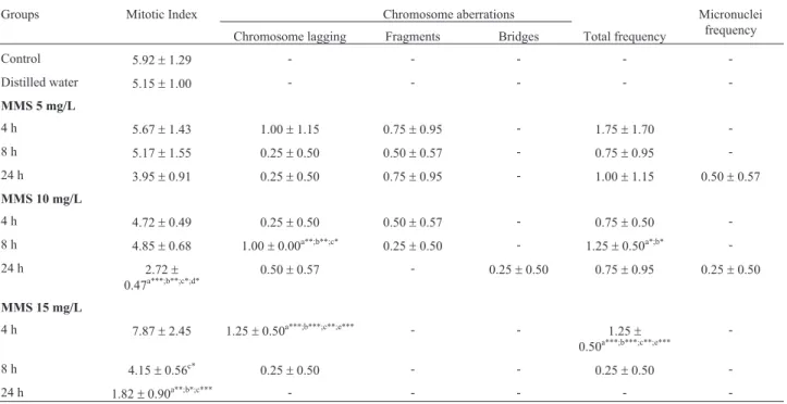
It should be noted that for seeds of accession GF004 of bean, lower MI values were already observed in interme-diate concentrations of MMS, differently from rice. At the highest concentration, of 15 mg/L, the period of 8 h was sufficient to reduce the MI, when compared to the MMS 15 mg/L group for 4 h. It may be suggested that for bean seeds the process of DNA synthesis and repair may occur in the initial stages of imbibition. Comparing the two bean acces-sions, the initial germination of the seeds of GF007 was 99%, well above that of GF004, which was 56%. This may have had an influence on the greater sensitivity of the seeds of accession GF004 to the deleterious effects of MMS.
Mutagenicity can also be analyzed by cytogenetic tests in meristematic cells of plant roots by using the fre-quencies of chromosome aberrations and MNs (Leme and Marin-Morales, 2009). MMS occasioned mutagenic effects by the significant increase in the frequency of chromosome aberrations in meristematic cells ofO. sativaroots at the
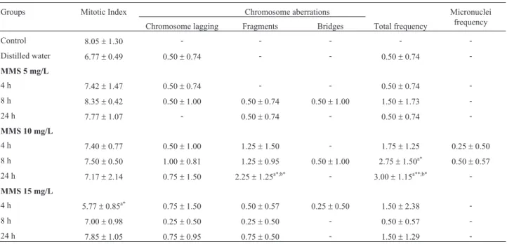
accession BGA008070 “Primavera” (Table 2), when com-pared with the control. In the latter accession, there was a significant increase in chromosome fragments for an expo-sure period of 24 h in relation to the control group and dis-tilled water. The significant increase in chromosome fragments induced by MMS in meristematic cells of O.
sativaroots corroborated what has been reported on the
clastogenic action of MMS (Kaina, 2004). The capacity of MMS to produce breaks seems to depend largely on gener-ating intermediaries of base excision repair (BER), since studies suggest that the intermediaries of BER generated by the removal of N-methylpurines are toxic and clastogenic (Wyatt and Pittman, 2006).
In accession GF004 ofP. vulgaris, MMS at the con-centration of 10 mg/L for 8 h induced an increase in the oc-currence of chromosome lagging in meristematic root cells of accession GF004, which differed statistically from the control, from the distilled water, and from the MMS 10 mg/L for 4 h group. There was also an increase in the total frequency of chromosome aberrations in the meristematic root cells for the MMS 10 mg/L for 8 h group, when com-pared with the control and distilled water. Similar effects were found for MMS at the concentration of 15 mg/L. In other words, there was an increase in the presence of chro-mosome lagging and total frequency of chrochro-mosome aber-rations after exposure for 4 h in relation to all the groups tested (Table 3). The fact that no significant increase in chromosome lagging and chromosome aberrations was
Table 2- Mitotic index, frequency of chromosome aberrations and of micronuclei obtained in the cytogenetic tests on cells from the meristematic region (n = 4000) in roots ofOryza sativaL. accession BGA008070 “Primavera” exposed to methyl methanesulfonate (MMS).
Groups Mitotic Index Chromosome aberrations Micronuclei
frequency Chromosome lagging Fragments Bridges Total frequency
Control 8.05±1.30 - - - -
-Distilled water 6.77±0.49 0.50±0.74 - - 0.50±0.74
-MMS 5 mg/L
4 h 7.42±1.47 0.50±0.74 - - 0.50±0.74
-8 h 8.35±0.42 0.50±1.00 0.50±0.74 0.50±1.00 1.50±1.73
-24 h 7.77±1.07 - 0.50±0.74 - 0.50±0.74
-MMS 10 mg/L
4 h 7.40±0.77 0.50±1.00 1.25±1.50 - 1.75±1.25 0.25±0.50
8 h 7.50±0.50 1.00±0.81 1.25±0.95 0.50±1.00 2.75±1.50a* 0.50
±0.57
24 h 7.17±2.14 0.75±1.50 2.25±1.25a*;b* - 3.00±1.15a**;b*
-MMS 15 mg/L
4 h 5.77±0.85a* 0.75
±1.50 0.50±0.57 0.25±0.50 1.50±2.38
-8 h 7.00±0.98 0.25±0.50 0.25±0.50 - 0.50±0.57
-24 h 7.85±1.05 0.75±0.95 0.75±0.50 - 1.50±1.29
-Control = seeds not exposed to MMS (methyl methanesulfonate) or to distilled water. (-) = not observed. The data are represented as means±SD;a
signifi-cant in relation to control;bsignificant in relation to the distilled water group; One-way ANOVA followed by Tukey test. (*
p<0.05;**
seen in periods later than those observed, at concentrations of 10 and 15 mg/L in accession GF004 of bean (p³0.05), may be explained as being due to mitotic inhibition for an exposure period of 24 h, because the chromosome aberra-tions evaluated in this study require cells that are undergo-ing division to be noticed (Rank, 2003). MMS is a clastogenic agent, and thus promotes chromosome breaks and the induction of chromosome lagging in plants, as pre-viously reported inCapsicum annumL. (Gulfishanet al., 2011; 2012) andVicia fabaL. (Sharmaet al., 2009) and also observed in the present study. In accession GF007, MMS did not produce a significant increase in the fre-quency of chromosome aberrations in the meristematic root cells, when compared with the control (Table 4).
Despite the significant increase in the frequency of chromosome aberrations in accessions BGA012099 “Ferrinho” and BGA008070 “Primavera” ofO. sativaand in GF004 ofP. vulgaris, MMS did not induce a significant increase in MN frequency in the meristematic root cells in the two tested accessions ofO. sativaandP. vulgaris, when compared with the control. Furthermore, all MNs found were very small, suggesting that these came from a clastogenic effect. Chromosome aberrations are the main mechanisms in MN formation, but they may not produce MNs in daughter cells, because chromosome lagging, chro-mosome bridges and chrochro-mosome fragments originating from bridges can be corrected, or have their effects mini-mized (Raoet al., 2008).
Comet test
Before carrying out the comet test, different voltages were checked for the electrophoresis stage. This was car-ried out with the purpose of determining which voltage was most appropriate for observing embryo nucleoids of O.
sativaand ofP. vulgaris. As described in the literature, in
the control the percentage of DNA found in the tail of nucleoids should be between 10 and 20% to avoid false positive results and statistical errors (Lovell and Omori, 2008). Only the voltage of 0.5 V/cm showed results be-tween 10 and 20%, and it was thus the most appropriate voltage (Figure 1). It should be emphasized that the neutral version of the test was used, in which the nucleoids of cells untreated with a genotoxic agent possess more DNA in the tail region of the comet when compared to nucleoids in the alkaline version of the test (Olive and Banáth, 2006). Therefore, the values of mean intensity from the nucleoid tails in the control experiment for the twoO. sativa acces-sions were close to the maximal limit established, which is 20%.
The results of the comet tests are represented in Fig-ures 2 and 3. MMS led to a significant increase in the tail in-tensity in the embryo nucleoids of the two accessions ofO.
sativaevaluated at all the tested concentrations, when
com-pared to the control (Figure 2). In the rice accession BGA012099 “Ferrinho”, MMS at the concentration of 15 mg/L also differed statistically from the distilled water group (Figure 2A) and in rice accession BGA008070 Table 3- Mitotic index, frequency of chromosome aberrations and of micronuclei obtained in the cytogenetic tests on cells from the meristematic region (n = 4000) in roots ofPhaseolus vulgarisL. accession GF004, exposed to methyl methanesulfonate (MMS).
Groups Mitotic Index Chromosome aberrations Micronuclei
frequency Chromosome lagging Fragments Bridges Total frequency
Control 5.92±1.29 - - - -
-Distilled water 5.15±1.00 - - - -
-MMS 5 mg/L
4 h 5.67±1.43 1.00±1.15 0.75±0.95 - 1.75±1.70
-8 h 5.17±1.55 0.25±0.50 0.50±0.57 - 0.75±0.95
-24 h 3.95±0.91 0.25±0.50 0.75±0.95 - 1.00±1.15 0.50±0.57
MMS 10 mg/L
4 h 4.72±0.49 0.25±0.50 0.50±0.57 - 0.75±0.50
-8 h 4.85±0.68 1.00±0.00a**;b**;c* 0.25±0.50 - 1.25±0.50a*;b*
-24 h 2.72±
0.47a***;b**;c*;d* 0.50±0.57 - 0.25±0.50 0.75±0.95 0.25±0.50
MMS 15 mg/L
4 h 7.87±2.45 1.25±0.50a***;b***;c**;e*** - - 1.25± 0.50a***;b***;c**;e***
-8 h 4.15±0.56c* 0.25
±0.50 - - 0.25±0.50
-24 h 1.82±0.90a**;b*;c*** - - - -
-Control = seeds not exposed to MMS (methyl methanesulfonate) or to distilled water. (-) = not observed. The data are represented as means±SD;a signifi-cant in relation to control;bsignificant in relation to the distilled water group;csignificant in relation to the time of 4 h;dsignificant in relation to the time
of 8 h;esignificant in relation to the time of 24 h. One-way ANOVA followed by Tukey test. (*
p<0.05;**
p<0.01;***
“Primavera” the MMS concentrations that differed statisti-cally from the distilled water group were 5 and 10 mg/L (Figure 2B).
In the accessions ofP. vulgaris, MMS also produced a significant increase in tail intensity of embryo nucleoids at all the tested concentrations (Figure 3), except at the con-centration of 5 mg/L in the accession GF004 ofP. vulgaris
(Figure 3A). In bean accession GF004, there was an in-crease in nucleoid tail intensity in the concentrations of 10 and 15 mg/L of MMS, when compared to the distilled water
group; the concentration of 15 mg/L also differed statisti-cally from the group exposed to MMS at the concentration of 5 mg/L, indicating a possible relationship between the concentration of MMS and the genotoxic effects observed in the comet test. In bean accession GF007, only the con-centration of 15 mg/L of MMS differed statistically from the distilled water group (Figure 3B). The profiles of nucleoids obtained in our laboratory for rice and common bean are shown in Figure 4. MMS, when used as positive control to evaluate two auxinic herbicides, at a
concentra-Figure 1- Mean percentage of tail intensity of nucleoids submitted to different voltages in the electrophoresis test in single cell gel.A) Embryo nucleoids fromO. sativaseeds.B) Embryo nucleoids ofP. vulgarisseeds. The data are represented as means±SEM.
Groups Mitotic Index Chromosome aberrations Micronuclei
frequency Chromosome lagging Fragments Bridges Total frequency
Control 7.90±1.60 - - - -
-Distilled water 5.97±1.21 0.25±0.50 - - 0.25±0.50
-MMS 5 mg/L
4 h 8.00±1.60 0.25±0.50 - 0.25±0.50 0.50±0.57 0.25±0.50
8 h 5.47±2.10 0.25±0.50 0.25±0.50 - 0.50±1.00
-24 h 6.37±2.65 0.25±0.50 - - 0.25±0.50
-MMS 10 mg/L
4 h 8.15±3.37 - - 0.25±0.50 0.25±0.50
-8 h 6.67±1.21 0.25±0.50 - 0.25±0.50 -
-24 h 5.17±1.42 1.50±1.73 0.25±0.50 - 1.75±1.70
-MMS 15 mg/L
4 h 8.35±1.27 0.50±1.00 0.25±0.50 0.25±0.50 1.00±1.41
-8 h 7.07±1.87 0.50±0.57 - 0.75±1.50 1.25±1.89
-24 h 4.52±0.95a*;b* 1.25±1.25 0.25±0.50 - 1.50±1.29
-Control = seeds not exposed to MMS (methyl methanesulfonate) or to distilled water. (-) = not observed. The data are represented as means±SD;a
tion of 10 ppm, demonstrated high genotoxic activity in a mild alkaline comet test (pH 12.3) in roots ofP. vulgaris
(Cenkciet al., 2010).
The neutral version of the comet test can detect breaks in single and double strands (Collinset al., 2008). Breaks in single strands seen during cell treatment with MMS are probable intermediaries of BER, while breaks in double strands are the result of replication forks that
en-counter methyl damage or strand breakage produced by in-termediaries of BER (Wyatt and Pittman, 2006). However, damage observed during the comet test can be repaired by cell repair mechanisms (Villelaet al., 2003). Therefore, the genotoxic damage occasioned by MMS in the two evalu-ated accessions ofO. sativaandP. vulgarismay be repara-ble.
Figure 2- Mean percentage of tail intensity of nucleoids fromOryza sativaL. exposed to different concentrations of methyl methanesulfonate (MMS).
A) accession BGA012099 “Ferrinho”.B) accession BGA008070 “Primavera”. The data are represented as means±SEM. Control = seeds not exposed to MMS or to distilled water.aSignificant in relation to control.bSignificant in relation to distilled water group. One-way ANOVA followed by Tukey test
(*p<0.05; **p<0.01; ***p<0.001).
Figure 3- Mean percentage of tail intensity of nucleoids fromPhaseolus vulgarisL. exposed to different concentrations of methyl methanesulfonate (MMS).A) accession GF004.B) accession GF007. The data are represented as means±SEM. Control = seeds not exposed to MMS or to distilled water.a
Significant in relation to control.bSignificant in relation to distilled water group.cSignificant in relation to MMS 5 mg/L group. One-way ANOVA
The comet test is a very sensitive bioassay that evalu-ates damage to DNA, and this sensitivity may have contrib-uted to detecting the damage occasioned by MMS at concentrations that did not produce significant effects in the cytogenetic tests. In addition, this test was carried out soon after exposing rice and bean seeds to MMS, but the seeds needed to have germinated for the cytogenetic tests after exposure to MMS. Germination and growth took 2 to 5 days for roots to be obtained at the ideal size for cytogenetic tests. This period may have been sufficient to repair damage to DNA (Villelaet al., 2003). It is worth
highlighting the value of cytogenetic tests together with the comet test, as together they provide a better understanding of the results obtained, because damage such as MN forma-tion, is irreversible in cells, whereas damage seen in the comet test can be repaired (Vasquez, 2010).
Conclusion
Rice and beans showed sensitivity to the cytotoxic, mutagenic and genotoxic effects of MMS. This study pro-vides contributions to the standardization of methodologies to evaluate cytotoxicity, mutagenicity and genotoxicity in rice and beans. It was shown that the comet test is more sen-sitive than the cytogenetic tests used. The comet test, in par-ticular, can be proposed for the measurement of genomic instability in accessions of rice and beans in gene banks. This may contribute to establishing sensitive tools to detect deterioration caused by accelerated aging and storage con-ditions in gene banks.
Acknowledgments
This project was supported by Embrapa Genetic Re-sources & Biotechnology, the Coordination for Training Higher Level Staff (CAPES), the National Council for
Sci-References
Barbosa L (2007) Feijão com arroz e arroz com feijão: O Brasil no prato dos brasileiros. Horiz Antropol 28:87-116.
Black M, Bewley JD and Halmer P (2006) The Encyclopedia of Seeds: Science, Technology and Uses. CABI, Wallingford, 900 pp.
Cenkci S, Yildiz M, Cigerci IH, Bozdag A, Terzi H and Terzi ESA (2010) Evaluation of 2,4-D and Dicamba genotoxicity in bean seedlings using comet and RAPD assays. Ecotoxicol Environ Saf 73:1558-1564.
Cerda H, Delincée H, Haine H and Rupp H (1997) The DNA `comet assay’ as a rapid screening technique to control irra-diated food. Mutat Res 375:167-181.
Collins AR, Oscoz AA, Brunborg G, Gaivão I, Giovannelli L, Kruszewski M, Smith CC and Stetina R (2008) The comet assay: Topical issues. Mutagenesis 23:143-151.
Endo M, Nakayama S, Umeda-Hara C, Ohtsuki N, Saika H, Umeda M and Toki S (2012) CDKB2 is involved in mitosis and DNA damage response in rice. Plant J 69:967-977. Guerra M and de Souza MJ (2002) Como Observar
Cromossomos: Um Guia de Técnicas em Citogenética Veg-etal, Animal e Humana. FUNPEC, Riberão Preto, 131 pp. Gulfishan M, Khan AH and Jafri IF (2011) Genotoxic effects
in-duced by methyl methane sulphonate in 2 cultivars of Capsi-cum annuumL. Cytologia 76:381-385.
Gulfishan M, Khan AH, Jafri IF and Bhat TA (2012) Assessment of mutagenicity induced by MMS and DES inCapsicum annuumL. Saudi J Biol Sci 19:251-255.
Hallak AMG, Davide LC and Souza IF (1999) Effects of sorghum (Sorghum bicolorL.) root exudates on the cell cycle of the bean plant (Phaseolus vulgaris L.) root. Genet Mol Biol 22:95-99.
Kaina B (2004) Mechanisms and consequences of methylating agent-induced SCEs and chromosomal aberrations: A long road traveled and still a far way to go. Cytogenet. Genome Res 104:77-86.
Khan AA, Khan HM and Delincée H (2002) Detection of radia-tion treatment of beans using DNA comet assay. Radiat Phys Chem 63:407-410.
Koppen G and Cerda H (1997) Identification of low-dose irradi-ated seeds using the neutral comet assay. Lebensm Wiss Technol 30:452-457.
Kwon YI, Abe K, Endo M, Osakabe K, Ohtsuki N, Nishizawa-Yokoi A, Tagiri A, Saika H and Toki S (2013) DNA replication arrest leads to enhanced homologous re-combination and cell death in meristems of rice OsRecQl4 mutants. BMC Plant Biol 13:62.
Leme DM and Marin-Morales MA (2009)Allium cepatest in en-vironmental monitoring: A review on its application. Mutat Res 682:71-81.
Liman R, Cigerci IH and Öztürk NS (2015) Determination of genotoxic effects of Imazethapyr herbicide inAllium cepa root cells by mitotic activity, chromosome aberration, and comet assay. Pest Biochem Physiol 118:38-42.
López A, El-Naggar T, Dueñas M, Ortega T, Estrella I, Hernández T, Gómez-Serranillos MP, Palomino OM and Carretero ME (2013) Effect of cooking and germination on phenolic com-Figure 4- Profiles of nucleoids observed in the comet assay in rice and
position and biological properties of dark beans (Phaseolus vulgarisL.). Food Chem 138:547-555.
Lovell DP and Omori T (2008) Statistical issues in the use of the comet assay. Mutagenesis. 23:171-182.
Macovei A and Tuteja N (2013) Different expression of miRNAs targeting helicases in rice in response to low and high dose rateg-ray treatments. Plant Signal Behav 8:1-11.
Macovei A, Garg B, Raikwar S, Balestrazzi A, Carbonera D, Buttafava A, Bremont JFJ, Gill SS and Tuteja N (2014) Syn-ergistic exposure of rice seeds to different doses ofg-ray and salinity stress resulted in increased antioxidant enzyme ac-tivities and gene-specific modulation of TC-NER pathway. Biomed Res Int 2014:676934.
Malini M, Marin-Morales MA, Mantovani MS, Jamal CM, Nati N, Passos T da S and Matsumoto ST (2010) Determination of the antimutagenicity of an aqueous extract ofRhizophora mangleL. (Rhizophoraceae), usingin vivoandin vitrotest systems. Genet Mol Biol 33:176-181.
Mei M, Deng H, Lu Y, Zhaung C, Liu Z, Qiu Q, Qui Y and Yang TC (1994) Mutagenic effects of heavy ion radianton in plants. Adv Space Res 14:363-372.
Mohanty S, Das AB, Das P and Mohanty P (2004) Effect of a low dose of aluminum on mitotic and meiotic activity, 4C DNA content, and pollen sterility in rice,Oryza sativaL. cv. Lalat. Ecotoxicol Environ Saf 59:70-75.
Muto S, Yamada K, Kato T, Wako Y, Kawasako K, Iwase Y and Uno Y (2015) Assessment of methyl methanesulfonate us-ing the repeated-dose liver micronucleus assay in young adult rats. Mutat Res 780-781:107-110.
Myung K and Kolodner RD (2003) Induction of genome instabil-ity by DNA damage inSaccharomyces cerevisiae. DNA Re-pair 2:243-258.
Olive PL and Banáth JP (2006) The Comet assay: A method to measure DNA damage in individual cells. Nat Protoc 1:23-29.
Peixoto AM, de Toledo FF, Reichardt K, Filho Molina J and de Sousa JSI (2000) Enciclopédia Agrícola Brasileira: E-H. Editora da Universidade de São Paulo, São Paulo, 511 p. Peixoto AM, de Sousa JSI, de Toledo FF, Reichardt K and Filho
JM (2006) Enciclopédia Agrícola Brasileira: S-Z, 6th vol-ume. Editora da Universidade de São Paulo, São Paulo, 632 p.
Plappert-Helbig U, Junker-Walker U and Martus HJ (2015) Eval-uation of methyl methanesulfonate, 2,6-diaminotoluene and 5-fluorouracil: Part of the Japanese center for the validation of alternative methods (JaCVAM) international validation study of the in vivorat alkaline comet assay. Mutat Res 786-788:120-124.
Rank J (2003) The methodAlliumanaphase-telophase chromo-some aberration assay. Ekologija 1:38-42.
Rank J and Nielsen MH (1993) A modified Allium test as a tool in the screening of the genotoxicity of complex mixtures. Hereditas 118:49-53.
Rao X, Zhang Y, Yia Q, Hou H, Xu B, Chu L, Huang Y, Zhang W, Fenech M and Shi Q (2008) Multiple origins of spontane-ously arising micronuclei in HeLa cells: Direct evidence from long-term live cell imaging. Mutat Res 646:41-49. Sharma M, Khan AH and Bhat TA (2009) Assessment of
mutagenicity of individual and combination treatments of gamma rays and MMS in broad bean (Vicia faba L.). Cytologia 74:235-241.
Shi JM, Guo JG, Li WJ, Zhang M, Huang L and Sun YQ (2010) Cytogenetic effects of low doses of energetic carbon ions on rice after exposures of dry seeds, wet seeds and seedlings. J Radiat Res 51:235-242.
Sousa JSI de, Peixoto AM and de Toledo FF (1995) Enciclopédia Agrícola Brasileira A-B. Editora da Universidade de São Paulo, São Paulo, 508 p.
Todoriki S and Toru H (1999) DNA comet assay for rice seeds treated with low energy electrons (“soft-electrons”). Food Irradiation Japan 34:9-15.
Vasquez MR (2010) Combining the in vivo comet and micronucleus assays: A practical approach to genotoxicity testing and data interpretation. Mutagenesis 25:187-199. Vaughan DA (1994) The Wild Relatives of Rice: A Genetic
Re-sources Handbook. International Rice Research Institute, Manila, 137 p.
Villela IV, Lau A, Silveira J, Prá D, Rolla HC and Silveira J de D (2003) Bioensaios para o monitoramento de genotoxicidade Ambiental. In: da Silva J, Erdtmann B and Henriques JAP (eds) Genética Toxicológica. Alcance, Porto Alegre, pp 147-163.
Walter M and Marchesan E (2011) Phenolic compounds and anti-oxidant activity of rice. Braz Arch Biol Technol 54:371-377. Wei LJ, Yang Q, XIA HM, Furusawa Y, Guan SH, Xin P and Sun YQ (2006) Analysis of cytogenetic damage in rice seeds in-duced by energetic heavy ions on-ground and after space-flight. J Radiat Res 47:273-278.
Wyatt MD and Pittman DL (2006) Methylating agents and dna re-pair responses: methylated bases and sources of strand breaks. Chem Res Toxicol 19:1580-1594.
Associate Editor: Catarina S. Takahashi