1. Department of Physical Medicine and Rehabilitation, Diyarbakir Training and Research Hospital, Diyarbakir, Turkey
2. Department of Physical Medicine and Rehabilitation, Bismil State Hospital, Diyarbakir, Turkey
3. Department of Physical Medicine and Rehabilitation, Dokuz Eylul University Hospital, Izmir, Turkey
4. Department of Physical Medicine and Rehabilitation, Dicle University Hospital, Diyarbakır, Turkey
5. Department of Cardiology, Dicle University Hospital, Diyarbakir, Turkey
Relation of asymmetric dimethylarginine
and cardiac involvement in systemıc sclerosis
ACTA REUMATOL PORT. 2014;39:228-235
AbsTRACT
Objectives: The heart is a commonly involved organ in
systemic sclerosis (SSc) and pulmonary hypertension is a commonly observed complication that is associated with poor prognosis in this disease. Asymmetric di-methylarginine (ADMA) is an endogenous inhibitor of nitric oxide synthases. In this study, we aimed to con-tribute to an early diagnosis of cardiac involvement by evaluating ADMA and tissue Doppler echocar dio -graphic findings in patients with SSc.
Methods: 30 SSc patients without clinical cardiac
symptoms and 30 controls were included. Plasma ADMA levels were measured and tissue Doppler echo-cardiography examination was carried out for all par-ticipants. Systolic and diastolic functions were asses-sed; pulmonary arterial systolic pressure and mean pul-monary arterial pressure were measured.
Results: The patient and control groups demonstrated
a significant difference with regard to right ventricular free wall tissue Doppler late diastolic wave, pulmona-ry arterial systolic pressure, right ventricular ejection fraction, and right ventricular diastolic dysfunction va-lues. ADMA levels were significantly higher in SSc pa-tients and also in active papa-tients compared to inactive patients. No significant relationship between ADMA and echocardiographic parameters was found.
Conclusion: Tissue Doppler echocardiography is
ca-pable of revealing impaired right ventricular functions and increased pulmonary arterial systolic pressure
be-Şevin Dağ1, Budulgan M2, Dilek B3, Batmaz I4, Arıtürk Z5, Nas K4, Çevik R4
fore the occurrence of any cardiac clinical symptoms in patients with SSc. Serum ADMA levels were increa-sed in SSc and in patients with active disease.
Keywords: Systemic sclerosis; Pulmonary
hyperten-sion; Asymmetric dimethylarginine.
INTRODUCTION
SSc is a multisystem disease characterized by collagen deposition in the skin and internal organs as well as microvascular obstruction and small artery involve-ment, the etiology of which is unknown1.
Cardiac involvement is a common event in SSc. Pe-ricardial, myocardial and conduction systems are af-fected from inflammation and fibrosis. Rhythmic and conduction system disorders are more frequently en-countered than congestive heart failure and myocardial infarction2-4. Many autopsy studies have revealed
patch--like myocardial fibrosis in ventricles despite the absence of a serious coronary lesion5,6.
Pulmonary vascular involvement presents with ele-vated pulmonary arterial pressure, ventricular dys-function, and tricuspid insufficiency. Pulmonary arte-ry pressure can either be measured indirectly by Dop-pler echocardiography using tricuspid insufficiency ve-locity or directly via cardiac catheterization. Tissue Doppler echocardiography can be used in the evalua-tion of right and left ventricular funcevalua-tions in SSc pa-tients. This is a recent non-invasive echocardiographic examination method allowing the measurement of myocardial velocity7.
Asymmetric dimethylarginine (ADMA) is a derivati-ve amino acid that is an analogue of L-arginine guani-tidine which is endogenously synthesized via methyla-tion of arginine residues in proteins by the protein ar-ginine methyltransferases (PRMTI). ADMA is an endo-genous inhibitor of nitric oxide synthases (NOS)8,9.
Since ADMA inhibits NOS activity, it decreases NO le-vels, which in turn generates endothelial dysfunction. Elevated ADMA concentration indicates endothelial dysfunction and is recognized as an important para-meter in determining cardiovascular mortality and morbidity9,10.
In this study, we aimed to investigate systolic and diastolic functions of the heart by tissue color Doppler echocardiography and estimate pulmonary artery sys-tolic pressure values in order to evaluate cardiac in-volvement and the relationship of these findings with disease activity and serum ADMA levels.
MeThODs
This study was performed as a joint effort between the Department of Physical Medicine and Rehabilitation and the Department of Cardiology, both at the Dicle University School of Medicine. Our trial included 30 patients who were diagnosed with SSc based on the criteria of American College of Rheumatology and follo-wed up at the Rheumatology Outpatient Clinic, De-partment of Physical Medicine and Rehabilitation, along with 30 controls that presented to the same clinic but were found to have no rheumatologic di -sease11.
The exclusion criteria for both the patient and the control groups were as follows: presence or history of coronary artery disease, diabetes mellitus, cerebrovas-cular disease, peripheral arterial disease, aortic aneu-rysm, chronic renal failure, cardiac arrhythmia, any collagen tissue disease other than SSc, rheumatic val-vular disease, prosthetic cardiac valve, Alzheimer’s di-sease, preeclampsia, hemorrhagic shock, use of drugs that may influence ADMA levels (L-Arginine, ACE in-hibitors, metformin and thiazolidinediones, estrogens, vitamin D, folic acid, all-trans retinoic acid, fenofibra-tes), and absence of consent.
A total of 60 subjects, 30 patients and 30 controls, wich did not display any of the exclusion criteria were enrolled in our study after obtaining their written in-formed consent. The study protocol was approved by the Local Ethics Committee at the Dicle University School of Medicine.
In all participants, venous blood samples were col-lected from the brachial vein of the antecubital fossa for the assessment of serum ADMA levels and routine bio-chemical parameters. An EDTA tube was used for CBC and plain tubes were used for serologic and
bioche-mical analyses.
All the SSc patients were subjected to modified Rod-nan score in order to determine the severity of skin in-volvement12. The disease activity was evaluated by
Va-lentini Disease Activity Criteria13.
Furthermore, for each patient, the ‘UK Functional Scoring’ questionnaire was completed to assess the abi-lity to carry out daily activities. This questionnaire com-prises 11 items and each item is scored between 0 (nor-mal ability) and 3 (complete inability). The total score is obtained from summation of the scores for each item14.
ADMA MeAsUReMeNT
The blood sample obtained for ADMA measurement was stored at -80°C until laboratory testing was carried out with an ADMA ELISA (Enzyme Immunoassay) kit (Immune Diagnostic). A micro Elisa method using a Dynex device was preferred. The dilution tubes were filled with 200μL standard, 200 μL control, and 50 μL sample. The derivitization reactive was prepared for the standard and control. The sample was pipetted and mixed before being left for incubation for 45 minutes at 45°C. Thereafter, 250 μL of dilution buffer was added to each tube and left for incubation at room tem-perature. We pipetted standard, control, and sample dilutions, respectively, into each well. Then we pipe tted 100 μL of ADMA antibody into each well and let the samples incubate for 15-20 hours. Thereafter, they were left in the device. Pod antibody of 250 μL was pi-petted into the plates five times. The plate was left for incubation in a shaker at room temperature. It was wa -shed 5 times with 250 μL wash buffer and left for in-cubation for 6-10 minutes. Stop solution at a volume of 100 µl was pipetted into each well and read at 450--620 nm. The results were obtained in μMol/L. eChOCARDIOgRAphIC exAMINATION
Each patient received transthoracic echocardiographic and tissue Doppler echocardiographic examinations using a transducer of 3.5 MHz frequency under ECG monitoring at the Echocardiography Unit, Department of Cardiology, Dicle University School of Medicine. The examination was performed with the patient lying down in supine or left recumbent position and by using the appropriate echocardiographic windows.
Each patient received a transthoracic echocardio-graphic examination along with ECG monitoring, accor ding to the criteria of the American College of
A pulmonary arterial pressure above 40 mmHg was recognized as high.
sTATIsTICAl ANAlyses
The statistical analyses were performed by SPSS 15.0 for Windows. The continuous variables were expres-sed by mean ± SD, whereas the categorical variables were expressed as percentage values. Intergroup com-parison of the normally distributed data was carried out with Student’s t-test, categorical variables were compared by Chi-square and Fisher exact tests, and the relations between the parameters were evaluated by Pearson’s correlation test. P<0.05 was recognized as statistically significant.
ResUlTs
The results of our study were obtained from 30 pa-tients with a definitive diagnosis of SSc based on ACR criteria and 30 controls. The characteristic and socio-demographic attributes of the study groups are shown in Table I. In both the SSc and control groups, 28 pa-tients were female and 2 were male. The mean age of the SSc and control groups was 42.5±12.8 and 40.5±9.3, respectively. There were no differences between the groups with regard to age and gender. The mean duration of disease for SSc was 8.1±6.1 years (Table I). Clinical characteristics and medications of patients with SSc are shown in Table II.
and M-mode recording with 2.5-3.25 MHz transducer at the Echocardiography Laboratory, Department of Cardiology, Dicle University School of Medicine12. The
examination was performed by making the patient take supine or left-recumbent position and using apical 4-chamber, apical 2-chamber, parasternal long and short axis, Doppler, color Doppler, tissue Doppler, and M-mode sections via proper windows. The following parameters were evaluated by the echocardiographic examination:
• Left ventricular ejection fraction (EF-left) • Right ventricular ejection fraction (EF-right) • Mitral valve early diastolic wave (MIT-E) • Mitral valve late diastolic wave (MIT-A)
• Mitral valve early diastolic wave / Mitral valve late diastolic wave (MIT-E / MIT A) ratio
• Left ventricular lateral wall tissue Doppler early diastolic wave (T-left-Em)
• Left ventricular lateral wall tissue Doppler late dias-tolic wave (T-left-Am)
• Left ventricular diastolic dysfunction (T-left-Em/T--left-Am)
• Right ventricular lateral wall tissue Doppler early diastolic wave (T-right-E)
• Right ventricular lateral wall tissue Doppler late diastolic wave (T-right-A)
• Right ventricular diastolic dysfunction (T-right-E/ /T-right-A)
• Pulmonary arterial systolic pressure (PASP) • Pulmonary arterial mean pressure (PAMP)
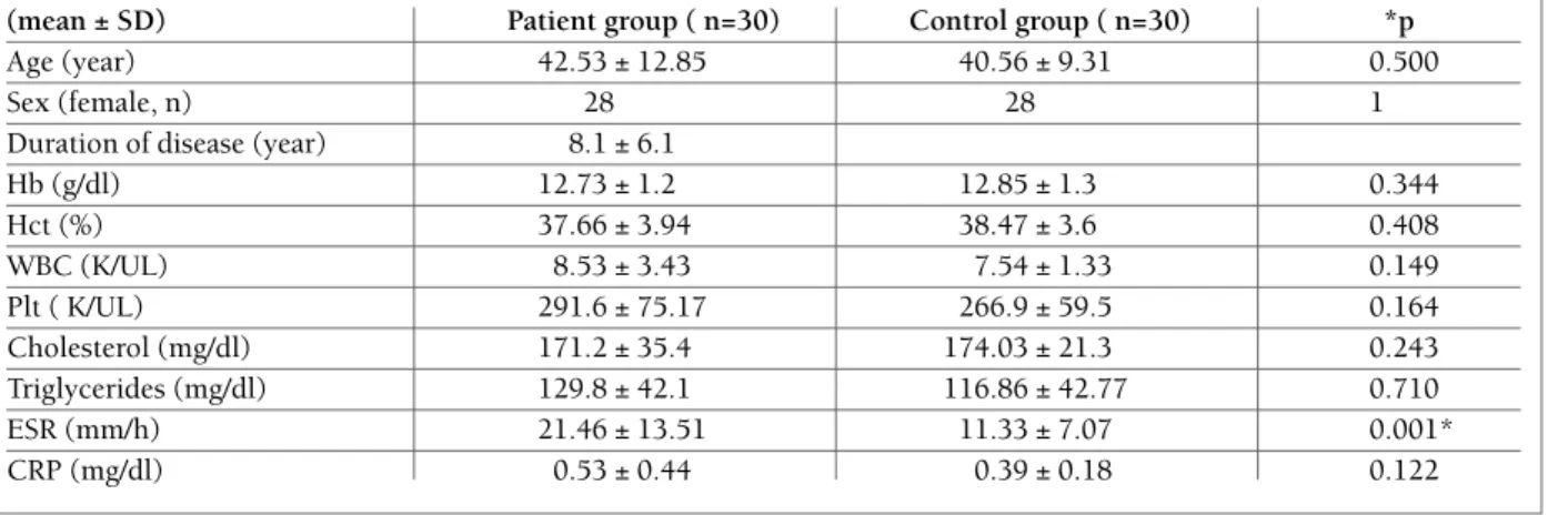
TAble I. COMpARIsION Of DeMOgRAphIC ATTRIbUTes AND lAbORATORy pARAMeTeRs beTweeN pATIeNTs AND CONTROls
(mean ± SD) Patient group ( n=30) Control group ( n=30) *p
Age (year) 42.53 ± 12.85 40.56 ± 9.31 0.500
Sex (female, n) 28 28 1
Duration of disease (year) 8.1 ± 6.1
Hb (g/dl) 12.73 ± 1.2 12.85 ± 1.3 0.344 Hct (%) 37.66 ± 3.94 38.47 ± 3.6 0.408 WBC (K/UL) 8.53 ± 3.43 7.54 ± 1.33 0.149 Plt ( K/UL) 291.6 ± 75.17 266.9 ± 59.5 0.164 Cholesterol (mg/dl) 171.2 ± 35.4 174.03 ± 21.3 0.243 Triglycerides (mg/dl) 129.8 ± 42.1 116.86 ± 42.77 0.710 ESR (mm/h) 21.46 ± 13.51 11.33 ± 7.07 0.001* CRP (mg/dl) 0.53 ± 0.44 0.39 ± 0.18 0.122
*p <0.05, SD – Standard deviation, ESR – Erythrocyte sedimentation rate, CRP – C-reactive protein, WBC – White blood cell count, Hb – Haemoglobin, Hct – Hematocrit, Plt – Platelets
rameters. Of the 30 scleroderma patients, 5 had ele-vated pulmonary arterial pressure, whereas there were 3 people with high pulmonary arterial pressure in the control group.
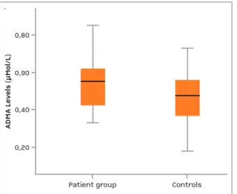
Serum ADMA levels were significantly higher in the patient group (0.545± 0.130) than in the control group (0.130± 0.155) (p = 0.034)) (Figure 1). Furthermore, in the patient group, ADMA levels were significantly higher in active patients (0.608± 0.128) than in inac-tive patients (0.494 ± 0.109) (p=0.016)) (Figure 2). There was no correlation found between ADMA and the echocardiographic parameters. PASP and T-left-Em demonstrated a significant positive correlation. There was also a significant correlation between PASP and MPAP. PASP and MIT-E displayed a significant negati-ve correlation. There was a significant relationship of PASP with T-left-Em and MPAP. A significant correla-tion between T-left-Am and T-right-Em was also found. In addition, there was a significant correlation between duration of disease and RVEF.
ADMA levels and MPAP exhibited a nearly signifi-cant relationship, with a negative correlation between the two parameters (p=0.067).
We determined a significant negative correlation between CRP and RVEF. The relationship of CRP with MIT-A and T-right-A was significant.
We did not find significant differences between pa-tient and control groups except for ESR (Table I). The-re was also no association between ADMA levels and inflammatory markers (CRP and ESR) in both the ac-tive and inacac-tive patient groups.
DIsCUssION
Since previous studies have shown contradictory fin-dings concerning the echocardiographic data of SSc patients, we aimed to evaluate the cardiac functions based on echocardiographic data enriched by measu-rement of pulmonary arterial pressure with various methods such as color and tissue Doppler imaging, while also trying to determine cardiac involvement at a preclinical stage and reveal its relationship with se-rum ADMA levels15-18.
In a study comprised of 54 SSc patients, 69% of the patients demonstrated abnormal findings in echocar-diography. The most common pathologic findings shown by echocardiography are elevated right ventri-cular systolic pressure, pericardial effusion, increased right ventricular diameter, and left atrial enlargement19.
TAble II. ClINICAl ChARACTeRIsTICs AND MeDICATIONs Of pATIeNTs N % Raynaud’s phenomenon 28 (93.3) Digital pitting 22 (73.3) Telangiectasia 10 (33.3) Arthritis 8 (26.7) Dyspnea 23 (76.7) Dysphagia 17 (56.7) Dyspepsia 11 (36.7) Lung involvement 22 (78.5) Calcinosis 1 (3.3)
Calcium channel blockers 22 (73.3) Corticosteroids 18 (60) Colchicine 11 (36.6) Pentoxifylline 1 (3.3) Azathioprine 2 (6.7) Methotrexate 2 (6.7) Cyclophosphamide 3 (10) Not use 5 (16.7)
Seventy percent of the patients were positive for ANA. However, positivity for anti-Scl 70 and ACA was low. Ery throcyte sedimentation rate (ESR) was signifi-cantly higher in the patient group than in the control group (p= 0.001); however, there was no significant difference in terms of CRP (p= 0.122). We found signi -ficant differences between inactive and active patients in terms of Modified Rodnan (p=0.041) and functional scores (p=0.003). Modified Rodnan and functional sco-res were significantly higher in active patients (20.50±7.75, 19.71±8.24 respectively) than inactive patients (14.86±6.19, 10.33±7.42 respectively). The comparison of Doppler echocardiographic parameters between the patient and control groups is shown in Ta-ble III. The patient group was found to have statisti-cally significantly higher T-right-Am, PASP, and T-right-E/ /T-right-A values than the control group. MPAP was nearly significantly higher in the patient group than in the control group (p= 0.054). There was a significant difference between the patient and control groups with regard to RVEF (p<0.05). No significant difference was determined in other parameters.
Doppler echocardiography results of the active and inactive SSc patients are shown in Table IV. There were no significant differences between the active and inac-tive patients regarding Doppler echocardiographic
pa-The patient may not exhibit clinicals symptoms of car-diac involvement if present during the early period, however progression may lead to significant cardiac involvement at later stages. Currently, clinical exami-nation and transthoracic echocardiography are re-commended for routine cardiac evaluation in SSc
ca-ses. However, it is inadequate for diagnosis at the pre-clinical stage20. Cardiac involvement can be detected at
the preclinical stage by evaluating right and left ven-tricular functions with tissue Doppler echocardio-graphy, which enables early treatment.
In our study, we enrolled SSc patients with no
TAble IV. COMpARIsON Of DOppleR eChOCARDIOgRAphIC pARAMeTeRs beTweeN ACTIVe AND INACTIVe pATIeNTs
aParameters (mean±SD) Active patients (n=14) Inactive patients (n=16) p*
T-right-E (m/s) 0.16 ± 0.15 0.18 ± 0.20 0.743 T-right-A (m/s) 0.18 ± 0.05 0.19 ± 0.08 0.868 PASP (mm/Hg) 27.78 ± 7.48 31.86 ± 10.03 0.224 PAMP (mm/Hg) 16.71 ± 5.36 18.66 ± 7.31 0.418 LVEF (%) 56.92 ± 7.26 62.26 ± 8.77 0.085 T-left-Em (m/s) 0.12 ± 0.04 0.13 ± 0.05 0.583 T-left-Am (m/s) 0.14 ± 0.18 0.14 ± 0.10 0.957 RVEF (%) 55.85 ± 8.75 57.33 ± 7.00 0.622 MIT-E (m/s) 5.96 ± 19.58 0.98 ± 0.29 0.359 MIT-A (m/s) 0.67 ± 0.22 0.78 ± 0.22 0.174
a. See Table 2 for acronym definitions * p<0.05, SD Standard deviation
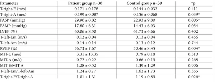
TAble III. COMpARIsON Of DOppleR eChOCARDIOgRAphIC pARAMeTeRs beTweeN pATIeNTs AND CONTROl gROUps
Parameter Patient group n=30 Control group n=30 *p
T-right-E (m/s) 0.171 ± 0.178 0.144 ± 0.032 0.411 T-right-A (m/s) 0.199 ± 0.087 0.156 ± 0.068 0.039* PASP (mmHg) 29.90 ± 8.82 22.93 ± 9.80 0.005* PAMP (mmHg) 17.80 ± 6.31 14.43 ± 6.93 0.054 LVEF (%) 60.06 ± 8.50 61.73 ± 6.66 0.402 T-left-Em (m/s) 0.12 ± 0.04 0.13 ± 0.04 0.456 T-left-Am (m/s) 0.14 ± 0.14 0.13 ± 0.12 0.744 RVEF (%) 56.73 ± 7.67 50.46 ± 8.45 0.004* MIT-E (m/s) 3.31 ± 13.35 0.79 ± 0.18 0.310 MIT-A (m/s) 0.72 ± 0.22 0.66 ± 0.19 0.268 MIT E/MIT A 1.28 ± 0.52 1.39 ± 1.29 0.906 T-left-Em/T-left-Am 1.24 ± 0.77 1.62 ± 1.73 0.355 T-right-E/T-right-A 1.01 ± 1.31 1.19 ± 0.89 0.026*
EF-left – Left ventricular ejection fraction, EF-right – Right ventricular ejection fraction, MIT-E – Mitral valve early diastolic wave, MIT-A – Mitral valve late diastolic wave, MIT-E / MIT A – Mitral valve early diastolic wave / Mitral valve late diastolic wave ratio, T-left-Em – Left ventricular lateral wall tissue Doppler early diastolic wave, T-left-Am – Left ventricular lateral wall tissue Doppler late diastolic wave, T-left-Em/T-left-Am – Left ventricular diastolic dysfunction, T-right-E Right – ventricular lateral wall tissue Doppler early diastolic wave, T-right-A – Right ventricular lateral wall tissue Doppler late diastolic wave, T-right-E/T-right-A – Right ventricular diastolic dysfunction, PASP – Pulmonary arterial systolic pressure, PAMP – Pulmonary arterial mean pressure
known cardiac complaint. Our patients demonstrated significant increases in parameters indicating right ven-tricular diastolic dysfunction: T-right-E/T-right-A, T-right-A, and PASP. There was a nearly significant in-crease of MPAP as well. Compared with the control group, significant or nearly significant increases in pul-monary arterial pressure may be beneficial in taking ti-mely precautions before the expected clinical deve-lopment of PH.
Dimitroulas et al. studied 52 SSc patients with no clinical cardiac involvement. They examined left and right ventricular functions by tissue Doppler echocar-diography and determined reduced right and left ven-tricular function in these asymptomatic patients20.
Si-milarly, in our study, right ventricular function was obser ved to be decreased.
Plazak et al. studied 46 SSc patients and found signi -ficantly higher pulmonary arterial pressure in SSc pa-tients than in controls21. In the present study, systolic
pulmonary artery pressure was significantly higher in the SSc patient group than in the control group. Fur -thermore, although not statistically significant, the SSc patients had higher MPAP values.
Hural et al. evaluated left ventricular systolic and diastolic functions by echocardiography in 31 SSc pa-tients and found depressed left ventricular function along with impaired systolic and diastolic functions22.
In another study, left ventricular dysfunction was as-sessed by conventional echocardiography in 7073 pa-tients and left ventricular dysfunction was observed in 5.4% of the patients. In the present study, we
evalua-ted the left ventricular function as well; however, no dysfunction was detected. We failed to determine left ventricular dysfunction, which was expected to be sta-tistically significant. This may have been due to the small number of patients. This may also be associated with the short duration of disease in our patients. Hu-ral et al. did not study right ventricular function22.
How ever, in our study, as expected, right ventricular diastolic dysfunction was detected. Furthermore, right ventricular systolic function was reduced in patients with active disease compared to patients with inactive disease. However, this decrease was not significant.
ADMA is an endogenous inhibitor of NO. It is pro-duced by the methylation of arginine. It is known to cause endothelial dysfunction by reducing the NO le-vel. ADMA levels have been observed to be high in sys-temic diseases with cardiac involvement accompanied by endothelial dysfunction. Many studies have found a correlation between ADMA levels and cardiac invol-vement23,24. In the present study, we found
significan-tly higher serum ADMA levels in the patient group than in the control group. We also determined a near-ly significant relationship between ADMA levels and MPAP values.
Perna et al. studied 125 SLE patients and found a re-lationship between elevated ADMA levels and cardio-vascular involvement. ADMA levels were particularly high in SLE patients with arterial wall thickening. The authors concluded that ADMA could be a biochemi-cal marker with regard to risk of cardiovascular invol-vement23.
Patient groupControls0,800,900,400,20
ADMA Levels (µMol/L)
Patient group Controls 0,80 0,90 0,40 0,20 A D M A L ev el s (µ M ol /L )
fIgURe 1.Comparison of ADMA levels of study groups (p<0.05)
Active disease groupInactive disease group0,800,900,700,600,500,400,30
ADMA Levels (µMol/L)
Active disease group Inactive disease group 0,80 0,90 0,70 0,60 0,50 0,40 0,30 A D M A L ev el s (µ M ol /L )
fIgURe 2.Comparison of ADMA levels in disease group according to the disease activity (p<0.05)
RefeReNCes
1. Black, CM. The aetiopathogenesis of systemic sclerosis: Thick skin-thin hypotheses. The Parkes Weber Lecture. J R Coll Phy-sicians Lond 1995.p.119-130.
2. Ferri C, Di Bello V, Martini A, et al. Heart involvement in sys-temic sclerosis: an ultrasonic tissue characterisation study. Ann Rheum Dis 1998;57:296–302.
3. Medsger TA. Systemic sclerosis (scleroderma), eosiniphilic fas-ciitis, and calcinosis. In: McCarty DJ. Arthritis and Allied Conditions: a Textbook of Rheumatology, 11th edition. Philadel -phia: Lea & Febiger. 1989.p.1118–1165.
4. James TN. De subitaneis mortibus. VIII. Coronary arteries and conduction system in scleroderma heart disease. Circulation 1974.p.844–856.
5. D’Angelo WA, Fries JF, Masi AT, et al. Pathologic observations in systemic sclerosis (scleroderma). A study of fifty-eight au-topsy cases and fifty-eight matched controls. Am J Med 1969;46:428–440.
6. Follansbee WP, Miller TR, Curtiss EI, et al. A controlled clini-copathologic study of myocardial fibrosis in systemic sclerosis (scleroderma). J Rheumatol 1990;17:656–662.
7. Güllülü S, Kaderli AA, Ekbul A, Ozdemir B, Baran I, Güllülü M, Ediz B, Cordan J, Yurtkuran M Tissue Doppler echocardio-graphy and myocardial performance index in patients with scle-roderma. J Int Med Res 2005;33(4):417-424.
8. Nijveldt RJ,Siroen MPC,Teerlink T, Leeuwen MV. Elimination of Asymetric Dimethylarginine by the Kidney and the Liver:A Link to the Development of Multiple Organ Failure..,The Ame-rican Society for Nutritional Sciences. J.Nutr 2004;134:2848--2852
9. Böger RH, aas R,Schulze F and Schwedhelm E. Elevated levels of ADMA as a marker of cardiovasküler disease and mortali-ty.Clin chem Lab med;2005;43(10):1124-1129
10. Nijveldt RJ, Siroen MPC, Teerlink T, et al. Elimination of Asy-metric Dimethylarginine by the Kidney and the Liver:A Link to the Development of Multiple Organ Failure.The American So-ciety for Nutritional Sciences J Nutr. 2004;134:2848-2852. 11. Subcommittee for Scleroderma Criteria of the American
Rheu-matism Association Diagnostic and Therapeutic Criteria Com-mittee. Preliminary criteria for the classification of systemic sclerosis (scleroderma). Arthritis Rheum 1980;23:581-90 12. Clements P, Lachenbruch P, Siebold J, White B, Weiner S,
Mar-tin R, Weinstein A, Weisman M, Mayes M, Collier D, et al In-ter and intraobserver variability of total skin thickness score (modified Rodnan TSS) in systemic sclerosis J Rheumatol. 1995;22:1281-5.
13. Valentini G, Della Rossa A, Bombardieri S, et al. European mul-ticentre study to define disease activity criteria for systemic sy-clerosis. II. dentification of disease activity variables and deve-lopment of preliminary activity indexes. Ann Rheum Dis. 2001;60:592-598.
14. Silman A, Akesson A, Newman J. Assessment of functional Abi-lity in patients with SSc: a proposed new disabiAbi-lity assessment instrument. JRheumatol. 1998;25:79-83.
15. G. P. Armsrong, G. A. Whalley, R. N. Doughty, G. D. Gamble, S. M. Flett, P. L. J.Tan and D. N. Sharpe. Left ventricular func-tion in Scleroderma. British Journal of Rheumatology 1996;35:983-988
16. Denton CP, Black CM. Scleroderma – clinical and pathological advances. Best Pract Res Clin Rheumatol 2004; 18: 271-290 17. Champion HC. The heart in scleroderma. Rheum Dis Clin
Sahin et al. conducted a study on patients with Beh-çet’s disease and detected higher ADMA levels, parti-cularly in patients with vascular involvement24.
ADMA levels are increased in diffuse, cutaneous subtypes of SSc disease and in patients with pulmonary hypertension25. Furthermore, a study by Dimitroulas
et al. consisted of 66 patients and 30 controls. Patients with and without PH, and controls were compared. They found significantly higher ADMA levels in SSc patients with PH25. In the present study, we
determi-ned a close relationship between MPAP and serum ADMA levels in SSc patients, a relationship that was nearly statistically significant. Dimitroulas et al. investigated the links between ADMA and echocardiogra -phic parameters of SSc patients with cardiac involve-ment in 56 patients and determined a considerably close relationship between ADMA levels and echocar-diographic measures of LV diastolic dysfunction25. In
our study, ADMA levels were observed to be high in SSc patients. In addition, ADMA levels were found to be higher in active patients than in inactive patients. However, no link was detected between ADMA levels and echocardiographic parameters. The small number of our study population and short duration of disease may explain this result.
There is no clear association between ADMA and systemic inflammation. ADMA has been associated with inflammatory indicators only in some studies in patients with arthritis26-27. We did not find a relation ship
between ADMA and inflammatory markers in the pre-sent study. Our results may be related to this unclear association with inflammatory indices and also may be attributed to mild inflammatory markers of SSc.
In conclusion, SSc patients demonstrate elevated ADMA levels which are correlated with the disease acti vity. This increase may be a precursor of possible cardiac and vascular complications in the future. In the present study, we detected impaired cardiac diastolic functions by applying advanced echocardiogra -phic methods on SSc patients exhibiting no symptom of known cardiac involvement. At the subclinical sta-ge, determination of the cardiac involvement may al-low taking precautionary steps and thus contribute po-sitively to the long-term prognosis of the disease.
CORRespONDeNCe TO
Banu Dilek
Dokuz Eylul University Hospital,
Department of Physical Medicine and Rehabilitation, Izmir, Turkey
North Am 2008; 34: 181-190.
18. Follansbee W, Zerbe T, Medsger TJ. Cardiac and skeletal mus-cle disease in systemic smus-clerosis (smus-cleroderma): a high risk as-sociation. Am Heart J 1993; 125: 194-203.
19. Fisher M, Mathai S, Champion H, et al. Clinical differences bet-ween idiopathic and scleroderma-related pulmonary hyper-tension. Arthritis Rheum. 2006; 54:3043-3052.
20. Dimitroulas T, Giannakoulas G, Papadopoulou K, et al. Early detection of cardiac involvement in systemic sclerosis assessed by tissue-Doppler echocardiography: relationship with neuro-hormonal activation and endothelial dysfunction. J Rheumatol. 2010;37:993-999.
21. Plazak W, Gryga K, Sznaid J, et al. Diastolic heart dysfunction, increased pulmonary capillary wedge pressure and impaired exercise tolerance in patients with systemic sclerosis. Kardiol Pol. 2011;69:250.
22. Hüral R, Özdemir A, Turhan S, et al. Assessment of left ventri-cular systolic and diastolic function myocardial performance in-dex with tissue Doppler echocardiography in patients with scle-roderma. MN cardiology. 2009;16:225-232.
23. Perna M, Roman MJ, Alpert DR, et al. Relationship of asym-metric dimethylarginine and homocysteine to vascular aging in systemic lupus erythematosus patients. Arthritis Rheum. 2010;62:1718-1722.
24. ahin M, Arslan Ç, Neziro lu M, et al. ADMA ve NO levels as a sign of endothelial disfunction in Behçet’s diseases, Ann.of Cli-nical lab Science. 2006;36:449-454.
25. T. Dimitroulas, G. Giannakoulas, T. Sfetsios, H. Karvounis, H. Dimitroula, G. Koliakos and L. Settas. Asymmetrical dimetlarginine in systemic sclerosis-related pulmonary arterial hy-pertension.Rheumatology 2008;47:1682–1685
26. Kemény-Beke, Á.; Gesztelyi, R.; Bodnár, N.; Zsuga, J.; Kerekes, G.; Zsuga, M.; Biri, B.; Kéki, S.; Szodoray, P.; Berta, A.; et al. In-creased production of asymmetric dimethylarginine (ADMA) in ankylosing spondylitis: Association with other clinical and la-boratory parameters. Joint Bone Spine 2011;78:184–187. 27. Kwa ny-Krochin, B.; Głuszko, P.; Undas, A. Plasma asymmetric
dimethyl-L-arginine (ADMA) in active rheumatoid arthritis: Links with oxidative stress and inflammation. Pol. Arch. Med. Wewn. 2012;122: 270–276.