Abstract
Submitted: September 20, 2016 Modiication: December 19, 2016 Accepted: December 29, 2016
Treatment of experimental periodontal
disease by laser therapy in
simvastatin-modiied rats
Low intensity laser can be used as a promising alternative in the treatment of periodontal disease. Objective: The aim of this study was to evaluate low-level laser therapy (LLLT) as an adjuvant treatment for scaling and root planing (SRP) for the treatment of induced periodontitis in
simvastatin-modiied rats. Material and Methods: A total of 180 rats were evenly divided
into two groups: Veh – receiving oral administration of polyethylene glycol (vehicle); S – receiving oral administration of Simvastatin. Periodontal disease
was induced in both groups at the irst mandibular molar. After seven days,
the ligature was removed and the animals were divided into subgroups according to the following local treatments: NT – no treatment; SRP – scaling and root planing and irrigation with saline solution; and LLLT ¬– SRP and laser irradiation (660 nm; 0.03 W; 4 J). Ten animals in each subgroup/local treatment were euthanized at 7, 15 and 30 days. Samples of gingival tissue were processed to analyze the tissue oxidative damage and radiographic analysis. Levels of oxidative stress were analyzed by the expressions of Tripeptideglutathione (TG), Malondialdehyde (MDA) and Carbonylated Proteins (CP). Results: The animals in S group had higher levels of TG and lower levels of MDA and CP compared with Veh group (p<0.05). Radiographically, in the intragroup analysis Veh and S, LLLT showed lower bone loss (BL) compared with NT and SRP, in all experimental periods (p<0.01). In addition, a lower BL was observed for the animals of Veh group treated with LLLT compared with treatment SRP in the S group, in all experimental periods. Conclusion: Within the limits of this study, we can conclude that LLLT was effective as adjuvant treatment for SRP protecting against the occurrence of oxidative tissue damages as well as for reducing alveolar bone loss in experimentally
induced periodontitis simvastatin-modiied rats.
Keywords: Alveolar bone. Laser therapy. Oxidative stress. Periodontal diseases. Simvastatin.
Andressa Araújo SWERTS1
Bianca Fernanda Espósito SANTOS1
Simone Ribeiro BRUZADELLI1
Maísa Ribeiro Pereira Lima
BRIGAGÃO2
Daniela Coelho de LIMA1
Leandro Araújo FERNANDES1
1Universidade Federal de Alfenas, Faculdade de Odontologia, Departamento de Clínica e Cirurgia,
Alfenas, MG, Brasil.
2Universidade Federal de Alfenas, Faculdade de Odontologia, Departamento de Bioquímica, Alfenas,
MG, Brasil. Corresponding address:
Leandro Araújo Fernandes Departamento de Clínica e Cirurgia - Faculdade de
Introduction
In inlammation, neutrophils are the irst cells
to be activated to defend the body together with
macrophages27. Those are chemotaxically attracted
by secretory cells, bacteria and other foreign bodies
to inlammation areas27. On this site, neutrophils
phagocyte the microorganisms, covered or not with
complement or speciic antibodies, which are killed by
cytotoxic proteins derived from cytoplasmic granules
and by oxygen and nitrogen reactive species such
as superoxide anion9, hydrogen peroxide28, hydroxyl
radical12 and peroxynitrite21.
There is evidence of a more aggressive destruction of tooth support tissues with elevated levels of oxidative stress markers during the development of periodontal disease (PD)24. Among the oxidants,
the superoxide anion9, in periodontal tissues, can be
involved in signaling of induction of bone resorption; the hydroxyl radical12 is extremely reactive and can
damage important biomolecules, such as proteins, lipids and nucleic acids, whereas hydrogen peroxide is
able to cross membranes, damaging adjacent cells and
increasing the oxidative cascade28. Thus, most studies
demonstrate that periodontitis is associated with increased lipid peroxidation29 and increased protein
carbonyls9 as well as decreased antioxidants, such as
reduced glutathione (GSH)11,29. In addition, there are
evidences of decreased oxidative injuries and changes in antioxidant system following periodontal treatment19.
The treatment of PD is based on the elimination of pathogenic subgingival microbiota by scaling and root planing (SRP)24. However, mechanical therapy used
alone may be defective in the elimination of pathogenic bacteria, since they are located within soft and hard tissues or in areas that are inaccessible to periodontal instruments7. In addition, an important component of
individuals themselves can lead to tissue destruction observed in the periodontitis. Therefore, therapeutic strategies performing the pharmacological modulation of host response have emerged as a new therapeutic approach13.
Simvastatin is an inhibitor of the 3-hydroxy-3-methyl-glutaryl-coenzyme A (HMG - CoA reductase) enzyme, which is responsible for the synthesis of cholesterol and therefore it is widely used for the systemic treatment of diseases related to hypercholesterolemia. This drug also has
anti-inlammatory, immunomodulator, antioxidant, and
angiogenic effects6,25, and it also promotes increased
osteoblast formation10,20. Such properties offer great
potential for statins to modify the course of chronic
inlammatory diseases such as periodontitis26.
In addition to drug therapy, the use of low intensity lasers associated with scaling and root planing for the local treatment of periodontal disease has been reported7. On the other hand, there are no studies
evaluating the local effects of low-level laser therapy (LLLT) associated with the systemic effects of statins.
Thus, the aim of this study was to evaluate LLLT as an adjuvant treatment for scaling and root planing
(SRP) in the treatment of induced periodontitis in
simvastatin-modiied rats.
Material and methods
Animals
This study was conducted on 180 adult male Wistar rats (200–250 g). The animals were kept in plastic cages with access to food and water ad libitum. Prior to surgical procedures, all animals were allowed to acclimatize to the laboratory environment
for a period of ive days. All protocols described below
were approved by the Ethics Committee on the Use of Animals (CEUA), following the standards adopted
by the Brazilian College of Animal Experimentation (COBEA), under protocol 472/2012.
Study design
Animals were numbered and randomly divided into two groups: Veh group (n=90) received Polyethylene Glycol 400 (All Chemistry; São Paulo, SP, Brazil) at
0.5 mg/kg body weight (vehicle), and S group (n=90)
received Simvastatin (Medley; Campinas, SP, Brazil) at 0.5 mg/kg body weight orally18. Administrations
were daily performed in a single dose, starting 24 h
before induction of PD and maintained until the end
of the respective periods of euthanasia. Animals were
weekly weighed throughout the experimental period to maintain the doses.
Simvastatin preparation was performed by diluting 400 mg of the drug in 400 mlL of Polyethylene Glycol
to reach a inal concentration of 1 mg/mL (Moss
Manipulation Pharmacy; Alfenas, MG, Brazil).
Induction of experimental periodontal disease
ketamine (0.4 mL/kg) (Fort Dodge Animal Health Ltda; Campinas, SP, Brazil) together with xylazine (0.2 mL/kg) (Coopers; São Paulo, SP, Brazil) via
intramuscular injection. The mandibular left irst
molar from each animal in both Veh and S groups was selected to receive a cotton ligature No. 10 (Coats; São Paulo, SP, Brazil) in a submarginal position to induce experimental periodontitis2.
Local treatment
After seven days of periodontal disease experimental induction, mandibular ligature was removed from the
left irst molar of all animals in groups Veh and S.
The animals were divided into subgroups according to the local treatments (performed only once): NT – no treatment; SRP – scaling and root planing and irrigation with saline solution; and LLLT – SRP and laser irradiation.
Left molars were subjected to SRP with manual
#1–2 micro mini ive curettes (Hu-Friedy; Chicago,
IL, USA) through 10 distal–mesial traction movements in both buccal and lingual aspects. The furcation and interproximal areas were scaled with the same curettes through cervical-occlusal traction movements. The entire SRP procedure was performed by the same experienced operator. The saline solutions were slowly deposited within the periodontal pocket using syringe
(1 mL) and insulin needle (13 mm x 0.04 mm) (Becton Dickinson; Curitiba, PR, Brazil).
The laser used in this study was gallium– aluminium–arsenide (GaAlAs) (Kondortech Equipment Ltd; São Carlos, SP, Brazil), with a wavelength of 660 nm and a spot size of 0.07 cm2. After 1 min of
saline solution application, LLLT was applied to three equidistant points at each buccal and lingual aspect of
the mandibular irst molar in contact with the tissue.
The therapeutic laser was released with a power of 0.03 W for 133 s/point, a power density of 0.428 W/ cm2, and energy of 4 J/point (57.14 J/cm2/point)7.
Experimental periods
Ten animals from each experimental subgroup/local
treatment were euthanized by exsanguination at 7, 15
and 30 days following local treatments. The jaws were removed and, in order to analyze the levels of oxidative stress, collections of the inserted and marginal gums
of the buccal faces of the irst left lower molars were
performed. Then, the jaws were split in the middle
and ixed in 10% buffered formalin for at least 48 h
for later radiographic analysis of the left side.
Preparation of gingival samples
Gingival tissue samples were homogenized with phosphate buffer (3 mL, pH 6.5). The homogenate was centrifuged at 3500 rpm for 10 min. The supernatant was separated into aliquots for subsequent biochemical analysis.
Determination of the total protein concentration
Protein concentrations were determined in allsamples of gingival homogenates by the method
of Bradford4, using bovine serum albumin (BSA)
as a standard calibration curve. The total protein
concentration is routinely determined for normalization
of biochemical results. Thus, the expression results in the mass in mg of tripeptideglutathione (TG), malondialdehyde (MDA) and carbonylated proteins (CP) per mg protein (units).
Determination of tripeptideglutathione (TG)
For veriication of TG in the gingival tissue of ratsalkylated derivatives of mBBr were separated by HPLC
in C-18 column (Shim-pack VP-ODS, 4.6 mm x 25 cm,
5 µm, connected in series with C-18 precolumn model
Shim-pack GVP-ODS, 4.6 mm x 10 mm), equilibrated with buffer A (14.2% ethanol and 0.25% acetic acid
m/v, 1 mL/min). Samples of tissue suspensions
derivatized with mBBr were injected and dilution
(buffer A) was performed for 30 min. Then, the column was washed with buffer B (90% methanol and 0.25%
acetic acid) for 8 min and re-equilibrated with buffer
A. Sulfosalicylic acid (internal reference standard),
cysteine and reduced glutathione were determined by comparing the retention time of authentic standards
(10-200 nmol) using a luorescence detector (model RF-10AXL) and λexc=394 nm and λemi=490 nm (ε=20
mM-1.cm-1), and the quantiication was performed by
counting the units in the respective areas (Software “LC-Solution Multi”)14.
Determination of malondialdehyde (MDA)
The MDA veriication through homogenized gum samples was performed by adding 250 μL of 1.22 M phosphoric acid, 450 μL of Milli-Q Water and 250 μL
of TBA reagent. Thereafter, aliquots are stirred for 30 s and the reaction is incubated for 1 h in water bath at 95°C and cooled in ice bath at 4°C. After this
of 1M NaOH are added to a 200 µL sample, in order to
neutralize the solutes and precipitate proteins before
injecting into the HPLC column, using a luorescence detector (model RF-10AXL) and λexc=515 nm and λemi=553 nm14.
Determination of carbonylated proteins (CP)
In order to determine these CP, 500 µL of the aliquots in phosphate buffer (pH 7.2) may be used,adding 500 µL of 2.4-dinitrophenylhydrazine solution
(10 mμ). After that, incubate for 1 h and then apply
(drops) 500 µL of 20% TCA solution until complete precipitation. Add 500 µL of Ethanol/Ethyl-Acetate
solution and centrifuge at 2000 rpm for 10 min.
Discard the supernatant and re-add 500 µL of
ethanol/ethyl-acetate solution and centrifuge it. Then, discard the supernatant and dissolve the precipitate
with 1000 µL of 6M guanidine, which should be
homogenized. The rate of change in absorbance was
spectrophotometrically measured at 370 nm14.
Radiograph and digital analysis
After ixation of the hemimandibules (HMs) in 10%
buffered formalin, the left side was submitted to X-ray procedure.
HMs were positioned on a table with the vestibular
surfaces facing the radiographic ilm (Eastman Kodak
Company; Rochester, Nova York, EUA), in such a way
that the right side stayed at the bottom, and the left side stayed at the top.
Standardization of radiographs was obtained as follows:
Use of an X-ray device Pampas - E (CDK X-ray Equipment; Diadema, SP, Brazil), with electric system of 65 kvp, 10 mA;
Central X-ray beam perpendicular to the ilm-object
plane, at a 90-degree angle in relation to the surface of the optical plate;
Focal length of 30 cm;
Exposure time of 0.8 seconds.
Radiographs were developed using solutions from
Kodak developer and ixer, using the climate-weather
development method.
They were scanned and the images were analyzed with the Imagelab software (Softium; São Paulo, SP, Brazil) using the tool distance and angle of measurement. With this feature, it was measured the distance from the cementoenamel junction to the alveolar bone crest on the mesial surface of the
irst left lower molars by drawing a line, and these
measurements were recorded in millimeters (mm). The mouse was positioned on the region corresponding to the cementoenamel junction. By left-clicking and dragging the mouse down to the level of alveolar bone crest the software automatically measured the distance.
Statistical analysis
Statistical analysis of oxidative stress and radiographic data was performed by BioEstat 3.0 software (Sonopress; Manaus, AM, Brazil). The
hypothesis of absence of a statistically signiicant
difference in the data obtained in the region of the
mandibular irst molars between different groups,
subgroups/treatments and periods in the teeth with induced periodontitis was tested. After the analysis of the normality of the data by the Shapiro-Wilk test, multiple comparisons among the variables were performed by two-way analysis of variance ANOVA
with supplementation by the Tukey test, with p<0.05 (oxidative stress data) and by the Bonferroni test, with p<0.01 (radiographic data).
Results
Expression of TG in gingival samples
In the intragroup comparison (Veh and S), TG
levels increased signiicantly (p<0.05) between the
experimental periods in all local treatments. Regarding
local treatments, LLLT showed a signiicant increase
(p<0.05) in GSH compared with NT and SRP at 7 and 30 days.
In the comparison between the groups (Veh and S), among the same local treatments, TG levels in
the S group were signiicantly higher than those in
the Veh group (p<0.05), for NT, SRP and LLLT in all experimental periods (Table 1).
Expression of MDA in gingival samples
In the intragroup comparison, levels of MDA in LLLT
decreased signiicantly (p<0.05) compared with NT,
in the S group in all experimental periods, and in the Veh group at 7 and 30 days.
In the comparison between the groups (Veh and S), among the same local treatments, MDA levels in S
group were signiicantly lower than those in Veh group
Group Veh
Periods 7 days 15 days 30 days
Treatments
NT 5.02±0.24#*´° 10.99±0.30#´° 22.17±1.21#*´°
SRP 8.52±1.00 #*´° 15.01±0.30#´° 40.76±1.98#*´°
LLLT 10.13±1.13#*´° 16.07±1.64#´° 52.49±0.58#*´
Group S
Periods 7 days 15 days 30 days
Treatments
NT 14.15±0.34#*´° 23.11±1.67#´° 43.87±0.56#*´°
SRP 17.43±0.02#*´° 26.79±1.02#´° 54.97±1.02#*´°
LLLT 23.29±0.17#*´° 24.32±1.09#´° 64.98±1.12#*´°
N 60 60 60
#Difference among same groups and local treatments (ANOVA and p<0.05)
*Difference between local treatments, same group and period (ANOVA and Tukey, p<0.05) ´Difference between groups, same local treatment and period (ANOVA and Tukey, p<0.05) °Difference between groups and local treatments, same period (ANOVA and Tukey, p<0.05)
Table 1- Means and standard deviations (M±SD) expression in TG units in the gingival tissue of the irst lower left molar, according to each group, local treatment and period
Group Veh
Periods 7 days 15 days 30 days
Treatments
NT 0.18±0.06´° 0.24±0.12° 0.30±0.03°
SRP 0.28±0.12´° 0.40±0.09 0.62±0.13*
LLLT 0.48±0.32*´° 0.47±0.43* 0.34±0.17°
Group S
Periods 7 days 15 days 30 days
Treatments
NT 0.34±0.07´° 0.36±0.16 0.44±0.08°
SRP 0.53±0.22´° 0.38±0.65 0.61±0.73*°
LLLT 0.72±0.54*´° 0.44±0.33*° 0.41±0.98°
N 60 60 60
#Difference among same groups and local treatments (ANOVA and p<0.05)
*Difference between local treatments, same group and period (ANOVA and Tukey, p<0.05) ´Difference between groups, same local treatment and period (ANOVA and Tukey, p<0.05) °Difference between groups and local treatments, same period (ANOVA and Tukey, p<0.05)
Table 2- Means and standard deviations (M±SD) expression in MDA units in the gingival tissue of the irst lower left molar, according to each group, local treatment and period
Group Veh
Periods 7 days 15 days 30 days
Treatments
NT 13.23±0.31+*´° 8.45±1.56+* 7.89±1.95*°
SRP 7.02±0.31+*´° 2.39±1.13+*° 3.92±0.06+*°
LLLT 4.71±0.23+*´° 1.99±0.94+*° 2.37±0.18+*°
Group S
Periods 7 days 15 days 30 days
Treatments
NT 9.78±.3+*´° 7.99±11.3+*° 5.21±0.04+*°
SRP 1.29±1.16+*´° 4.80±1.12+*° 3.92±1.00+*°
LLLT 1.57±0.95+*´° 3.41±1.04+*° 1.09±0.32+*°
N 60 60 60
#Difference among same groups and local treatments (ANOVA and p<0.05)
*Difference between local treatments, same group and period (ANOVA and Tukey, p<0.05) ´Difference between groups, same local treatment and period (ANOVA and Tukey, p<0.05) °Difference between groups and local treatments, same period (ANOVA and Tukey, p<0.05)
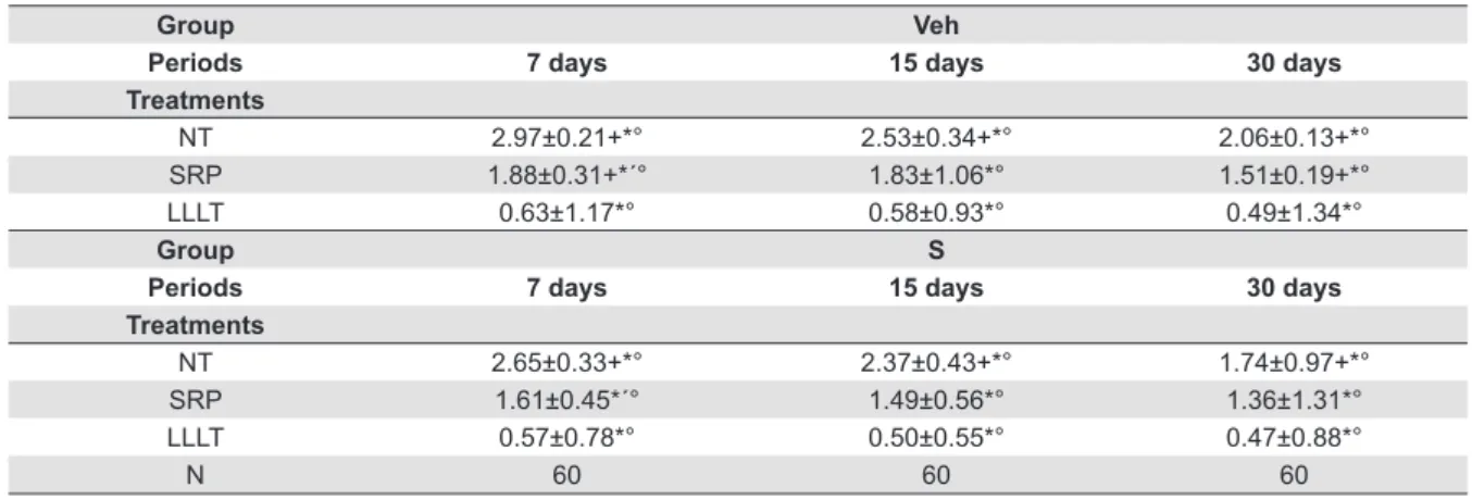
Expression of CP in gingival samples
In the intragroup comparison (Veh and S), levels of
CP in LLLT decreased signiicantly (p<0.05) in relation
to NT in all the experimental periods.
In the comparison between Veh and S groups, among the same local treatments, CP levels in the S
group were signiicantly lower than those in the Veh
group (p<0.05), for NT, SRP and LLLT at 7 days. In addition, CP levels in the Veh group, treated by LLLT,
were signiicantly lower than those in the S group
(p<0.05), which did not receive local treatment (NT) at 7 and 15 days (Table 3).
Radiographic analysis
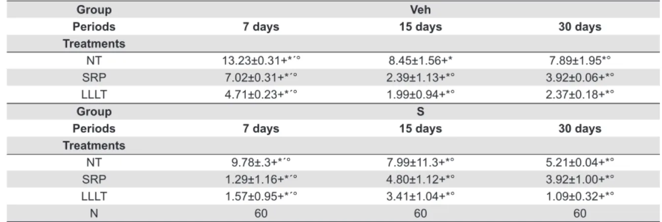
In the intragroup analysis between Veh and S, LLLT showed lower BL compared with NT and SRP, in all experimental periods (p<0.01).
In the intergroup analysis between the same local treatments, the SRP presented a lower BL (p<0.01) in
Group Veh
Periods 7 days 15 days 30 days
Treatments
NT 2.97±0.21+*° 2.53±0.34+*° 2.06±0.13+*°
SRP 1.88±0.31+*´° 1.83±1.06*° 1.51±0.19+*°
LLLT 0.63±1.17*° 0.58±0.93*° 0.49±1.34*°
Group S
Periods 7 days 15 days 30 days
Treatments
NT 2.65±0.33+*° 2.37±0.43+*° 1.74±0.97+*°
SRP 1.61±0.45*´° 1.49±0.56*° 1.36±1.31*°
LLLT 0.57±0.78*° 0.50±0.55*° 0.47±0.88*°
N 60 60 60
#Difference among periods, same groups and local treatments (ANOVA and Bonferroni, p<0.01) *Difference between local treatments, same group and period (ANOVA and Bonferroni, p<0.01) ´Difference between groups, same local treatment and period (ANOVA and Bonferroni, p<0.01) °Difference between groups and local treatments, same period (ANOVA and Bonferroni, p<0.01)
Table 4- Means and standard deviations (M±SD) of the distances between the cementoenamel junction and alveolar bone crest (mm) on
the mesial surface of the irst lower left molar, according to each group, local treatment and period
Figure 1- Radiographic images of the hemimandibules of animals of the Veh group – A: NT 7 days; B: NT 15 days; C: NT 30 days; D: SRP
7 days; E: SRP 15 days; F: SRP 30 days; G: LLLT 7 days; H: LLLT 15 days; and I: LLLT 30 days
Figure 2- Radiographic images of the hemimandibules of animals of the S group – A: NT 7 days; B: NT 15 days; C: NT 30 days; D: SRP
the animals of the S group (1.61±0.45 mm) compared with those in the Veh group (1.88±0.31 mm) in the 7-day period.
In the S group, LLLT values (0.57±0.78 mm, 0.50±0.55 mm, 0.47±0.88 mm) showed lower BL (p<0.01) compared with animals of the Veh group treated with SRP (1.88±0.31 mm, 1.83±1.06 mm,
1.51±0.19 mm) in all experimental periods. In addition, a lower BL was observed for the animals
of the Veh group treated with LLLT (0.63±1.17 mm;
0.58±0.93 mm; 0.49±1.34 mm) compared with SRP treatment in the S group (1.61±0.45 mm; 1.49±0.56
mm; 1.36±1.31 mm), in all experimental periods
(Table 4, Figure 1 and Figure 2).
Discussion
Periodontal disease is a chronic inflammatory p r o c e s s c h ara c t e r i ze d by g i n g i va l b l e e d i n g ,
formation of periodontal pockets and destruction of
periodontal supporting tissues, through the release of
lipopolysaccharide and proteases27 of bacteria present
in the dental bioilm, a key etiological factor of this change. This tissue inlammation is associated with
increased release of oxygen reactive species (ORS) by neutrophils, and also with the activation of several
inlammatory mediators, such as interleukins (IL-1β, 6 and 8) and tumor necrosis factor (TNF-α)11, which
promotes the imbalance in bone homeostasis, resulting
in destruction of alveolar bone tissue, increased activity of matrix metalloproteinases and connective
tissue degradation17, due to exacerbated immune
response and localized osteoclastogenesis12.
Periodontal treatment is used to paralyze the destruction of the supporting tissues of the teeth in order to avoid their loss5. However, there are cases
where isolated periodontal mechanical therapy is ineffective, suggesting that systemic factors, not yet understood, could interfere with the development of the disease22. Thus, the use of antioxidants could be
an adjuvant therapy to conventional treatment17.
Simvastatin is a drug with hypolipidemic function, but it stands out for other minor effects, including
anti-inlammatory25, immunomodulatory, and antioxidant
effects, and the promotion of angiogenesis and
increased differentiation of osteoblasts, inducing bone formation10,20. Such properties offer great potential for
statin to modify the course of chronic inlammatory
diseases26 such as chronic periodontitis.
Ligature-induced alveolar bone loss may occur due to abnormal activation of the host’s immune system27.
This will result in an imbalance, leading to an excessive production of oxidants and inhibiting the formation of antioxidants, which will cause the development of oxidative stress11. The association between systemic
oxidative stress and periodontal disease in human and animal studies has been described in a review of the literature by Tomofuji, et al.28 (2011). This
author mentioned that there is a correlation between the production of oxidants in sites with periodontal disease and the development of lesions in various organs of the body.
In animals, the association between ORS/
nitrogen reactive species (NRS) in sites of induced periodontitis is well established. With the use of an
experimental model induced by topical application of
lipopolysaccharide and speciic proteases to the gums,
Tomofuji, et al.28 (2011) showed a clear correlation
between the severity of periodontal disease and
oxidative lesions in the liver tissues and descending
aorta. This oxidative stress can be evaluated in several ways such as by measuring the ORS levels, damages
to nucleic acids, proteins and lipids, and detecting the
levels of antioxidants3.
In this context, the proposed experimental model evaluated the concentration of TG, an endogenous antioxidant, and our results demonstrated that in
both groups (Veh and S) NT showed signiicantly
reduced levels compared with SRP and LLLT. This fact demonstrated that the development of oxidative damage occurred as well as in another study described in the literature using experimental periodontitis in rats11.
Another widely used method for determining the occurrence of oxidative damage mediated by ORS in tissues is the measurement of MDA3. Studies have
demonstrated an association between periodontitis
and increased MDA in samples of gingival luid, saliva
and gingival tissue11,28. In the intragroup comparison
we observed that levels of MDA in NT were signiicantly
higher (p<0.05) compared with LLLT, in the S group in all experimental periods, and in the Veh group at 7 and 30 days. These results demonstrate that MDA is a good marker of oxidative stress. Our results corroborate other studies11,23, which report that lipid peroxidation is
role in the progression of periodontal destruction3.
The CP analysis is another way to verify the presence of oxidative tissue damage with changes
mediated by inlammatory processes. In this model we observed a signiicant decrease in CP in the LLLT
in the S group compared with NT and SRP in Veh group (p<0.05). This is probably due to the
anti-inlammatory effects of LLLT and simvastatin. A study
carried out in 200316 reinforced this hypothesis, since
it demonstrated that simvastatin was able to inhibit the secretion of matrix metalloproteinases. Thus, they
could reduce the inlammatory response, providing
protection against the destruction of periodontal tissue. In humans, some studies have established that chronic periodontitis is directly correlated with the high occurrence of oxidative damage to proteins, determined by the measurement of serum CP as well as the increase in total oxidant status and lipid
peroxidation products, quantiied as MDA15,30. Whereas
regarding LLLT, it was observed that it inhibited the
production of inlammatory mediators1.
In the intragroup analysis between Veh and S
groups, LLLT showed lower BL compared with NT and SRP in all experimental periods (p<0.01). These results
demonstrate that SRP was not effective in controlling
bone loss in the furcation areas of animals. Clinically,
it is proved that SRP with hand tools provides the best results for the treatment of periodontal disease.
However, several factors may limit the success of SRP
such as root concavities, dental crowding, deep areas,
and areas of bifurcation that hinder the access of hand tools in the periodontal pocket. Due to these limiting
anatomic factors, therapies to support conventional
treatment have been proposed15.
Therefore, in this study simvastatin was chosen, since statins have different effects on bone, such as
increased bone formation, whereas lovastatin and
pravastatin have a smaller effect than simvastatin,
atorvastatin and cerivastatin10. In addition, it
stands out for acting in important events during an
exacerbated inlammatory response. In the intergroup analysis between the same local treatments, the SRP presented a lower BL (p<0.01) in the animals of the S group compared with those in Veh group in the 7-day period. In S group, LLLT values showed lower BL (p<0.01) compared with animals of Veh group treated with SRP in all experimental periods. This result is in agreement with other studies3,10,15. According to
Luan, et al.16 (2003) statins decrease the production
of many pro-inlammatory cytokines and have also
been described as promoting decreased secretion of MMP-1 (matrix metalloproteinase - 1), MMP-2, MMP-3 and MMP-9 in vitro. Thus, they could reduce strong immune response, protecting against the destruction of periodontal tissue.
In addition, a lower BL was observed for the animals of the Veh group treated with LLLT compared with SRP
treatment in the S group, in all experimental periods,
conirming previous studies5,7 that demonstrate a
better outcome of periodontal treatment with this combination, by stimulating bone formation7,8,22. These
studies have reported that the use of this light source
inhibits the production of inlammatory mediators
by cells of the periodontal ligament, promotes cell chemotaxis, and promotes local angiogenesis
and vasodilation, and therefore there could be an
increase in tissue oxygen diffusion, promoting the
repair process, because the secretion of collagen
by ibroblasts in the extracellular space only occurs
in the presence of high rates of oxygen pressure1.
However, in a meta-analysis study23, the results
showed no difference when comparing the treatment of periodontal disease through SRP or in combination
with lasers. These conlicting results may be due to
methodological differences, mainly in relation to the protocols of laser used and to the different irradiation parameters used.
Among the limitations of the study, we can mention the fact of being carried out in animals, and it is not prudent to extrapolate the results to the human species, for further studies in the literature would be necessary.
Conclusion
Within the limits of this study, we can conclude that LLLT was effective as adjuvant treatment for SRP protecting against the occurrence of oxidative tissue damages as well as reducing alveolar bone loss in experimentally induced periodontitis
simvastatin-modiied rats.
Conlict of interest
The authors declare no conlict of interest in this
References
1- AlGhamdi KM, Kumar A, Moussa NA. Low-level laser therapy: a useful technique for enhancing the proliferation of various cultured
cells. Lasers Med Sci. 2012;27:237-49.
2- Almeida J, Ervolino E, Bonietti LH, Novaes VC, Theodoro LH,
Fernandes LA, et al. Adjuvant therapy with sodium alendronate for the treatment of experimental periodontitis in rats. J Periodontol.
2015;86:1166-75.
3- Baltacıoglu E, Yuva P, Aydın G, Alver A, Kahraman C, Karabulut E,
et al. Lipid peroxidation levels and total oxidant/antioxidant status in serum and saliva from patients with chronic and aggressive
periodontitis. Oxidative stress index: a new biomarker for periodontal disease? J Periodontol. 2014;85:1432-41.
4- Bradford MM. A rapid and sensitive method for the quantitation of microgram quantities of protein utilizing the principle of proteins-dye
binding. Anal Biochem. 1976;7:248-54.
5- Cappuyns I, Cionca N, Wick P, Giannopoulou C, Mombelli A.
Treatment of residual pockets with photodynamic therapy, diode laser, or deep scaling. A randomized, split-mouth controlled clinical trial.
Lasers Med Sci. 2012;27:979-86.
6- Fentoğlu O, Köroğlu BK, Hiçyılmaz H, Sert T, Özdem M, Sütçü R, et al. Pro-inlammatory cytokine levels in association between periodontal
disease and hyperlipidaemia. J Clin Periodontol. 2011;38:8-16.
7- Garcia VG, Fernandes LA, Macarini VC, Almeida JM, Martins TM, Bosco AF, et al. Treatment of experimental periodontal disease with
antimicrobial photodynamic therapy in nicotin-modiied rats. J Clin
Periodontol. 2011;38:1106-14.
8- Giannelli M, Formigli L, Lorenzini L, Bani D. Combined photoablative and photodynamic diode laser therapy as an adjunct to non-surgical
periodontal treatment. A randomized split-mouth clinical trial. J Clin Periodontol. 2012;39:962-70.
9- Greabu M, Totan A, Miricescu D, Radulescu R, Virlan J, Calenic B.
Hydrogen sulide, oxidative stress and periodontal diseases: a concise
review. Antioxidants (Baesel). 2016;5(1). pii: E3. doi: 10.3390/ antiox5010003.
10- Houshmand B, Hassanizade R, Eslami B, Amouei S, Dashti G, Morad G, et al. Simvastatin and lovastatin induce ectopic bone formation in
rat subcutaneous tissue. J Periodontol Impl Dent. 2011;2:12-6. 11- Kara A, Akman S, Ozkanlar S, Tozoglu U, Kalkan Y, Canakci
CF. Immune modulatory and antioxidant effects of melatonin in experimental periodontitis in rats. Free Radic Biol Med. 2013;55:21-6.
12- Karim S, Pratibha PK, Kamath S, Bhat GS, Kamath U, Dutta B, et al. Superoxide dismutase enzyme and thiol antioxidants in gingival
crevicular luid and saliva. Dent Res J. 2012;9:266-72.
13- Killeen AC, Rakes PA, Schmid MJ, Zhang Y, Narayana N, Marx DB,
et al. Impact of local and systemic alendronate on simvastatin-induced new bone around periodontal defects. J Periodontol. 2012;83:1463-71.
14- Kohen R, Nyska A. Oxidation of biological systems: oxidative stress phenomena, antioxidants, redox reactions, and methods for
their quantiication. Toxicol Pathol. 2002;30:620-50.
15- Liu S, Bertl K, Sun H, Liu ZH, Andrukhov O, Rausch-Fan X. Effect
of simvastatin on the osteogenetic behavior of alveolar osteoblasts and periodontal ligament cells. Human Cell. 2012;25:29-35.
16- Luan Z, Chase AJ, Newby AC. Statins inhibit secretion of
metalloproteinases-1, -2, -3, and -9 from vascular smooth muscle cells and macrophages. Arterioscler Thromb Vasc Biol. 2003;23:769-75.
17- Meisel P, Kroemer HK, Nauck M, Holtfreter B, Kocher T. Tooth loss, periodontitis, and statins in a population-based follow-up study.
J Periodontol. 2014;85:160-8.
18- Monteiro LO, Macedo AP, Shimano RC, Shimano AC, Yanagihara
GR, Ramos J, et al. Effect of treatment with simvastatin on bone microarchitecture of the femoral head in an osteoporosis animal model.
Microsc Res Tech. 2016;79:684-90.
19- Önder C, Kurgan Ş, Altıngöz SM, Bağış N, Uyanık M, Serdar
MA, et al. Impact of non-surgical periodontal therapy on saliva and serum levels of markers of oxidative stress. Clin Oral Investig.
2017;21(6):196-9.
20- Qi Y, Zhao T, Yan W, Xu K, Shi Z, Wang J. Mesenchymal stem
cell sheet transplantation combined with locally released simvastatin enhances bone formation in a rat tibia osteotomy model. Cytotherapy.
2013;15:44-56.
21- Razzouk S. Regulatory elements and genetic variations in
periodontal diseases. Arch Oral Biol. 2016;72:106-15.
22- Saglam M, Kantarci A, Dundar N, Hakki SS. Clinical and biochemical
effects of diode laser as an adjunct to nonsurgical treatment of chronic periodontitis: a randomized, controlled clinical trial. Lasers Med Sci.
2014;29:37-46.
23- Sgolastra F, Severino M, Gatto R, Monaco A. Effectiveness of
diode laser as adjunctive therapy to scaling root planning in the treatment of chronic periodontitis: a meta-analysis. Lasers Med Sci.
2013;28:1393-402.
24- Singh N, Chander Narula S, Kumar Sharma R, Tewari S, Kumar
Sehgal P. Vitamin E supplementation, superoxide dismutase status, and outcome of scaling and root planing in patients with chronic
periodontitis: a randomized clinical trial. J Periodontol. 2014;85:242-9. 25- Subramanian S, Emami H, Vucic E, Singh P, Vijayakumar J, Fifer
KM, et al. High-dose atorvastatin reduces periodontal inlammation: a
novel pleiotropic effect of statins. J Am Coll Cardiol. 2013;62:2382-91.
26- Surve SM, Acharya AB, Thakur SL. Eficacy of subgingivally
delivered atorvastatin and simvastatin as an adjunct to scaling and
root planing. Drug Metab Pers Ther. 2015;30:263-9.
27- Tebloeva LM, Gurevich KG. Osteoimmunnology and periodontitis.
Patol Fiziol Eksp Ter. 2014;3:67-72.
28- Tomofuji T, Ekuni D, Irie K, Azuma T, Tamaki N, Maruyama T, et
al. Relationships between periodontal inlammation, lipid peroxide and
oxidative damage of multiple organs in rats. Biomedical Research.
2011;32:343-9.
29- Tonguç MÖ, Özturk O, Sütçü R, Ceyhan BM, Kılınç G, Sönmez Y,
et al. The impact of smoking status on antioxidant enzyme activity and malondialdehyde levels in chronic periodontitis. J Periodontol. 2011;82:1320-8.
30- Tymkiw KD, Thunell DH, Johnson GK, Joly S, Burnell KK, Cavanaugh
JE, et al. Inluence of smoking on gingival crevicular luid cytokines