IntroductIon
Although studies have demonstrated clinical benefits of the enamel matrix protein derivative (EMD) during the treatment of intra-bony defects (1-3), it has also been shown extensive variability with regard to predictability. This variability has been attributed to many factors, such as patient characteristics, variations in anatomy defect, and design of the surgical approach. With regard to design of the surgical procedure, specific surgical techniques have been suggested during the treat-ment of intra-bony defects (4). A minimally invasive surgical technique (MIST) for periodontal regeneration has been proposed, maintaining the preoperative gingi-val architecture, and creating a minimal wound (1). In addition, this approach was performed with the use of operating microscopes, since this may promote a less
Use of Enamel Matrix Protein Derivative
with Minimally Invasive Surgical Approach
in Intra-bony Periodontal Defects: Clinical and
Patient-Centered Outcomes
Fernanda Vieira RIBEIRO Francisco Humberto NOcITI JÚNIOR
Enilson Antonio SALLUM Antonio Wilson SALLUM Márcio Zaffalon cASATI
Department of Prosthodontics and Periodontics, Division of Periodontics, Piracicaba Dental School, University of Campinas, Piracicaba, SP, Brazil
This case series evaluated the clinical performance and patient-centered outcomes after a minimally invasive surgical technique (MIST) associated with enamel matrix protein derivative (EMD), for the treatment of intra-bony defects. Twelve patients presenting teeth
with probing depth ≥ 5 mm and bleeding on probing associated with radiographic evidence of intra-bony defect were treated by MIST
associated with EMD. clinical parameters were measured at baseline, 3 and 6 months. Patient perception during the intraoperative period and during the first postoperative week was evaluated. The use of MIST with EDM promoted significant improvements in clini-cal parameters, minimal pain/discomfort and maximum esthetics satisfaction. Within of limits of the present study, it could be shown that MIST combined with EMD for the treatment of intra-bony defects promotes satisfactory clinical and patient-centered outcomes.
Key Words:minimally invasive procedures,periodontal diseases, periodontal regeneration, microsurgery, osseous defects.
invasive surgery, and a more comfortable postoperative
period (1,3,5).
The success of periodontal therapy has usually been demonstrated with clinical parameters such as bleeding on probing (BOP), probing depth (PD), and clinical attachment level (cAL). However, there has been considerable discussion regarding the use of these traditional outcome indicators, as these parameters are surrogate markers and do not reflect the real patient-cen-tered outcomes, such as the consequences of periodontal treatment on patients’ daily routine. Furthermore, in ad-dition to the psychological well-being of patients during and after the procedure, esthetics is an important aspect for consideration. In this context, patient satisfaction is an essential and desirable outcome of periodontal care and should be one of the main objectives of health care providers, and is considered a relevant component in the
assessment of health care quality (6). Much attention has been focused on exploring the relationship between patient satisfaction with medical care and behavior, but little research has been done on this topic in dentistry. No information is available, however, regarding patient satisfaction after MIST associated with the application of EMD in the treatment of intra-bony defects. The purposes of this case series were to evaluate the clinical performance of MIST associated with the use of EMD for the treatment of intra-bony defects, and to examine the perception and satisfaction of patients 6 months after the surgical procedure.
MAtErIAL And MEtHodS
Twelve patients displaying a total of 12 intra-bony
defects were recruited and signed informed consent forms were obtained. Subjects who were invited to participate met the following inclusion criteria: 1)
diag-nosis of chronic periodontitis; 2) at least one anterior or single-rooted premolar with PD and CAL loss ≥ 5 mm
with BOP, after nonsurgical therapy; 3) associated with radiographic evidence of an intra-bony defect of depth
of at least 4 mm and width at least 2 mm; 4) full-mouth plaque (FMPS) and bleeding score (FMBS) < 20%.
Exclusion criteria were as follows: 1) medical disorders;
2) scaling and root planing in the 6 months preceding
patient recruitment; 3) consumption of drugs known to
affect periodontal status; 4) pregnancy or lactation; 5)
smoking. Patients were submitted to an initial periodontal treatment phase consisting of hygiene instructions and non-surgical scaling and root planing. The patients were maintained in periodontal supportive therapy. After 6 months, the patients that fulfilled the inclusion criteria were incorporated in the study.
Clinical Measurements
The following clinical measurements were evaluated at baseline immediately before regenerative therapy, and at the 3 and 6 months follow-up visits, by a calibrated examiner. FMPS (7),FMBS (8),and BOP were dichotomously evaluated at the site presenting intra-bony defect. An individually-manufactured stent was made to standardize the location of periodontal probes. The position of the gingival margin (PGM) was measured from the stent to the gingival margin, and the relative cAL (RcAL) from the stentto the bottom of
the periodontal pocket. The PD was calculated based on RcAL and PGM. All parameters were measured using a periodontal probe with 1-mm markings (carolina do Norte; Hu Friedy do Brasil, Rio de Janeiro, RJ, Brazil). Measurements were taken intra-surgically by the sur-geon: distance between the cementoenamel junction and the bottom of the defect (cEJ-BD); distance between the bone crest and the bottom of the defect (Bc-BD); total depth of the intra-bony component of the defectand width of defect at the coronal portion (WD). The defects were also classified according to Goldman and cohen (9) as three-, two-, or one-wall or a combination thereof.
Surgical Procedure
One hour before surgery, the patients received a
dose of 4 mg dexamethasone (São Bernardo do Campo, São Paulo, SP, Brazil). All operative procedures were
performed by the same operator under an operating
microscope (DF Vasconcelos, São Paulo, SP, Brazil).
The experimental sites were accessed by the MIST(1) and the incisions were made using papilla preservation flap techniques (4). Only the papilla associated with intra-bony defect was accessed and vertical-releasing incisions were not made. A full-thickness flap was
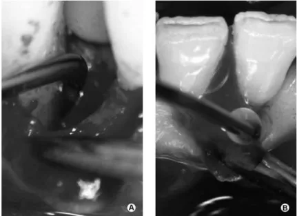
minimally elevated (Figs. 1 and 2A). The defect area
was carefully debrided and the EMD (Emdogain; In-stitute Straumann AG, Switzerland)was applied (Fig.
2B). The flaps were then repositioned and microsutures
were made to obtain primary closure of the inter-dental tissues.At the end of the procedure, patients received
8 analgesic paracetamol medications (Tylenol 750 mg; Janssen; Cilag Farmacêutica Ltda., Butantã São Paulo,
SP, Brazil) and were instructed to take them every 6 h, only in case of pain. The patient was instructed to mark the number of medications taken. Plaque control was
provided by rinsing with a 0.12% chlorhexidine solution
during 30 days (Fig. 3).
Patient Satisfaction and Perception about the Surgical Procedure
Figure 1. Preoperative view (A) and probing depth of the left central incisor (B).Intra-bony defect on the mesial aspect (c).
Figure 2. Intraoperative view of scaling and root planing at an intra-bony defect (A). Placement of the enamel matrix protein derivative
into the periodontal intra-bony defect (B).
visual analog scale (VAS). The anchors for each end of the scales were designated as “none” and “extreme”
(2).Patients were also instructed to quantify analgesic medication taken. In addition, extent of discomfort, root hypersensitivity, edema, hematoma, high fever, and interference with daily activities during the first post-operative week were evaluated in the same way. After 6 months, another questionnaire was given to the patients to determine their perception of the therapy outcomes and their level of satisfaction with treatment. The ques-tionnaire used a simplified scale to record satisfaction in terms of esthetic appearance of the treated teeth by selecting one of the following choices: very satisfied, satisfied, neutral, moderately satisfied or unsatisfied (10). In addition, the patients were asked to describe their perception of the therapy outcome at the treated tooth in terms of improvement of gingival bleeding, redness and gingival edema and hygiene ability. These parameters were also measured using VAS, with 0 = no improvement and 100 = maximum improvement.
Statistical Analysis
Repeated-measures analysis of variance (ANO-VA) was used to detect differences regarding the time of evaluation in clinical parameters (PGM, PD, RcAL). When statistical difference was found, analysis of the difference was determined using the Tukey test. The Friedman test was used to detect differences in FMPS and FMBS among all periods. The number of intra-bony sites with or without BOP was analyzed using the McNemar test and the SAS 9.01 program (SAS Institute
Inc.) was used. An experimental p value of 5% was set
to demonstrate significance for all statistical analyses.
rESuLtS
Patients’ characteristics (7 females, 5 males) at
baseline were as follows: Mean age was 47.4 ± 7.0 years, and acceptable oral hygiene was achieved before the
study, as seen from the FMPS (17.21 ± 2.61) and FMBS (12.95 ± 3.35). The distributions of intra-bony defects, according to the teeth were: 41.67% incisor, 16.66% canine and 41.67% premolar. With regard to the clinical
parameters at baseline, the mean PD, PGM and RcAL
at intra-bony defect sites were 7.21 ± 1.67, 5.0 ± 1.89 and 12.15 ± 2.19 mm, respectively. With regard to the surgical parameters, CEJ-BD was 7.88 ± 1.57 mm, the
intra-bony component of the defects (BC-BD) was 5.25 ± 1.76 mm and WD was 3.54 ± 1.05 mm. Evaluating
the number of walls present intra-surgically at angular
defects, 50% of them presented a combination of 1 and 2 walls, 41.67% of defects presented a combination of 2 and 3 walls, and only one defect (8.33%) presented just 2 walls, with no combination.
Clinical Parameters
The FMPS and FMBS means, as well as the percentages of BOP at surgical site are shown in Table 1. The FMPS and FMBS were maintained at lower
than 20% throughout the study. At the 3- and 6-month
evaluations, decreases in FMPS were observed, with statistically significant difference from baseline for both periods. With regard to FMBS, a statistical reduction in the percentage from baseline could be observed at the 6-month evaluation. The BOP at baseline was present
in all 12 sites at baseline (100%); however, during the third month there was a decrease in this percentage (0%)
and this reduction was maintained until the end of the evaluation, with statistical difference from baseline for both groups (Table 1).
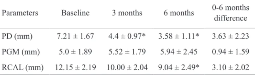
PGM, PD and RcAL means are shown in Table
2. After 6 months, the PD reduction was 3.63 ± 2.23
mm. At 3 and 6 months, a significant improvement in
PD (p<0.05) from baseline was obtained, without dif -ference between the re-evaluation periods. Although a discreet increase in the PGM was observed after 6
months (0.94 ± 1.59), no statistically significant differ
-ences (p>0.05) were noted at any time interval. With
regard to RcAL, at 6 months the gain from baseline
was 3.10 ± 2.02 mm (p<0.05). Although an increase in
RcAL was seen at 3 months, no statistically significant
differences (p>0.05) were observed at this time. In ad
-dition, after 6 months, 75% of the sites showed more than a 2 mm gain in clinical attachment (Table 3). In all treated sites (100%), primary closure was obtained
upon completion of the surgical procedure. At 1-week
follow-up, primary closure was noted in 83.33% of the sites. However, at the 2-week evaluation, 100% of the
sites presented interdental primary closure.
Postoperative Period and Patient Perception about the Surgical Procedure
nec-essary in 58.33% of the cases. The mean number of medications taken was 1.42 ± 1.62. With regard to patient perception about intraoperative surgery, 58.33%
of patients related some discomfort and/or pain during the procedure. However, this discomfort and/or pain
was negligible (5.68 ± 7.33 VAS units, with 0 = none
and 100 = extreme). When the patient perception about the postoperative period was evaluated at 1 week after
surgery, 16.67% of patients related experiencing discreet discomfort (1.86 ± 5.01 VAS units); 33.33% of patients reported feeling slight root hypersensitivity (6.45 ± 11.29 VAS units), and 41.67% of patients described presenting some edema (6.71 ± 11.72 VAS units). No
patient reported hematoma or high fever. Only one
pa-tient (8.33%) reported interference with daily activities
during the first postoperative week, however this was minimal (0.98 ± 3.38 VAS units).
Patient Satisfaction and Perception of Outcome after 6 months
With regard to patient satisfaction in terms of
the esthetic appearance of the treated teeth, 100% of
patients selected the option “very satisfied”. The evalu-ation of the patient perception of the outcome of the therapy for the treated tooth, in terms of improvement in gingival bleeding, redness and gingival edema and
hygiene ability, showed that 100% of the subjects related
an important improvement for all parameters. The best improvements were observed in gingival bleeding,
red-ness and gingival edema (92.27 ± 9.06 and 97.04 ± 6.47
VAS units, respectively). The improvement in hygiene
ability related was 62.48 ± 17.45 VAS units.
dIScuSSIon
Several factors may contribute to the success of regenerative therapy in intra-bony defects, these include the surgical techniques. In addition to being clinically efficient, the surgical approach needs to promote pa-tient satisfaction, especially in terms of morbidity and aesthetics, since these aspects are considered
impor-tant factors in the assessment of health care quality (6). This study evaluated the clinical performance of MIST associated with EMD in the treatment of intra-bony defects, and examined patient-centered outcomes, such as intra and postoperative patient perception and satisfaction after the surgical procedure. The results obtained in this study demonstrate that the application of EMD, associated with MIST, was clini-cally effective in promoting statisticlini-cally significant cAL gain and PD reduction, without statistically significant changes in PMG at 6 months from baseline. The successful clinical results obtained in this study along with those of other trials (1,3) confirmed the clinical use of MIST, associated with EMD in the treatment of intra-bony defects.
Although the present data show statistically significant clinical benefits, the mean in cAL gain
Table 1.Percentage of FMPS, FMBS, PI and BOP at the different assessment times.
Parameters Baseline 3 months 6 months
FMPS (%) 17.21 ± 2.61 13.82 ± 4.52* 13.39 ± 4.21* FMBS (%) 12.95 ± 3.35 10.45 ± 3.69 8.87 ± 4.07*
BOP 100% (100) 0% (0) 0% (0)
FMPS: full-mouth plaque score; FMBS: full-mouth bleeding
score; BOP: bleeding on probing at surgical site. *Statistically significant difference from baseline (p<0.05; Friedman test). Statistically significant difference from baseline (p<0.05; Mc
Nemar test).
Table 2.Means ( ± SD) of PD, PGM and RcAL at baseline, 3 and 6 months.
Parameters Baseline 3 months 6 months 0-6 months
difference
PD (mm) 7.21 ± 1.67 4.4 ± 0.97* 3.58 ± 1.11* 3.63 ± 2.23
PGM (mm) 5.0 ± 1.89 5.52 ± 1.79 5.94 ± 2.45 0.94 ± 1.59
RcAL (mm) 12.15 ± 2.19 10.00 ± 2.04 9.04 ± 2.49* 3.10 ± 2.02
SD: standard deviation; PD: probing depth; PMG: position of the gingival margin;
RCAL: relative clinical attachment level. *Statistically significant difference from baseline (p<0.05; ANOVA/Tukey’s test).
Table 3.Frequency distribution of clinical attachment level gain at 6 months.
Differences in clinical attachment level (mm)
Loss 0-2 2-4 4-6 ≥6
(3.1 ± 2.02) and PD decrease observed in the present study (3.63 ± 2.23 mm) were slightly lower than those of other similar trials that reported average cAL gains ranging from 4.4 ± 1.4 mm to 4.9 ± 1.7 and average PD
reduction ranging 4.6 ± 1.3 mm to 5.2 ± 1.7 mm, after
12 months from baseline (3,11). Even though a proper comparison cannot be made across studies (3,12,13),
these differences could be explained by some aspects, such as baseline clinical measurements and time of longitudinal of follow-up.
The baseline PDs and depth of the intra-bony defect component in the present study were less pro-nounced than those of other investigations of the clini-cal effects of EMD after MIST or papilla preservation
technique (3,12,13). This is important to note that the
greater the baseline PDs and depth of the intra-bony defect component, the better the potential for reduction of PD, cAL gain and bone fill in intra-bony defects
(20,21).Other important morphological characteristics that could interfere in results include the baseline width of the intra-bony component of the defect and the number of residual bony walls, since narrower and contained defects have been associated with increased amounts of cAL gain and bone fill (9,14). Furthermore, since some additional clinical benefits could be expected along with longitudinal assessment after the regenerative procedure
with EMD (5), the difference longitudinal follow-up
duration could also explain the variation in the results. An important aspect of the design of the pres-ent experimpres-ent is the time of indication of regenera-tive procedure. In general, patients scheduled for the regenerative approach are first submitted to an initial periodontal treatment phase, including cause-related therapy. Re-evaluation of results following cause-related therapy is important for adequate indication and selection of regenerative therapy and is associated with particular response ability. The healing following nonsurgical
therapy occurs within at least 3 months (15), although
it may still continue for a period of 9 months (16). In this context, in the present study, only the patients that
present CAL loss ≥ 5 mm with BOP 6 months after
completion of cause-related therapy were submitted to regenerative surgery. According to published evidence (17,18), the re-evaluation of clinical changes occurring in the periodontal tissues following nonsurgical therapy should not be performed earlier than or at 4 weeks after therapy, as performed in other studies (13), because clinical parameters have demonstrated major changes
after this period (16,18).
In the present study, no difference was observed in the PGM after 6 months. This result could be attributed to some aspects, such as the employment of MIST and the use of an operative microscope. The reduced flap reflection may prevent post-surgical bone resorption and, in consequence, soft tissue recession. This result is in accordance with reports from the literature that have demonstrated that a preservation of soft tissues, including the gingival margins of the adjacent teeth and the interdental papillae, could benefit clinical results in terms of position of the gingival margin (1,3).
This positive clinical outcome may be associated with the very high levels of patient satisfaction obtained in the present study in terms of esthetic appearance. These clinical and patient-centered results could also be a consequence of the maintenance of the integrity of the flap achieved in this study. In addition, these data were consistent with the assessment of the wound in terms of primary closure during the healing phase. These observa-tions could suggest that MIST used in combination with EMD results in fewer scarce complications that could be related i.e. with the use of membranes.These aspects are critical to the outcomes since wound dehiscence and contamination of the regenerative material have been associated with reduced clinical results and subsequent soft tissue recession (19). Moreover, the use of a surgical microscope may have contributed to the results obtained, since the use of magnification and optimal illumina-tion improves the visual acuity, promoting a minimally increased gingival recession and, in consequence, very high patient satisfaction.
Studies on regenerative periodontal therapy have always been interested in the clinical outcomes of treatments and have developed various parameters to measure these outcomes. However, most of these objective measures only reflect the clinical end points of the therapy and they give no indication of the impact of the treatment on the psychosocial well-being of the patient, which have a considerable impact on the patient’s
day-to-day life or life quality (2,20). In this context, the
intra-operative and postintra-operative pain and morbidity, and only in a minority of patients, according to previous studies (1,3,11). Moreover, the results of the assessments, which were performed after 6 months to understand the impact of the surgical approach on patients, showed high levels of improvement in terms of gingival bleeding, hygiene ability, and especially redness and gingival edema. Since patient-focused issues have been recognized as a
research priority area (2), a larger study is necessary to
confirm the reported findings.
In addition to the positive biological effects of EMD and MIST, it should be noted that the psychological benefits of the therapy on patients may have influenced these outcomes. Patients who were informed about the beneficial effects of the therapy may feel that they are being treated in the best manner and receiving a high-quality treatment technique that may subsequently lead to better perceptions and trust for the provided therapy. In consequence, such information could influence the patient perception with regard to both intraoperative and postoperative evaluations.
Although this study has demonstrated favorable clinical and patient-centered outcomes, it is difficult to determine, from these data, the proportion of results that may be attributed to the use of MIST and the proportion of the results that may be attributed to the use of EMD. Both of these items have been reported to improve peri-odontal support and soft tissue maintenance, promoting, in consequence, reduced patient morbidity and favorable satisfaction. Thus, a blinded and controlled clinical trial is necessary to establish which factor is most respon-sible for the improved outcome observed in the present study. In addition, since the choice of treatment strategy depends on the understanding of patient perceptions and satisfaction, more research must be focused on patient-centered outcomes.
In conclusion, the present study showed clinical benefits in terms of PD reduction, cAL gain and no significant changes in PGM after 6 months. In addition, all patients reported minimal pain or discomfort associ-ated with intra and postoperative surgical procedures and maximum satisfaction in terms of esthetics.
rESuMo
O objetivo deste estudo foi avaliar os resultados clínicos e centrados no paciente após abordagem cirúrgica minimamente
invasiva (CMI) associada à aplicação das proteínas derivadas
da matriz do esmalte (PDE) no tratamento de defeitos
infra-ósseos. Doze pacientes apresentando um sítio com profundidade
de sondagem ≥ 5 mm e sangramento à sondagem , associado à
evidência radiográfica de defeito infra-ósseo, foram tratados com
CMI e aplicação das PDE. Os parâmetros clínicos foram avali -ados imediatamente antes do procedimento e após 3 e 6 meses.
A percepção de dor e desconforto do paciente durante o período
trans-cirúrgico e ao longo da primeira semana de pós-operatório,
bem como a satisfação estética 6 meses após o tratamento, foram
avaliadas por meio de questionários. Os resultados mostraram que
o uso da CMI associada à aplicação de PDE promoveu melhoras estatisticamente significantes nos parâmetros clínicos, mínima dor e desconforto e máxima satisfação estética aos pacientes. Dentro dos limites do estudo, foi demonstrado que a associação de CMI e PDE, no tratamento de defeitos infra-ósseos, é capaz de
promover satisfatórios resultados clínicos e centrados no paciente.
rEFErEncES
1. cortellini P, Tonetti MS. A minimally invasive surgical technique with an enamel matrix derivative in the regenerative treatment of intra-bony defects: a novel approach to limit morbidity. J clin Periodontol 2007;34:87-93.
2. Tonetti MS, Fourmousis I, Suvan J, Cortellini P, Bragger U, Lang NP. Healing, postoperative morbidity and patient perception of outcomes following regenerative therapy of deep intrabony de-fects. J Clin Periodontol 2004;31:1092-1098.
3. cortellini P, Pini-Prato G, Nieri M, Tonetti MS. Minimally inva-sive surgical technique and enamel matrix derivative in intrabony defects: 2. Factors associated with healing outcomes. Int J Peri -odontics Restorative Dent 2009;29:257-265.
4. cortellini P, Pini Prato G, Tonetti MS. The simplified papilla preservation flap. A novel surgical approach for the management of soft tissues in regenerative procedures. Int J Periodontics Re-storative Dent 1999,19:589-599.
5. Wachtel H, Schenk G, Bohm S, Weng D, Zuhr O, Hurzeler M. Microsurgical access flap and enamel matrix derivative for the treatment of periodontal intrabony defects: a controlled clinical study. J Clin Periodontol 2003;30:496-504.
6. Locker D, Dunt D. Theoretical and methodological issues in so-ciological studies of consumer satisfaction with medical care. Soc Sci Med 1978;12:283-292.
7. Ainamo J, Bay I. Problems and proposals for recording gingivitis and plaque. Int Dent J 1975;25:229-235.
8. Muhlemann HR, Son S. Gingival sulcus bleeding - A leading symptom in initial gingivitis. Helv Odontol Acta 1971;15:107-113. 9. Goldman H, cohen DW. The infrabony pocket: classification and
treatment. J Periodontol 1958;29:272-291.
10. Wismeyer D, van Waas MA, Vermeeren JI. Overdentures support-ed by ITI implants: a 6.5-year evaluation of patient satisfaction and prosthetic aftercare. Int J Oral Maxillofac Implants 1995;10:744-749.
11. cortellini P, Nieri M, Prato GP, Tonetti MS. Single minimally invasive surgical technique with an enamel matrix derivative to treat multiple adjacent intra-bony defects: clinical outcomes and patient morbidity. J Clin Periodontol 2008;35:605-613. 12. Cortellini P, Tonetti MS. Clinical performance of a regenerative
strategy for intrabony defects: scientific evidence and clinical experience. J Periodontol 2005;76:341-350.
14. Tonetti MS, Pini-Prato G, cortellini P. Periodontal regeneration of human infrabony defects. IV Determinants of the healing response. J Periodontol 1993;64:934-940.
15. Beck JD. Periodontal implications: older adults. Ann Periodontol 1996;1:322-357.
16. Badersten A, Nilveus R, Egelberg J. Effect of non-surgical periodontal therapy. II. Severely advanced periodontitis. J clin Periodontol 1984;11:63-76.
17. caton JG, Proye M, Polson AM. Maintenance of healed periodon-tal pockets after a single episode of root planing. J Periodontol 1982;53:420-424.
18. Kaldahl WB, Kalkwarf KL, Patil KD, Dyer JK, Bates RE. Evalua-tion of four modalities of periodontal therapy. Mean probing depth, probing attachment level and recession changes. J Periodontol 1988;59:783-793.
19. De Sanctis M, Zucchelli G, clauser c. Bacterial colonization of bioabsorbable barrier material and periodontal regeneration. J Periodontol 1996;67:1193-1200.
20. Cunha-Cruz J, Hujoel PP, Kressin NR. Oral health-related quality of life of periodontal patients. J Periodontal Res 2007;42:169-176.