INHERITED NEUROMUSCULAR DISEASES IN THE MOUSE
A R E V I E W OF THE LITERATURE
LUIZ FERNANDO BLEGGI TORRES
SUMMARY — There are several neuromuscular disorders affecting the human being. Most of these are poorly understood and lack an effective treatment. Due to the limitation of experimental manipulation in «anima nobili», inherited neuromuscular diseases in laboratory animals constitute a valuable source of scientific information. Amongst several animal species affected by neuromuscular disorders the house mouse is of particular interest because of its small size, short pregnancy and low costs of maintanence. In the present review 20 murine mutants with diseases affecting peripheral nerves, skeletal muscles and motor end-plates are tabulated. Genetic, clinical and pathological aspects are discussed aiming to provide infor-mation about these mutants which might be of great interest as animal models for human neuromuscular diseases.
Doenças neuromusculares hereditárias em camundongos: revisão da literatura
RESUMO — Existem inúmeras doenças neuromusculares que acometem seres humanos. A grande maioria delas é insuficientemente conhecida quanto a mecanismos fisiopatológicos e tratamentos adequados. A limitação na manipulação experimental em «anima nobili» faz-nos procurar meios alternativos para o estudo dessas doenças, tais como animais experimentais com distúrbios neuromusculares geneticamente transmitidos. Estes mutantes constituem fonte inesgotável e valiosa de informações quanto a mecanismos fisiopatogênicos e processos pato-lógicos básicos em doenças neuromusculares. Entre as diversas espécies animais afetadas por distúrbios neuromusculares o camundongo é de particular interesse devido ao seu baixo custo de manutenção, rápida reprodutividade e pequeno tamanho, o que permite amplos estudos morfológicos a custos acessíveis. Nesta revisão analisamos 20 camundongos mutantes com distúrbios afetando nervo periférico, músculo esquelético ou junção neuromuscular. Aspectos genéticos, clínicos e patológicos são discutidos na intenção de oferecer informação atualizada sobre essas mutações animais, muitas das quais de. grande interesse como modelos experimentais de doenças neuromusculares humanas.
In recent years the presence of a close relationship and interdépendance between skeletal muscle and peripheral nerves became obvious. Several studies exemplified the essential role of the nervous system in the maturation, function and full expression of skeletal muscle fibres 10,21,24,28,43,50,66,100. There are many inhe-rited human diseases affecting peripheral nerves, skeletal muscles or both. Most of these conditions differ from each other in a number of ways and are classified according to the distribution and characteristics of the wasting process, the pattern of inheritance, age of onset, rate of progression of weakness. However, in most of the situations, there is little understanding of the pathogenesis of the diseases and they lack an effective way of treating or controlling their symptons and evolution. The scarcity of biopsy material and the limitation of experimental manipulation in man make it difficult to study the aetiology and molecular mechanisms of many
* MD, PhD, Neuropathologist, Hospital de Clinicas and Hospital Nossa Senhora das Gra-ças, Curitiba.
inherited neuromuscular diseases. It is therefore necessary to consider the possibility of obtaining relevant information from the study of animal mutants with neuro-muscular diseases 6
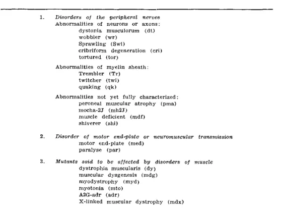
° . If features are discovered wnich are siminar to those expressed in human disease, the mutant might become a useful model for therapeutic trials and experimental investigation which are not possible in man 52. Apart from man, several animal species are known to be affected by inherited disorders of muscle, motor end-plate and nerve. These include birds (chicken, turkey and duck), small rodents (mouse and hamster) and other larger mammals (goat, pig, horse, cattle, sheep, dog, cat and mink) 48,51,52,60,62,102. One of the most extensively studied labo-ratory animals is the mouse due to its small size, short pregnancy and relatively low costs of maintanence. These advantages allow the investigator to employ a wide range of techniques aiming to gain an accurate idea of how the disease begins, the primary sites of pathology and its evolution from the earliest clinical and patholo-gical manifestation through to its full expression. From a survey of recent publi-cations concerning murine mutations (Mouse News Letter, 1986) 20 out of about 1000 genetically determined disorders affect peripheral nerves, neuromuscular junction and/or skeletal muscles (Table 1 ) . The great majority of the mutations listed on table 1 are spontaneous and transmitted as an autosomal recessive trait. In the present tabulation the criteria used for classification were the structures predominantly affected by the mutant gene. The clinical syndromes and pathological abnormalities are distinct in most of these three main groups as will be discussed below. However in a few mutants more than one set of structures might be involved.
1. Mutant mice with disorders of peripheral nerves
a ) Those with pathological abnormalities in nerve cells or axonal profiles:
Dystonia musculorum (dt) — This spontaneous mutation is transmitted by an
autosomal recessive gene and expressed as a severe and progressive sensory neuro-pathy. Affected animals, by the end of the first postnatal week, have abnormal posture and muscle spasms. Although no paralysis or tremor are noted there is
1. Disorders of the peripheral nerves Abnormalities of neurons or axons:
dystonia musculorum (dt) wobbler (wr)
Sprawling (Swl)
cribriform degeneration (cri) tortured (tor)
Abnormalities of myelin sheath: Trembler (Tr)
twitcher (twi) quaking (qk)
Abnormalities not yet fully characterized: peroneal muscular atrophy (pma) mocha-2J (mh2J)
muscle deficient (mdf) shiverer (shi)
2. Disorder of motor end-plate or neuromuscular transmission motor end-plate (med)
paralyse (par)
3. Mutants said to be affected by disorders of muscle dystrophia muscularis (dy)
muscular dysgenesis (mdg) myodystrophy (myd) myotonia (mto) A2G-adr (adr)
X-linked muscular dystrophy (mdx)
progressive incoordination of movements and the growth is severely retarded 35,42. The morphological abnormalities consist of widespread axonal degeneration of the main sensory pathways with degeneration of peripheral nerves and atrophy of the muscle spindles and skeletal muscle fibres 35,53,63. it has been suggested that a defect in axoplasmic flow with the eventual formation of axonal swellings could be the underlying mechanism of this disorder 53. However, segmental demyelination probably due to a primary Schwann cell dysfunction occurs prior to any identifiable swelling of the axons 84-85.
Wobbler (wr) — This mutant mouse, initially identified by its abnormal
wobbling gait, presents with proximal muscle weakness affecting mainly neck and forelimb musculature. The disease progresses from one to six months of age and may be fatal. The pathological process consists of widespread vacuolation of motor nerve cells in brainstem and most levels of spinal cord, the result of dilatation of rough endoplasmic reticulum 3,4,12,23,36,38,83,87. Morphological and physiological evi-dence of denervation of skeletal muscle has been observed. The former is represented by atrophy of fibres and conspicuous branching of preterminal axons 38 and the latter by spontaneous fibrillations, absence of miniature end-plate potentials and increased extra-junctional sensitivity to acetylcholine 57. Therefore this mutant could be considered as a potential model for human spinal muscular atrophy 30.
Sprawling (Swl) — The sprawling mouse was identified in subsequent
gene-rations of mice previously used in irradiation experiments. Affected animals are usually identifiable by 7 to 10 days of age by abnormal resting posture more marked in the hindlimbs. Morphological studies showed that the sensory neurons are the site of primary pathology. Deficiency of myelinated axons in the ascending tracts, spinal roots and peripheral nerves and a reduction in number of muscle spindles were also observed 29. It seems that a genetically-induced abnormality of ganglia could be responsible for the failure of induction of sensory receptors thus accounting for the marked reduction of peripheral sensory receptors including muscle Spindles 33,34,95,96.
In the cribriform degeneration 49 and tortured 55 mutant mice the morphological changes have not been so extensively studied. They both present clinically with incoordination, ataxia and behaviour abnormalities. Some preliminary observations showed intracellular vacuolar changes in spinal cord and brainstem of the cribriform while in the tortured mutant progressive degeneration of cerebellum, spinal cord and sensory ganglia has been described. In both mutants as might be expected peripheral nerves are also affected 103.
b) Mice with disorders of myelin of peripheral nerves:
Trembler mouse ( T r ) — This mutation is transmitted by a autosomal dominant
gene and is expressed by spastic paralysis, convulsions and tremor 45. Preliminary electroencephalographic studies suggested a possible cerebellar malfunction but no morphological confirmation was obtained 19. It is now accepted that an hereditary demyelinating neuropathy constitutes the major abnormality in the trembler mouse 5,6, 72,73,89. The electrophysiological consequences of this hypomyelinated neuropathy are the inability to conduct rapid trains of impulse with marked reduction of motor conduction velocities 74,75. The pathogenesis of this disorder has been initially attri-buted to a primary defect in the synthesis of myelin 73,74 but elegant nerve grafting experiments demonstrated conclusively that the neuropathy is due to a primary Schwann cell disorder 1.2. The pathological abnormalities in skeletal muscle and motor end-plates consist of atrophy of fibres, abundant axonal sprouting and electro-physio-logical evidences of denervation affecting predominantly slow-twitch muscles. It has been suggested that the more severe involvement of slow fibres could be due to its innervation by motor units subiected to prolonged activity and therefore predominantly affected by conduction block 39,46.
Twitcher mouse (twi) — Special interest has been raised since this mutant was
The quaking mouse is another mutant of value for the study of myelin loss in central and peripheral nerves 94,101,108,113. Several etiological factors have been postulated such as deficiency of mielyn-specific proteins 26; a plasmalemma molecular
defect in oligodendrocytes 109 and a Schwann cell dysfunction i.
c) Peripheral nerve disorders not yet characterized:
The animals described in this group show variable degrees of abnormalities in the peripheral nervous system as part of more complex pleiotropic genetic defects. They lack, however, adequate morphological and developmental studies and will be discussed only briefly. The peroneal muscular atrophy shows agenesis of common peroneal nerve which induces secondary atrophy of skeletal muscles of the l e g4 4
. In other mutations such as the mocha-2], muscle deficient and shiverer, peripheral neuropathy and shortening of internodal lengths have been reported 25,68,103.
2 . Functional and morphological disorders of motor end-plate:
Both mutants belonging to this group have devastating clinical signs of pro-gressive muscle weakness, paralysis and premature death. In the motor end-plate mutant mouse there is striking atrophy of muscle fibres and complex terminal axonal sprouting at motor end-plates a l
>3 7
. It was suggested that these changes were induced by a progressive loss of functional innervation 32,57 but other authors considered that the pathological process in the med mouse is a m y o p a t h y1 1 4
or peripheral neuro-pathy 92,93. The paralyse mutant shows progressive atrophy of motor nerve terminals with a reduction of neuromuscular transmission and eventual denervation of end-plates. The cause of such morphophysiological abnormalities remains uncertain 40.
3 . Mutants said to show primary muscle pathology:
Dystrophia muscularis (dy) — The dystrophic mouse presents with progressive
weakness of axial and limb muscles and a reduced life-span. It was initially thought to be affected by a primary myopathic disorder 7,9,71,82,88,112 A few years later McComas et a l .7 8
"8 1
suggested that muscle degeneration could be the result of a primary disorder of the motor neurons. Furthermore Gallup and Dubowitz47
demons-trated in tissue culture that motor nerve cells of the dystrophic mouse were unable to support functional regeneration of both normal and dystrophic muscle fibres. These observations suggesting the influence of a neural factor upon muscle degeneration led to more extensive studies of the nervous system of this murine mutant with the eventual characterization of its abnormalities of myelin sheath in roots, cranial and peripheral nerves 13,15,16,17,104. These abnormalities consist of large clusters of naked axons packed closely together and not invested by Schwann cell cytoplasm, myelin or even basal lamina. Such neural abnormalities are attributed to a genetically--determined impairment of Schwann cell differentiation, proliferation and survival together with a possible disorder in the interaction between axons, Schwann cells and fibroblasts 1.20,100. Many other abnormalities in the peripheral nerves such as deficiency in basal lamina™, widening of nodes of Ranvier with retraction of Schwann cell cytoplasm from the paranodal areas 18 and shortening of internodal lengths n have been reported. The electrophysiological disturbances originated from this insu-lation defect of roots and nerves are a possible lateral transmission of action poten-tials between neighboring bared axons (cross-talk) with a reduction in motor con-duction velocities 14.61,67. it has been suggested that muscle fibres of the dystrophic mouse might be functionally denervated 7
o >7 7
. Further studies, however, demonstrated no extra-junctional sensitivity to acetylcholine, no tetrodotoxin-resistant action poten-tials and normal muscle action potential in response to nerve stimulation in the dystrophic strain. Such observations indicate normal functional innervation of skeletal muscle fibres 56,58,59. Although some authors point out that the muscle abnormalities in the dy mouse do not seem to be due to neural abnormalities 67,115 at present it is
not known whether the neural or muscular changes are the first to appear and whether they are causally related. Until this problem is resolved it would be unwise to use this mutant as a model for primary muscular disease.
have been reported 55, The myotonic and the A2G-adr mutant mice lack compre-hensive investigations of the skeletal muscles and nervous system. The first was assessed only electrophysiologically and showed widespread myotonic discharges while the second was reported to have a possible biochemical defect of muscle fibres 110,111.
X-linked muscular dystrophy (mdx) — The mdx mutant mouse was first
observed during a survey of genetic variations of pyruvate kinase in the m o u s e2 2
. Further studies showed that the gene defect in the mdx mutant is in the X-chromosome in a region similar to human muscular dystrophy of the Duchenne type. Affected mice have high serum levels of this enzyme and although showing little disability they have widespread and severe muscle disease. Preliminary morphological studies were restricted only to the analysis of muscle pathology and showed that muscle necrosis was followed by regeneration with little impairment of function 22,27,64,105. Recent studies by Torres and Duchen 106,107 demonstrated that significant ultrastruc-tural abnormalities were present in the mdx as early as 1 day of age consisting of streaming of Z-line material followed by segmental necrosis and regeneration. Further-more the disease was progressive, initially affecting proximal muscle groups but in 2-3 months most muscles were abnormal. At this stage affected muscles showed marked variation in fibre size and diameter with little increase in endomysial connec-tive tissue or fat infiltration. There was no inflammation and the heart and eye muscles were normal. Quantitative and qualitative studies of peripheral nerves showed no abnormalities and the nerve terminals were unaffected but there was a reduction in the number and depth of post-synaptic folds at motor end-plates. These authors concluded that the mdx mouse has a primary muscle disease with a total absence of structural pathology in either central or peripheral nervous systems.
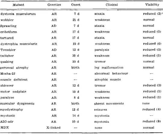
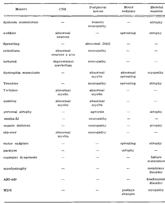
A summary of the main genetic and clinical characteristics of all the murine mutants which have been described above is presented in table 2 while in table 3 their main pathological changes are tabulated.
Mutant Genetics Onset Clinical Viability
dystonia musculorum A R 7 d ataxia reduced (2)*
wobbler A R 21 d weakness normal
Sprawling AD 7 d ataxia normal
cribriform A R 17 d weakness reduced (3)
tortured AR 17 d ataxia normal
dystrophia muscularis AR 10 d weakness reduced (6)
Trembler AD 12 d paralysis reduced (3)
twitcher A R 30 d weakness reduced (3)
quaking A R 10 d tremor normal
peroneal atrophy A R birth leg malformation normal
Mocha-2J A R
—
abnormal behaviour-muscle deficient A R
—
atrophic muscle—
shiverer A R 12 d tremor reduced (3)
motor endplate A R 10 d weakness reduced (1)
paralyse A R 8 d weakness reduced (1)
muscular dysgenesis AR birth absent movements none
myodystrophy AR 12 d seizures reduced (4)
myotonic AR 14 d myotonia
—
A2G-adr A R 10 d myotonia reduced (9)
MDX X-linked
—
none normalMutant CNS Peripheral nerves
Motor endplate
dystonia musculorum
wobbler
Sprawling cribriform
tortured
dystrophia muscularis
Trembler Twitcher
quaking
peroneal atrophy mocha-2j muscle deficient shiverer
motor endplate paralyse
muscular dysgenesis
abnormal neurons
abnormal neurons + glia
degeneration cerebellum
abnormal myelin abnormal
myelin
abnormal myelin
sensory neuropathy
abnormal DRG neuropathy
neuropathy
abnormal myelin neuropathy
abnormal myelin abnormal
myelin agenesis neuropathy neuropathy neuropathy
sprouting
abnormal sprouting sprouting
sprouting atrophy
atrophy
atrophy
atrophy
myodystrophy
A2G-adr
MDX postsyn
changes
Table S — Main pathological abnormalities in murine mutants.
Conclusions — It is relevant to note the wide variability of inherited diseases of skeletal muscle and nerves affecting the house mouse with clinical manifestations, evolution and pathological features in many ways similar to those of human disorders. However it seems unrealistic to look at animal models as exact counterparts of human neuromuscular diseases. A more wise approach would be to study those features of animal models which are shared with inherited human disorders aiming to find a fundamental abnormality at cellular level and to understand the common manifes-tations that arise from these genetic abnormalities.
REFERENCES
1. Aguayo AJ, Bray GM, Perkins SC — Axon-Schwann cell relationships in neuropathies of mutant mice. A n n New York Acad Sci 317 : 512, 1979.
2. Aguayo AJ, Attiwell M, et al — Abnormal myelination in transplanted mouse Schwann cells. Nature (Lond) 265 : 73, 1977.
3. Andrews JM — The fine structure, of the cervical spinal cord, ventral root and brachial nerves in the wobbler (wr) mouse. J Neuropathol E x p e r Neurol 34 : 12, 1975. 4. Andrews JM, Maxwell DS — Ultrastructural features of anterior horn cell degeneration
in wobbler (wr) mouse. Anat Records 157 : 206, 1967.
5. Ayers MM, Anderson R McD — Onion bulb neuropathy in the trembler mouse: a model for hypertrophic interstitial neuropathy (Dejerine-Sottas) in man. Acta Neuropathol
(Berlin) 25 : 54, 1973.
6. Ayers MM, Anderson R McD — Development of onion bulb neuropathy in the trembler mouse — comparison with normal nerve maturation. Acta Neuropathol (Berlin) 32 : 43, 1975.
7. Banker BQ — A phase and electron microscopic study of dystrophic muscle: II. The pathological changes in the newborn Bar Harbor 129 dystrophic mouse. J Neuropathol Exper Neurol 27 : 183, 1968.
8. Banker BQ — Muscular dysgenesis in the mouse (mdg/mdg) : I. Ultrastructural study of skeletal and cardiac muscle. J Neuropathol Exper Neurol 36 : 100, 1977.
9. Banker BQ, Denny-Brown D — A study of denervated muscle in normal and dystrophic mice. J Neuropathol E x p e r Neurol 18 :517, 1959.
10. Bennett M R — Development of neuromuscular synapses. Physiological Rev 63 : 915, 1983. 11. Beuche W , Friede R L — A quantitative assessment of myelin sheaths in the peripheral nerves of Dystrophic, Quaking and Trember mutants. A c t a Neuropathol (Berlin) 66 : 29, 1985.
12. Bird MT, Shuttleworth EC, Koestner A, Reinglass J — The wobbler mouse mutant: an animal model of hereditary motor system disease. Acta Neuropathol (Berlin) 19 : 39, 1971.
13. Biscoe TJ, Caddy K, Pallot DJ, Pehrson UMM — Investigation of cranial and other nerves in the mouse with muscular dystrophy. J Neurol Neurosurg Psychiat 38 : 391, 1975.
14. Biscoe TJ, Headley PM, Martin MR, Stirling CA — Electrophysiological observations on the spinal cord of normal and dystrophic mouse. J Neurol Sci 31 : 51, 1977. 15. Biscoe TJ, Caddy K, Pallot D, Pehrson M, Stirling CA — The neurological lesion in
the dystrophic mouse. Brain Res 76 : 534, 1974.
16. Bradley WG, Jenkison M — Abnormalities of peripheral nerves in murine muscular dystrophy. J Neurol Sci 18 : 227, 1973.
17. Bradley WG, Jenkison M — Neural abnormalities in the dystrophic mouse. J Neurol Sci 25 : 249, 1975.
18. Bradley WG, Jaros E, Jenkison M — The nodes of Ranvier in the, nerves of mice with muscular dystrophy. J Neuropathol E x p e r Neurol 36 : 797, 1977.
19. Braverman IM — Neurological actions caused by the mutant gene «trembler» in the house mouse. J Neuropathol Exper Neurol 12 : 64, 1953.
20. Bray G, Perkins S, Peterson AC, Aguayo AJ — Schwann cell multiplication deficit in nerve roots of newborn dystrophic mice. J Neurol Sci 32 : 203, 1977.
21. Buchthal F, Schmalbruch H — Motor unit of mammalian muscle. Physiological Rev 60 : 90, 1980.
22. Bulfield G, Siller WG, Wight PA, Karen JM — X-chromosomelinked muscular dystrophy (mdx) in the mouse. Proc Nat Acad Sci 81 : 1189, 1984.
23. Campbell MJ — Ultrastructural observations on the wobbler mouse. J Neuropathol Exper Neurol 31 : 190, 1972.
24. Carlson BM, Gutman E — Contractile and histochemical properties of sliced muscle grafts regenerating in normal and denervated rat limbs. Exper Neurol 50 : 319, 1976. 25. Chernoff GF — Shiverer: an autosomal recessive mutant mouse with myelin deficiency.
J Hereditary 72 : 128, 1981.
26. Constantino-Ceccarini E, Morell P — Quaking mouse: in vitro studies of brain sphingolipid biosynthesis. Brain Res 29 : 75, 1971.
27. Dangain J, Vrbova G — Muscle, development in mdx mutant mice. Muscle and Nerve 7 : 700, 1984.
29. Duchen L W — «Sprawling»: a new mutant mouse with failure of myelination of sensory axons and a deficiency of muscle spindles. Neuropathol Appl Neurobiol 1 : 89, 1975. 30. Duchen L W — Motor neuron diseases in man and animals. Invest Cell Pathol 1 : 249, 1978. 31. Duchen L W , Searle AG — Hereditary motor end-plate disease in the mouse: light and
electron microscopic studies. J Neurol Neurosurg P s y c h 33 : 238, 1970.
32. Duchen LW, Stefani E — Electrophysiological studies of neuromuscular transmission in hereditary motor end-plate disease of the mouse. J Physiology 212 : 535, 1971.
33. Duchen L W , Scaravilli F — The structure and composition of peripheral nerves and nerve roots in the Sprawling mouse. J Anatomy 123 : 763, 1977.
34. Duchen LW, Scaravilli F — Quantitative and electron microscopic studies of sensory ganglion cells of the Sprawling mouse. J Neurocytol 6 : 465, 1977.
35. Duchen LW, Strich SJ, Falconer DS — Clinical and pathological studies of an hereditary neuropathy in mice (Dystonia musculorum). Brain 87 : 367, 1964.
36. Duchen LW, Falconer DS, Strich SJ — Hereditary progressive neurogenic muscular atrophy in the, mouse. J Physiology 183 : 53, 1966.
37. Duchen L W , Searle AG, Strich SJ — An hereditary motor end-plate disease in the mouse. J Physilology 189 : 4, 1967.
38. Duchen LW, Strich SJ, Falconer DS — An hereditary motor neurone disease with progressive denervation of muscle in the mouse: the mutant «woobler». J Neurol Neurosurg Psychiat 31 : 535, 1968.
39. Duchen L W , Gale AN, Gomez S — Motor end-plate morphology and neuromuscular transmission in the mutant Trembler mouse. J Physiology 308 : 18, 1980.
40. Duchen L W , Gomez S, Guenet J-L, Love S — Paralyse: a new neurological mutant mouse with progressive atrophy and loss of nerve terminals. J Physiology 345 : 166P, 1983.
41. Duchen LW, Eicher EM, Jacobs JM et al — Hereditary leucodystrophy in the mouse : the new mutant twitcher. Brain 103 : 695, 1980.
42. Eldridge R, Fahn S — Advances in Neurology. Raven Press, New York, 1976. 43. Engel W K , Karpati G — Impaired skeletal muscle maturation following neonatal
neu-rectomy. Develop Biol 17 : 713, 1968.
44. Esaki K, Yasuda Y, Nakamura M et al — A new mutant in the mouse: peroneal muscular atrophy. Exper Animals 30 : 151, 1981.
45. Falconer DS — Two new mutants «Trembler» and «Reeler» with neurological actions in the house mouse (Mus musculus L ) . J Genetics 50 : 192, 1951.
46. Gale AN, Gomez S, Duchen L W — Changes produced by a hypomyelinating neuropathy in muscle and its innervation. Morphological and physiological studies in the Trembler mouse. Brain 105 : 373, 1982.
47. Gallup B, Dubowitz V — Failure of «dystrophic» neurons to support functional regene-ration of normal and dystrophic muscle in culture. Nature (London) 243 : 287, 1973. 48. Green EL — Biology of the Laboratory Mouse. Mc Graw Hill, New York, 1966. 49. Green MC, Sidman RL, Pivetta O — Cribriform degeneration (cri) : a new recessive
neurological mutation in the mouse. Science 176 : 800, 1972.
50. Grinnell AD, Herrera A — Specificity and plasticity of neuromuscular connections: long-¬ -term regulation of motor neuron function. Progress in Neurobiology 17 : 203, 1981. 51. Gruneberg H — The Genetics of the Mouse. Martinus Nijhoff, The Hague, 1952. 52. Gruneberg H — An annotated catalogue of the mutant genes of the house mouse.
HMSO, London, 1956.
53. Hanker JS, Peach R — Histochemical and ultrastructural studies of primary sensory neurons in mice with dystonia musculorum : I. Acetylcholinesterase and lysosomal hydrolases. Neuropathol Appl Neurobiol 2 : 79, 1976.
54. Harris AJ — Embryonic growth and innervation of rat skeletal muscles. Philosophical Trans R o y Soc London 293 : 257, 1981.
55. Harris JB — Muscular dystrophy and other inherited diseases of skeletal muscle in animals. New York Academy of Sciences, New York, 1979.
56. Harris JB, Marshall M — A study of action potential generation in murine dystrophy with reference to functional denervation. Exper Neurol 41 : 331, 1973.
57. Harris JB, Ward MR — A comparative study of denervation in muscles from mice with inherited progressive neuromuscular disorders. Exper Neurol 42 : 169, 1974.
58. Harris JB, Montgomery A — Some mechanisms and electrical properties of distal hind limb muscles of genetically dystrophic mice (C75B1/6J dy2j/dy2j). Exper Neurol 48 : 568, 1975.
60. Harris JB, Slater C — Animal models: what is their relevance to the pathogenesis of human muscular dystrophy? Brit Med Bull 36 : 193, 1980.
61. Huizar P, Kuno M, Miyata Y — Electrophysiological properties of spinal motorneurones of normal and dystrophic mice. J Physiology 248 : 231, 1975.
62. Innes JR — Myopathies in animals. Brit Veterinary J 107 : 131, 1951.
63. Janota I — Ultrastruetural studies of an hereditary sensory neuropathy in mice (Dystonia musculorum). Brain 95 : 529, 1972.
64. Karpati G, Carpenter S, Prescott S — Small-caliber skeletal muscle fibres do not suffer necrosis in X-linked Muscular Dystrophy (MDX) of mice. Neurology 36 : 240, 1986. 65. Kobayashi T, Yamanaka T, Jacobs JM et al — The twitcher mouse: an enzymatically
authentic model of human globoid cell leucodystrophy (Krabbe disease). Brain Res 202 : 479, 1980.
66. Kugelberg E — Adaptive transformation of rat soleus motor units during growth. Histochemistry and contraction speed. J Neurol Sci 27 : 269, 1976.
67. Kuno M — Physiologic consequences of neural abnormalities in murine dystrophy. Ann New York Acad Sci 317 : 143, 1979.
68. Lane PW, Deol M — Mocha, a new coat color and behaviour mutation on chromosome 10 of the mouse. J Hereditary 65 : 362, 1974.
69. Lane PW, Beamer TC, Myers D — Myodystrophy, a new myopathy on chromosome 8 of the mouse. J Heredity 67 : 135, 1976.
70. Law P, Atwood H, McComas AJ — Functional denervation in the soleus muscle of dystrophic mice. E x p e r Neurol 51 : 434, 1976.
71. Locke S — Modern Neurology. Little-Brown, Boston, 1969.
72. Low PA — Hereditary hypertrophic neuropathy in the trembler mouse: 1. Histopa-¬ thological studies : light microscopy. J Neurol Sci 30 : 327, 1976.
73. Low PA — Hereditary hypertrophic neuropathy in the trembler mouse: 2. Histopa-¬ thological studies: electron microscopy. J Neurol Sci 30 : 343, 1976.
74. Low PA, McLeod JG — Hereditary demyelinating neuropathy in the trembler mouse. J Neurol Sci 26 : 565, 1975.
75. Low PA, McLeod JG — Refractory period, conduction of trains of impulses and effect of temperature on conduction in chronic hypertrophic neuropathy: electrophysiological studies on the trembler mouse. J Neurol Neurosurg Psychiat 40 : 434, 1977.
76. Madrid RE, Jaros E, Cullen MJ et al — Genetically determined defect of Schwann cell basement membrane in dystrophic mouse. Nature (London) 257 : 319, 1975.
77. McComas AJ, Mrozek K — Denervated muscle fibres in hereditary mouse dystrophy. J Neurol Neurosurg Psychiat 30 : 526, 1967.
78. McComas AJ, Sica RE — Muscular dystrophy: myopathy or neuropathy? Lancet 1 : 1119, 1970.
79. McComas AJ, Sica RE, Currie S — Muscular dystrophy: evidence for a neural factor. Nature (London) 226 : 1263, 1970.
80. McComas AJ, Sica RE, Campbell MJ — «Sick motoneurones» : a unifying concept of muscle disease. Lancet 1 : 321, 1971.
81. McComas AJ, Sica RE, Currie S — An electrophysiological study of Duchenne dystrophy. J Neurol Neurosurg Psychiat 34 : 461, 1971.
82. Michelson AM, Russell ES, Harman PJ — Dystrophia muscularis: a hereditary primary myopathy in the house mouse. Proc N a t Acad Sci 41 : 1079, 1955.
83. Mitsumoto H et al — Vacuolated anterior horn cells in wobbler mouse motor neuron disease: peripheral axons and regenerative capacity. J Neuropathol Exper Neurol 46 : 214, 1987.
84. Moss T H — Segmental demyelination in the peripheral nerves of mice affected by a hereditary neuropathy (Dystonia musculorum). Acta Neuropathol (Berlin) 53 : 51, 1981. 85. Moss T H — Schwann cell involvement in the neurological lesion of the dystonic mutant
mouse. J Neurol Sci 49 : 207, 1981.
86. Pai AC — Developmental genetics of a lethal mutation, muscular dysgenesis (mdg) in the mouse. Develop Biol 11 : 82, 1965.
87. Papapetroupolos TA, Bradley W G — Spinal motor neurons in murine muscular dystrophy and spinal muscular atrophy. J Neurol Neurosurg Psychiat 35 : 60, 1972.
88. Pearce GW, Walton JN — A histological study of muscle from the Bar Harbor strain of dystrophic mice. J Pathol Bacteriol 86 : 25, 1963.
89. Perkins S, Mizuno K, Aguayo AJ et al — Increased Schwann cell proliferation in trembler mouse nerves — quantitative microscopy and radioautography. Neurology 27 : 377, 1977. 90. Platzer A — Embryology of two murine muscle diseases: muscular dystrophy and
91. Platzer A, Gluecksohn-Waelsch S — Fine structure of mutant (Muscular dysgenesis) embryonic mouse muscle. Develop Biol 28 : 242, 1971.
92. Rieger F, Pincon-Raymond M — The motor innervation in motor end-plate disease (med) in the mouse: evidence for nerve abnormalities, prior to muscle damage. Inserm Symposium 14 : 163, 1980.
93. Rieger F, Pincon-Raymond M et al — Paranodal dysmyelination and increase in tetro-¬ dotoxin binding sites in the sciatic nerve of the motor end-plate disease (med/med) mouse during postnatal development. Develop Biol 101 : 401, 1984.
94. Samorajski T, Friede RL, Reimer P R — Hypomyelination in the quaking mouse: a model for the analysis of disturbed myelin formation. J Neuropathol Exper Neurol 29 : 507, 1970.
95. Scaravilli F, Duchen L W — The development of sensory ganglion cells in normal and Sprawling mutant mice. J Neurocytol 9 : 363, 1980.
96. Scaravilli F, Duchen L W — Electron microscopic and quantitative studies of cell necrosis in developing sensory ganglia in normal and Sprawling mutant mice. J Neurocytol 9 : 373, 1980.
97. Scaravilli F, Jacobs JM — Peripheral nerve grafts in hereditary leukodystrophy mutant mice (twitcher). Nature (London) 290 : 56, 1981.
98. Scaravilli F, Jacobs JM — Improved myelination in nerve grafts from the leucodystrophy twitcher into trembler mice : evidence for enzyme replacement. Brain Res 237 : 163, 1982. 99. Scaravilli F, Suzuki K — Enzyme replacement in grafted nerve of twitcher mouse. Nature
(London) 305 : 713, 1983.
100. Schotland DL — Disorders of the Motor Unit. Wiley Medical, New York, 1982. 101. Sidman RL, Dickie MM, Appel SH — Mutant mice (quaking and jimpy) with deficient
myelination in the central nervous system. Science 144 : 309, 1964.
102. Sidman R L , Green MC, Appel SH — Catalog of the Neurological Mutants of the Mouse. Harvard University Press, Massachusetts, 1965.
103. Sidman RL, Cowen JS, Eicher EM — Inherited muscle and nerve diseases in mice: a tabulation with commentary. Ann New York Acad Sci 317 : 497, 1979.
104. Stirling CA — Abnormalities in Schwann cell sheaths in spinal nerve roots of dystrophic mice. J Anatomy 119 : 169, 1975.
105. Tanabe Y, Esaki K, Nomura T — Skeletal muscle pathology in X-chromosome linked Muscular Dystrophy (mdx) Mouse. A c t a Neuropathol (Berlin) 69 : 91, 1986.
106. Torres L F B — Muscle, motor end-plate and nerve in hereditary and experimental myopathies in the mouse. PhD Thesis, University of London, London, 1986.
107. Torres LFB, Duchen L W — The mutant m d x : inherited myopathy in the mouse: morphological studies of nerves, muscles and end-plates. Brain 110 : 269, 1987.
108. Valsamis P, Wisniewski H, Morell P — Dysmyelogenesis in the quaking mouse: an ultrastructural study. J Neuropathol Exper Neurol 31 : 190, 1972.
109. Watanabe I, Bingle GJ — Dysmyelination in «quaking» mouse: electron microscopic study. J Neuropath Exper Neurol 31 : 352, 1972.
110. Watkins J, Watts DC — Biological features of the new A2G-adr mouse mutant with abnormal muscle function. Laborat Animals 18 : 1, 1984.
111. Watkins J, Watts R L , Watts D — Deficiency of fructose 1,6-biphosphate aldolase in type 2 muscle fibres of the A2G-adr mouse mutant with abnormal muscle function. Biochemical Soc Trans 7 : 907, 1979.
112. West W T , Murphy E D — Histopathology of hereditary progressive muscular dystrophy in inbred strain 129 mice. Anatomical Rec 137 : 279, 1960.
113. Wisniewski H, Morell P — Quaking mouse: ultrastructural evidence for arrest of myelinogenesis. Brain Res 29 : 63, 1971.
114. Zacks I, Sheff M, Rhodes M et al — MED myopathy: a new hereditary myopathy of mice. Laboratory Invest 21 : 143, 1979.