INTRODUCTION
There is a controversy on the best definition of lymphangioma, a condition that was first described by Redenbacher in 1828 (1). Some authors consider it as a congenital malformation of the lymphatic system, others as a hamartoma, and others as a vascular benign tumor (2-7). It is a rare lesion and is mainly located at the dorsal surface and lateral border of the tongue. Its occurrence on the palate, gingiva, oral mucosa and lips is very rare (2,3). The condition may be reported under several names, including: lymphangioma, cyst lymphangioma, lymphatic malformation, hygroma, hygroma cystic and cyst hygroma (8). The lesion is often asymptomatic
Surgical Treatment of Oral Lymphangiomas with
CO
2
Laser: Report of Two Uncommon Cases
Gilberth Tadeu dos Santos AcIole1
Jouber Mateus dos Santos AcIole1
luiz Guilherme Pinheiro SoAReS1
Nicole Ribeiro Silva SANToS1
Jean Nunes dos SANToS2
Antônio luiz Barbosa PINheIRo1,3,4
1Center of Biophotonics, Dental School, Federal University of Bahia, Salvador, BA, Brazil 2Laboratory of Surgical Pathology, Dental School, Federal University of Bahia, Salvador, BA, Brazil
3National Institute of Optics and Photonics, Physics Institute of São Carlos, University of São Paulo, São Carlos, SP, Brazil
4Institute of Biomedical Engineering, Unicastelo, São José dos Campos, SP, Brazil
This paper reports the treatment of oral lymphangiomas with carbon dioxide co2 laser. lymphangiomas are rare congenital lymphatic malformations. These lesions are most frequently diagnosed during childhood, are most commonly located in the head and neck region, and are extremely rare in the oral cavity. oral lymphangiomas are of complex treatment due to the difficulty in performing a complete excision. co2 laser is the most often used laser in the oral cavity due to its affinity with water and high absorption by the oral mucosa. Several benefits of the use of co2 laser have been reported for surgical oral procedures. The cases reported herein were biopsy-proven lymphangiomas of the oral cavity. The surgical procedures were carried out under local anesthesia and a focused co2 laser beam (l10.600 nm, f ~2 mm, cW/RSP) was used. At the end of the surgery, the laser beam was used on a defocused mode to promote better hemostasis. Neither sutures nor dressings were used after surgery. No medication and only mouthwashes were prescribed to all patients on the postoperative period. There were no postsurgical complaints from the patients and no relapses of the conditions were observed after follow-up periods of 12 and 18 months. The use of co2 laser was practical, easy to carry out and effective on the treatment of oral lymphangiomas, with no lesion recurrence.
Key Words: laser surgery, co2 laser, oral lymphangioma.
and is detected with the appearance of a slow growing painless cystic mass covered by healthy mucosa (9).
Despite being a congenital benign lesion, lymphangioma may cause severe esthetic deformities, and surgical excision is the main treatment. The technique is of complicated execution due to the difficulty to dissect the very thin cystic membrane, being associated with high incidence of relapses, possibly causing both functional and esthetic impairments (5). These aspects demand the search for other treatments for this lesion (10).
Several alternative techniques to the gold-standard surgical excision have been reported namely aspiration, incision and drainage, use of steroids,
oral procedures, namely coagulation of small blood and lymphatic vessels, making the surgical field drier, and reducing the risk of metastasis; decreasing postoperative pain and discomfort due to the formation of thermal neuromas at the nerve endings; immediate sterilization of wound surface due to the high temperature generated during the irradiation; and minimal or no wound contraction and scarring due to the presence of small amount of myofibroblasts; no need of sutures or wound dressings, and others (12).
on the other hand, few disadvantages are also reported and are mainly related to a slightly delay on healing that occurs due to the thermal damage around the irradiation site, high cost of the equipment, and need of surgeon training on laser use (12). This paper reports 2 cases of surgical removal of oral lymphangiomas with the co2 laser.
CASE REPORT
General Procedures
All patients were seen at the laser clinic of the center of Biophotonics of the Dental School of the Federal University of Bahia with histopathological diagnosis of oral lymphangioma. The patients were routinely prepared for surgery under local anesthesia with 2% lidocaine. The surgical procedures were carried out at the oral Maxillofacial Surgery center of the Dental School of the Federal University of Bahia. All procedures were undertaken with a co2 laser (Sharplan 20c; laser Industries, Israel, l10.600 nm, f=2 mm, cW/ RSP). Power output varied according to the lesion. All safety procedures were observed during the surgical procedures. The beam was used focused to delimit each lesion and then lesions were either excised and the removed specimens were sent for histopathological analysis to assess if the surgical margins were clean or vaporized. The choice of the technique was based on
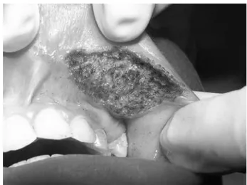
A 10-year-old caucasian boy presented with history of a fall at 1 year of age. Some time after this trauma, a painful irregular growth developed at the left upper lip. The lesion was surgically removed at that time and sent for histopathological examination and diagnosed as a lymphangioma. The lesion relapsed 4 times following the surgery (Fig. 1). After 10 years of the initial trauma, the patient was examined at the laser clinic of the Biophotonics center of the Dental School of the Federal University of Bahia. The lesion was removed with co2 laser (8W). The lesion was vaporized until complete removal under local anesthesia and was left to heal by second intention. Neither suture or dressings were used (Fig. 2). The patient was followed up during 1 year without signs of relapse of the lesion (Fig. 3).
Case 2
A 37-year-old male patient complaining of a painful lesion on the hard palate was examined at the laser clinic of the center of Biophotonics of the Dental School of the Federal University of Bahia. clinical examination revealed an irregular non-ulcerated lesion about 3 cm long. A biopsy was carried out and the lesion was diagnosed as a lymphangioma (Fig. 4A). Under local anesthesia, the lesion was excised with a focused co2 laser beam (10 W) (Fig. 4B). At the end of the surgery the beam was used on a defocused mode to promote better hemostasis. The patient has been followed up for 18 months without signs of relapse (Fig. 5).
Histopathological Findings
Figure 1. clinical aspect of the lesion after four conventional surgical procedures (case 1).
Figure 2. clinical aspect of the area following surgical removal of the lesion with the co2 laser (case 1).
Figure 3. clinical aspect of the operated area 12 months after surgery, with no signs of lesion relapse (case 1).
Figure 5. clinical aspect of the operated area 18 months after surgery. No signs of relapse were seen (case 2).
Figure 4. oral lymphangioma. A= Intraoral aspect of the lesion on the hard palate. B = clinical aspect of the lesion after surgical removal with the co2 laser (case 2).
Figure 6. histological aspect of the lesions showing numerous lymphatic vessels.
at birth and 90% developed around the second year of life (9,11). These aspects corroborate our findings on regarding the rarity of the reported cases.
Small lesions usually do not demand treatment. however, large ones may need surgical removal. The treatment of lesions on the hard palate is difficult due to anatomic constraints (11). Several reports suggested that total excision with safety margins is the best option for its treatment (13). Partial or incomplete removal of the lesion is associated to high recurrence rates; around 50% of the cases will relapse within 2 years following surgery. The recurrence rate following total removal is around 7% (14). The reported relapses of the lesion may be considered high and around 40% of these cases followed excision with (3). In our cases no relapse was seen at a minimal follow up of 12 months. This may be indicative that the use of the co2 laser surgery should be preferred to the conventional excision.
Medium output power laser was used in both cases. The use of small output power has been shown to cause less damage and be effective on the removal of oral mucosal lesions. The less is the thermal damage the better is the repair. It may be questioned why the use of different outputs on these cases, this is explained by the fact that the location and clinical characteristics be different. Thick tissues demanded higher energy for an efficient cut and less accessory thermal damage (12).
Another aspect that needs clarification is the technique used for the removal of the lesions. In one case we opted to excise the lesion and in another we decided to vaporize it. Both methods are acceptable for the removal of lesions; however, the excision is always preferred to vaporization as it allows additional histopathological examination. however, some conditions may not allow the use of excision. one of these conditions is the location of the lesion. In one of the reported cases, the damaged area was on the lip, a site of aesthetic importance.
The most interesting features of co2 laser surgery are local hemostasis, cauterization of nerve endings, and
the two cases reported. Another important aspect is the reduction of the surgical time and the stress to both patient and surgeon in the postoperative period (12).
cauterization of the nerve endings is important as it causes formation of thermal neuromas causing less pain after surgery and avoiding the use of painkillers. No analgesics were prescribed to our patients and they did not complain of pain in the postoperative course even when extensive areas were excised (12).
Due to the high temperatures caused by the laser beam on the tissue, it shows the capability of sterilizing the site, reducing the risk of post-surgical infection and avoiding the use of antibiotic therapy. We prescribed no antibiotic to any of the patients and none showed signs of local infection on follow up time (12).
Additionally, scar formation is minimized due to the sparse presence of myofibroblasts. This late feature is very important on the treatment of oral lesions especially on areas in which scar tissue may cause impairment of the function. It has been shown that the amount of myofibroblasts on co2 laser wound is three times less than that the found on scalpel wounds (15). Neither sutures nor dressings are used on co2 laser wounds and the healing occur by second intention. These aspects also influence on the cost of the procedure. In our cases, we were unable to observe any scarring of the operated area even when large areas were removed.
The use of laser surgery has increased largely over the last 10 years. however, the cost of the equipment and need for qualification on surgical specialties have limited the access of dentists to its benefits. The correct indication of this surgical technique is both cost effective and reliable on the treatment of oral lesions, including premalignant ones. The use of co2 laser does not reduce the risk of lesion relapse, but it is an easy technique to use by trained professionals, which results on both quick surgical procedure and silent postoperative period, and can be safely used on the dental practice (12). In both cases reported in this paper. The use of co2 laser was practical, easy to carry out and effective on the treatment of oral lymphangiomas, with no lesion recurrence within the follow-up periods.
RESUMO
o objetivo deste trabalho é relatar o tratamento de linfangiomas orais com o laser de co2. os linfangiomas são raras malformações linfáticas congênitas que geralmente são diagnosticados na infância. São localizados preferencialmente na região de cabeça e pescoço, mas são extremamente raros na cavidade oral. As lesões da cavidade oral são de tratamento complexo, devido à dificuldade em exercer uma completa remoção. o laser de co2 é o laser mais usado na cavidade oral devido à sua afinidade com a água e alta absorção pela mucosa oral. Diversos benefícios da utilização deste aparelho são relatados na literatura sobre a relação de procedimentos cirúrgicos realizados na cavidade oral. os casos relatados são de linfangiomas comprovados por biópsia prévia. os procedimentos cirúrgicos foram realizados sob anestesia local com um feixe de laser de co2 no modo focado (20c Sharplan laser Indústrias Israel, l10.600 nm, f ~2 mm, cW/RSP). Ao final da cirurgia foi utilizado um feixe de laser no modo desfocado, para promover uma melhor hemostasia. Nem suturas e curativos foram realizados após a cirurgia. Nenhuma medicação foi utilizada, somente anti-sépticos bucais foram prescritos para os pacientes no período pós-operatório. Não houve queixas no pós-operatório dos pacientes e nem recidivas após acompanhamentos de 12 e 18 meses. A utilização do laser de co2 é um método prático, fácil e eficaz no tratamento de linfangiomas orais, sem recidiva nos períodos de acompanhamento.
ACKNOWLEDGEMENTS
The authors shall thank cNPq for financial support to this study.
REFERENCES
1. Souza RJSP, Tone lG. Treatment of lymphangioma with alpha-2a-interferon. J Pediatr 2001;77:139-142.
2. Motaharry P, Sarrafpour B, Abdirad A. Bilateral symmetrical lymphangiomas of the gingiva: case report. Diagnostic Pathol 2006;1:1-3.
3. harashima T, hossain M, Walverde AD, Yamada Y, Matsumoto K. Treatment of lymphangioma with Nd:YAG laser Irradiation: A case report. J clin laser Med Surg 2001;19:189-191. 4. Smith RJh. lymphatic malformations. lymph Res Biol
2004;2:25-31.
5. White JM, chaudhry SI, Kudler JJ, Sekandari N, Schoelch Ml, Silverman S. Nd:YAG and co2 laser therapy of oral mucosa
lesions. J clin laser Med Surg 1998;16:299-304.
6. Pinheiro AlB, Neves Jc, castro JFl, Santos JZlV, De Senna KXFR, Brugnera Junior A, et al.. comparison of the effects of co2 laser and chlorhexidine on the decontamination of infected
cutaneous wounds: A histologic study in rats. J clin laser Med Surg 2002;20:123-127.
7. lapidoth M, Ackerman l, Amital DB, Raveh e, Kalish e, David M. Treatment lymphangioma circumscriptum with combined radiofrequency current and 900 nm diode laser. Dermatol Surg 2006;32:790-794.
8. ethunandan M, Mellor TK. haemangiomas and vascular malformations of the maxillofacial region - A review. Br J oral Maxillofac Surg 2006;44:263-272.
9. Kalpidis cDR, lysitsa SN, Kolokotronis Ae, Samson J, lombardi T. Solitary superficial microcystic lymphatic malformation (lymphangioma circumscriptum) of the gingiva. J Periodontol 2006;77:1797-1801.
10. Mello-Filho FV, Tone lG, Kruschewsky lS. The use of Picibanil (oK-432) for treatment of lymphangioma in the head and neck. Rev Braz otorrinolaringol 2002;68:552-556.
11. lourenço SV, Fernandes JD, Nico MMS. An ulcerated plaque on the hard palate. clin exp Dermatol 2007;34:429-430.
12. Gama SKc, Araújo TM, Pinheiro AlB. Benefits of the use of the co2 laser in orthodontics. lasers Med Sci 2008;23:459-465.
13. Treharne Jl, Murison ScM. co2 laser ablation of lymphangioma
circumscriptum of the scrotum. lymph Res Biol 2006;4:101-103. 14. FitzGerald K, Barry S, Fleming P. Alveolar lymphangioma in infants: report of two cases. J Irish Dent Assoc 2009;55:144-145. 15. Brandon MS, Strauss RA. complications of co2 laser procedures
in oral and maxillofacial surgery. J oral Maxillofac Surg 2004;16:289-299.
16. Pinheiro AlB. comparison of tissue damage and healing in scalpel and co2 laser mucosal wounds. PhD thesis, University of
Birmingham; 1992. 257p.
17. Wang X, Ishizaki NT, Matsumoto K. healing process of skin after co2 laser ablation at low irradiance: A comparison of
continuous-wave and pulsed mode. Photomed laser Surg 2005;23:20-26. 18. Strauss RA, Fallon SD. lasers in contemporary oral and
maxillofacial surgery. Dent clin North Am 2004;48:861-888. 19. Paes Junior TA, Niccoli Filho W. clinical comparison between
conventional suture and vaporization with carbon dioxide laser in rat’s skin. J clin laser Med Surg 2001;19:319-324.
20. Romeo U, Del Vecchio A, Palata G, Tenore G, Visca P, Maggiore c. Bone damage induced by different cutting instruments - an in vitro study. Braz Dent J 2009;20:162-168.