D
ABSTRACT
A SURVEY OF ORAL AND MAXILLOFACIAL BIOPSIES IN
CHILDREN. A SINGLE-CENTER RETROSPECTIVE STUDY
OF 20 YEARS IN PELOTAS-BRAZIL
Giana da Silveira LIMA1, Silvia Terra FONTES1, Lenita Maria Aver de ARAÚJO2, Adriana ETGES2, Sandra Beatriz Chaves TARQUINIO2, Ana Paula Neutzling GOMES2
1- DDS, MSc, PhD, Graduate student, Postgraduate Program in Dentistry, Department of Operative Dentistry, Dental School, Federal University of Pelotas, Pelotas, RS, Brazil.
2- DDS, MSc, PhD, Associate Professor, Department of Semiology and Clinic, Dental School, Federal University of Pelotas, Pelotas, RS, Brazil.
Corresponding address: Dra. Ana Paula N. Gomes - Centro de Diagnóstico das Doenças da Boca (CDDB) - Departamento de Semiologia e Clínica, Faculdade de Odontologia, Universidade Federal de Pelotas - Rua Gonçalves Chaves, 457, Pelotas, RS, Brasil. 96015-560 - Tel/Fax: +55-53-3222-6690 - e-mail: [email protected].
Received: April 01, 2008 - Modification: May 19, 2008 - Accepted: June 19, 2008
espite the large number of published cases about oral and maxillofacial pediatric lesions, the literature is scarce on epidemiological studies regarding the prevalence of these entities. This study retrieved oral and maxillofacial pediatric lesions from the Center of Diagnosis of Oral Diseases (CDDB) at the Dental School of the Federal University of Pelotas (UFPEL), comprising a 20-year period (1983-2002). From the total of 9,465 biopsies received in this period, 625 (6.6%) were from children aged 0 to 14 years. Regardless of the histopathological diagnosis, patient data referring to lesion location, sex and age were collected. Diagnoses were grouped in 13 categories. As much as 89% of the cases occurred in patients aged 7 to 14 years (53% in females and 47% in males). Mucocele (17.2%) was the most common type of lesion, followed by dentigerous cyst (8.6%). In the category of odontogenic tumors, odontoma was the most frequent lesion (64.2%). Malignant lesions were observed in a small section of the sample (1.2%). Generally, the results of the present study are in line with those reported in the literature concerning the most prevalent lesions in the pediatric population. Most lesions were benign, and malignant lesions were diagnosed in a very small part of the sample.
Keywords:Oral and maxillofacial diseases. Pathology. Epidemiology. Children.
INTRODUCTION
Despite the vast literature reporting the prevalence of oral and maxillofacial diseases in the last decades, few studies have focused on biopsied lesions in the pediatric population.
When comparing the occurrence of lesions in the pediatric population, variations regarding the age, prevalence and geographic distribution have been found. In Brazil, only two studies carried out in the states of Minas Gerais7 and São Paulo10 reported data about biopsied oral
pediatric lesions in Brazil. However, they represent exclusively the southeastern part of the country. Since that geographic distribution is a source of variation, the occurrence of lesions in pediatric patients in other parts of the country may be a relevant topic to be investigated.
In this study, we performed a survey of oral and maxillofacial biopsies from children aged 0 to 14 years in a southern city of Brazil. Also, the epidemiological data were
compared to previously published literature.
MATERIAL AND METHODS
Sample Selection and Collection of Clinical Data
This study had the approval of the Research Ethics Committee of the Federal University of Pelotas. The biopsies files from the Center of Diagnosis of Oral Diseases (CDDB) of the Dental School of the Federal University of Pelotas, were retrieved and those related to pediatric patients aged 0 to 14 years, during the period comprised between January 1983 and December 2002, were selected. For the oral and maxillofacial lesions detected, data regarding to location, histopathological features, age and gender were evaluated. Biopsies were then ranked under 13 categories, as follow:
1) Hyperplastic/reactionary lesions: Pyogenic
hyperplasia, gingival hyperplasia, pulp polyp, parulis.
2) Benign neoplasms: Fibroma, squamous papilloma,
nevus, hemangioma, giant cell fibroma, lymphangioma, neurofibroma, congenital epulis, vascular hamartoma, lipoma, common wart.
3) Oral mucosa lesions: Unspecific inflammatory
process, acanthosis, hyperkeratosis, hematoma, foreign body granulomatous inflammation.
4)Infectious diseases: Actinomycosis, toxoplasmosis,
paracoccidioidomycosis.
5) Cystic lesions: Periapical cyst, paradental cyst,
residual cyst, dentigerous cyst, eruption cyst, unclassified cysts of odontogenic origin, epidermoid cyst, median lingual cyst, pilar cyst, incisive canal cyst.
6)Periapical inflammations: Periapical granuloma, sinus tract, fibrous scar.
7) Odontogenic tumors: Odontoma, keratocystic
odontogenic tumor, ameloblastoma, myxoma, ameloblastic fibrodentinoma.
8) Bone pathologies: Giant cell central granuloma,
aneurysmal bone cyst, traumatic bone cyst, fibrous dysplasia, exostosis, central ossifying fibroma, massive osteolysis, ossifying periostitis, cherubism, reaction bone tissue, bone sequestrum.
9) Salivary gland lesions: Mucocele, ranula,
sialolithiasis, sialocyst, adenomatoid hyperplasia, pleomorphic adenoma, myoepithelioma.
10) Malignant neoplasms: Rhabdomyosarcoma,
Langerhans cell histiocytosis, Burkitt’s lymphoma, neuroblastoma, nevoid basal cell carcinoma.
11) Healthy tissues and teeth: Pericoronal follicle,
healthy tissue, natal tooth.
12)Dental alterations:Dens in dente, fusion, mesiodens, dental malformation, external dental resorption, pulp nodules, pulp necrosis, ankylosis.
13) Inconclusive diagnoses
Statistical analysis
Data were recorded and analyzed by descriptive statistics using the SPSS statistical package (SPSS Inc.Chicago,IL,
USA).
RESULTS
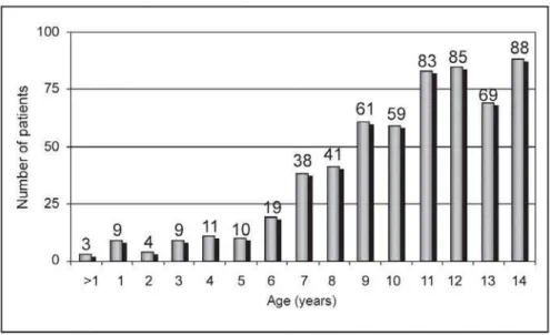
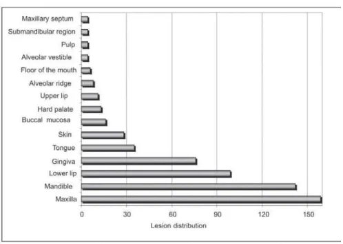
Throughout the study period, 9,465 biopsies were performed, 625 (6.6%) being from pediatric patients. Figure 1 shows the patient distribution for age groups and the prevalence of cases in ages between 7 and 14 years (89.0%). Similar prevalence was observed between females (53.0%) and males (47.0%). Maxilla was the most commonly affected site, followed by the mandibular region, and the lower lip (Figure 2). The most common conditions diagnosed individually were mucocele (17.2%), pericoronal follicle (9.1%), and dentigerous cyst (8.6%) (Figure 3).
Regarding the diagnosis categories, the largest number of cases represented the salivary gland lesions, cystic lesions, and healthy tissues/teeth groups, in this order (Table 1). Mucocele was also the most common condition (89.2%) in the salivary gland lesion category. Dentigerous cyst was the most frequent condition in the cystic lesion group (46.1%), followed by periapical cyst (36.7%).
Fibroma was the most prevalent lesion in the benign neoplasm group, followed by squamous papilloma, nevus, and hemangioma (Figure 4). Odontoma accounted for 64.2% of the odontogenic tumors. Malignant lesions were rarely diagnosed, corresponding to 1.2% of the total sample size (Table 2).
FIGURE 3- Most frequent diagnoses
FIGURE 2- Lesion distribution by location
Categories
1. Hyperplastic/reactionary lesions
Pyogenic granuloma
Inflammatory fibrous hyperplasia Giant cell peripheral granuloma Peripheral ossifying granuloma
2. Benign neoplasms
Fibroma
Squamous Papilloma Nevus
Hemangioma Giant cell fibroma
3. Oral mucosa lesions
Unspecific inflammatory process
4. Infectious diseases
Actinomycosis Toxoplasmosis
Paracoccidioidomycosis
5. Cystic lesions
Dentigerous cyst Periapical cyst Eruption cyst
Unclassified cysts of odontogenic nature
6. Periapical inflammations
Periapical granuloma
7. Odontogenic tumors
Odontoma
Keratocystic odontogenic tumor
8. Bone pathologies
Central giant cell granuloma Aneurysmal bone cyst Traumatic bone cyst Fibrous dysplasia
9. Salivary gland lesions
Mucocele
10. Malignant neoplasms
Rhabdomyosarcoma Neuroblastoma
Langerhans cell histiocystosis Burkitt’s lymphoma
Nevoid basal cell carcinoma
11. Healthy tissues and teeth
Pericoronal follicle Healthy tissue
12. Dental alterations
13. Inconclusive diagnoses
Total
Total number of biopsies
69 16 14 14 14 50 14 9 8 6 5 50 44 3 1 1 1 117 54 43 6 5 33 29 42 27 10 30 9 3 3 3 121 108 8 2 2 2 1 1 78 57 20 8 16 625
Percentage of total biopsies (%) 11.04 2.56 2.24 2.24 2.24 8.00 2.24 1.44 1.28 0.96 0.80 8.00 7.04 0.48 0.16 0.16 0.16 18.72 8.64 6.88 0.96 0.80 5.28 4.64 6.72 4.32 1.60 4.80 1.44 0.48 0.48 0.48 19.36 17.28 1.28 0.32 0.32 0.32 0.16 0.16 12.48 9.12 3.20 1.28 2.56 100
DISCUSSION
Previous studies2,5,8 investigating the occurrence of oral
and maxillofacial lesions in pediatric patients showed that the number of pediatric biopsies generally accounts for less than 10% of all cases referred to histopathology services. Similar prevalence was observed, in the present study, since 6.6% of the total number of biopsies was conducted in children aged 7 to 14 years.
Recently, Jones and Franklin4 performed a retrospective
investigation of oral and maxillofacial pathologies within a 30-year period. Those authors verified that biopsies in patients aged between 0 and 16 years represented 8.2% of all cases reported. Other authors found percentages of pediatric biopsies varied between 11 and 13% of the total number of cases3,6,9. These higher values probably appear
as consequence of setting a higher upper age threshold for the referred casuistic.
In the present findings, a higher concentration of specimens from older children was observed, corresponding to 89% of all pediatric biopsies. Sousa, et al.10 also observed
a higher occurrence of lesions in children between 9 and 14 years of age. Regarding the prevalence in genders, there was a similar distribution of cases between females and males. This result is in accordance with the data published elsewhere4,10.
The evaluated material comprised a wide spectrum of lesions, which ranged from inflammatory processes to malignant tumors. Additionally, a considerable part of the sample was formed by healthy tissues, especially by the pericoronal follicle (9.2%). The files reviewed in this study showed that mucocele as the most common of all lesions (17.2%), followed by dentigerous cyst (8.6%) and unspecific inflammatory process (7.0%). Taken together, these lesions accounted for 42.1% of the total number of biopsies in children. Likewise, several authors2,9,10 have considered the
mucus extravasation phenomenon as the most frequent lesion in children. Mucocele also appears among one of the main conditions diagnosed in pediatric populations in a previous review of the most common labial lesions1. Maia, et al.7
ranked follicular cyst, fibrous hyperplasia, mucocele, and pericoronal follicle as the most frequent conditions diagnosed in their study.
Another point that that should be highlighted is the characterization of the difference between dentigerous cyst and pericoronal follicle. Regarding this topic, many studies are based on radiographic investigations, which are susceptible to observer’s bias, sparking off diagnostic controversies. Also, the classification criteria are usually different for each study, which leads to discrepant results.
Considering the salivary gland lesion group, mucocele represented 89.2% of all biopsies, revealing a considerable occurrence in pediatric populations. Odontoma accounted for 64.2% of the odontogenic tumors. When all neoplastic lesions are considered, odontoma occurrence was higher than that observed of fibroma, which represented 28% of all non-odontogenic benign neoplasms. However, if non-odontogenic origin of benign neoplasms is taken in account as a parameter for tumor classification, odontoma would still be ranked ahead of all other tumors recorded in the present study. In this aspect, our data are corroborated by those of Tomizawa, et al.11, who performed a retrospective study of lesions in
Japanese children. In their analysis, the authors registered 39 odontoma cases, most of them linked to eruptive dysfunctions.
Regarding benign neoplasms, fibroma was the most prevalent diagnosed condition, followed by squamous papilloma, nevus, and hemangioma. It is important to underline the fact that hemangioma may not always be biopsied. Therefore, hemangioma occurrence might be even higher than the number of cases reported herein. On the other hand, the number of malignant lesions was very small in our study, corresponding to only 8 cases (1.2% of the biopsies). Similar results have been reported by Jones and Franklin4, who detected malignity in 1% of the all analyzed
biopsies. Additionally, the most commonly affected sites were the maxilla, mandible, lower lip, and gingiva. Maaita, et al.6 mentioned the mandible and the lip as the most
prevalent sites, while Das and Das3 found a higher
prevalence in the periodontium and the lip.
Another essential aspect to be addressed while analyzing our data is the existence of variations in the prevalence of typical lesions of any given population group. Moreover, it is worth mentioning that the data retrieved in the present study do not reproduce the prevalence of oral and maxillofacial lesions diagnosed by dentists in the clinical
Lesion Rhabdomyosarcoma Rhabdomyosarcoma Burkitt’s lymphoma Neuroblastoma Neuroblastoma
Langerhans cell histiocystosis Langerhans cell histiocystosis Nevoid basal cell carcinoma
Sex Female Female Male Male Male Female Male Male Age 10 years 3 years 7 years 1 year 6 months 1 year 1 year 10 years Location buccal mucosa subcutaneous tissue face gingiva orbit frontal-parietal region mandible
gingiva, skin face
practice, since some pathologies, like herpes, aphthous ulcerations and even hemangioma, are diagnosed based on clinical features, while the present study included biopsied lesions exclusively.
CONCLUSION
Generally, the results of the present study are in line with those reported in the literature concerning the most prevalent lesions in the pediatric population. Most detected lesions were benign, and malignant lesions were diagnosed in a very small part of the whole sample. The epidemiologic survey of oral and maxillofacial lesions biopsied in children is important to determine lesion prevalence in different geographic areas. This type of study also contributes with the characterization of lesion specificities in the pediatric population, providing to general dentists and pediatric dentists a solid background for diagnosis and treatment of the entities.
REFERENCES
1- Bentley JM, Barankin B, Guenther LC. A review of common pediatric lip lesions: herpes simplex/recurrent herpes labialis, impetigo, mucoceles, and hemangiomas. Clin Pediatr (Phila). 2003;42(6):475-82.
2- Chen YK, Lin LM, Huang HC, Lin CC, Yan YH. A retrospective study of oral and maxillofacial biopsy lesions in a pediatric population from southern Taiwan. Pediatr Dent. 1998;20(7):404-10.
3- Das S, Das AK. A review of pediatric oral biopsies from a surgical pathology service in a dental school. Pediatr Dent. 1993;15(3):208-11.
4- Jones AV, Franklin CD. An analysis of oral and maxillofacial pathology found in children over a 30-year period. Int J Paediatr Dent. 2006;16(1):19-30.
5- Keszler A, Guglielmotti MB, Dominguez FV. Oral pathology in children. Frequency, distribution and clinical significance. Acta Odontol Latinoam. 1990;5(1):39-48.
6- Maaita JK. Oral tumors in children: a review. J Clin Pediatr Dent. 2000;24(2):133-5.
7- Maia DM, Merly F, Castro WH, Gomez RS. A survey of oral biopsies in Brazilian pediatric patients. ASDC J Dent Child. 2000;67(2):128-31.
8- Sato M, Tanaka N, Sato T, Amagasa T. Oral and maxillofacial tumours in children: a review. Br J Oral Maxillofac Surg. 1997;35(2):92-5.
9- Skinner RL, Davenport WD Jr, Weir JC, Carr RF. A survey of biopsied oral lesions in pediatric dental patients. Pediatr Dent. 1986;8(3):163-7.
10- Sousa FB, Etges A, Correa L, Mesquita RA, de Araujo NS. Pediatric oral lesions: a 15-year review from Sao Paulo, Brazil. J Clin Pediatr Dent. 2002;26(4):413-8.