ABSTRACT
Pituitary duplication is a rare malformation commonly associated with other major neural/craniofacial anomalies, easily shown by magnetic resonance imaging. The authors describe two girls with duplication of the pituitary gland and thickening of the hypothalamus, facial dysmorphism and precocious pubertal development. The pathogenesis of pituitary duplication and its relationship with precocious pubertal development are discussed. (Arq Bras Endocrinol Metab 2005;49/2:323-327)
Keywords:Pituitary duplication; Precocious puberty; Embryogenesis
RESUMO
Duplicação de Hipófise Associada Com Puberdade Precoce: Apresentação de Dois Casos e Revisão da Embriogênese Hipofisária.
Duplicação pituitária é uma malformação rara muitas vezes associada a anomalias neurais/craniofaciais, facilmente demonstradas por ima-gens em ressonância magnética. Os autores descrevem duas crianças do sexo feminino com duplicação da glândula pituitária e espessamen-to do hipotálamo, dismorfismo facial e desenvolvimenespessamen-to puberal pre-coce. Discute-se a etiopatogenia da duplicação hipofisária e sua relação com o quadro de puberdade precoce. (Arq Bras Endocrinol Metab 2005;49/2:323-327)
Descritores:Duplicação hipofisária; Puberdade precoce; Embriogênese
P
ITU ITARY D U PLICATIO N IS Arare malformation, reported previously inapproximately 23 patients (1-8), three of them presenting with pre-cocious or delayed puberty (2,6,8). Most of the cases are associated with other major neural/ craniofacial anomalies which are easily shown by magnetic resonance imaging (MRI) and the majority of patients do not survive beyond infancy. O ther pituitary hormone abnormalities are not usually present.
The formation of the hypophysis depends on interaction of the embryonic primordium with normal growth processes in the prechordal region of the head. The prechordal plate and the rostral portion of the notochord are closely related to the development of the pituitary gland. The duplication of the rostral end of the notochord may act as the main factor that leads to duplication of the pituitary primordium, with resultant formation of two morphologically normal glands (8).
The authors describe two girls with duplication of the pituitary gland and thickening of the hypothalamus, facial dysmorphism and preco-cious pubertal development.
apresentação de casos
and R evi ew of Pi tui tar y Embr yogenesi s
Gustavo Cancela e Penna
Már ci a Por to Pi menta
Juli ana B. D r ummond
Mar ta Sarqui s
José Carlos T. Mar ti ns
R odri go C. de Campos
Eduardo Pi mentel D i as
Serviço de Endocrinologia, H ospital Felício R ocho (GCP, EPD), Serviço de Endocrinologia, H ospital Mater Dei (MPP, JBD), A xial Centro de Im agens (JCTM), Departamento de Clínica
Médica, Universidade Federal de Minas Gerais – UFMG (MS) e Serviço de N eurologia Infantil, H ospital das Clínicas, UFMG (R CC), Belo H orizonte, MG.
CASE 1
A 7-year-old white female presented with facial dys-morphism and pubertal development starting at age six. Physical exam revealed hypertelorism, microre-trognathia, cleft nose and pubertal development at P3B3 according to Tanner. Laboratory evaluation showed responsive GnRH -st im ulat ed LH levels (7.89U I/ L), normal thyroid function, prolactin and I G F-I levels. A pelvic u lt raso n o graph y (U S) disclosed stimulated uterus (11mL) and ovaries (2.8 and 4.2mL with several follicles greater than 5mm). Bone age was advanced (10 years compared to 7.8 years of chronologic age). Computed tomography (CT) and MRI (figures 1 to 4) showed duplication of the hypophysis with two small and independent glands, each with a stalk, two median eminences, a wide hypothalamus with two infundibular recesses and other malformations. This thickening of the hypothalamus associated with hypophysis duplica-tion has been named pseudohamartoma. This inter-posed abnormal hypothalamic mass, which consists of arrested cells that normally migrate laterally to form the hypothalamic nuclei, represents the most common associated intracranial abnormality and is probably related to splitting of the end of the notochord (4). The bright posterior pituitary signal was preserved in both glands. Persistence of the -cranio-pharyngeal canal, associated with a naso-coanal mass, adherent to the base of the sphenoid was also noted.
The child underwent excision of the nasopha-ryngeal mass and pathological examination revealed hyperplasia of salivary glands. Treatment with a GnRH agonist was started and 6 months later new lab o rat o ry assessm en t sh o wed n o n -respo n sive GnRH -stimulated LH levels, significant improve-ment of the pelvic U S measureimprove-ments (uterus of 4.2mL, ovaries of 1.2 and 1.6mL with no visible fol-licles) and no advance in bone age.
CASE 2
A 6.8-year-old white female presented with a history of strabismus, cleft and uvula, dental abnormalities and esophageal fistula, which had been previously treated, and precocious pubertal development. Physical exam revealed right breast development at B2 according to Tanner and growth velocity above the 97th percentile
(9cm/ year). Laboratory evaluation showed responsive GnRH -stimulated LH levels (7.5U I/ L) and a bone age of 6.6 years. Serum measurements of prolactin, thyroid function and IGF-I were within normal limits. MRI (figures 5 to 8) showed duplication of the hypophysis and thickening of the hypothalamus. The patient was reevaluated 6 months later, presenting with no further progression of pubertal development, although persis-tence of increased growth velocity (8cm/ year) was confirmed, associated with advance in bone age (increase of 1.4 years in 6 months), stimulated uterus (6mL) and multiple bilateral ovarian primordial follicles on pelvic U S. Treatment with GnRH analog was started and follow-up evaluations showed normalization of growth velocity (6cm/ year), uterus volume (3mL) and no progression of Tanner pubertal stage.
DISCUSSION
H ypophysis duplication is a rare phenomenon. Its association with true precocious puberty has been sug-gested by Burke et al. (6) when they described an 11-year-old female with hypertelorism and pituitary dupli-cation who achieved menarche at 8.5 years of age. The
Figure 1.Coronal T1 weighted image before (A) and after
(B) contrast, showing pituitary (arrow-heads) and infundibu-lum duplication (arrows).
Figure 3.Sagital (A), and coronal (B) T1 weighted sections, and coronal T2 weighted (C)showing hypothalamic pseudohamartoma (arrow-heads) and duplication of the infundibular recess of the third ventricle (arrows).
Figure 4.Axial (A)and coronal (B)T2 weighted sections showing cranial duplication of basilar artery (arrows).
exact mechanism responsible for the early increase in frequency and amplitude of GnRH pulses causing precocious puberty in these patients is still unknown, but may be related to the well-documented associa-tion of hypothalamic hamartomas and true preco-cious puberty. Burke et al (6). proposed that the development disorder leading to duplication may
have caused precocious secretion of LH RH as a con-sequence of nuclear derangement and failure of regu-lation. Indeed, delayed puberty has also been described in association with hypophysis duplication (2), emphasizing that the disruption of the hypothala-mic hormonal millieu can occur associated with this embryologic abnormality. Taking into consideration
Figure 8.Axial (A)and coronal (B)T2 weighted sections showing duplication of basilar artery(arrows).
Figure 6.CT – axial (A), coronal (B and C)and MRI coronal T1 (D)and T2 (E)weighted sections showing palate cleft (arrow-head), persistence of nasopharingeal channel (black arrows) and nasopharingeal mass (white arrows) in continuity with the sphenoid.
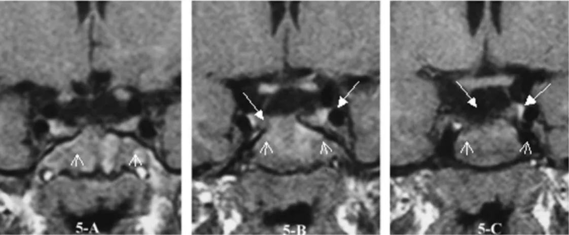
Figure 7.Sagital (A) and coronal (B) T1 weighted sections; coronal T2 weighted section (C)
that many of the cases of hypophysis duplication reported thus far were diagnosed in very young children, precocious and delayed puberty should probably be far more frequent.
H ypophysis duplication is usually associated with the median cleft face syndrome. Several malfor-mations have been described, including (4): facial dysmorphism, development abnormalities of the tongue, hydrocephalus, abnormalities of the circle of Willis, posterior cranial fossa abnormalities, agenesis of the corpus callosum, spinal abnormalities, thickening of the hypothalamus, cleft palate, nasopharyngeal masses, absence of the anterior commissure, absence of the olfactory bulbs and tracts, and basilar artery duplication The embryogenesis of pituitary duplication is still controversial. The rostral end of the notochord and the prechordal plate are closely related to the primordium of the pituitary gland and stalk, which is initially distinguished at about 22 days of gestation. It begins as an adhesion of neural and stomodeum ectoderm. O vergrowth of surrounding mesenchyme leads to elongation and incorporation of the gland to its normal anatomic position. An alternative view to the classic concept is that the anterior lobe may be of neuroectodermal origin (9). In fact, the anterior neural ridge develops into the oral epithelium after head fold turning and delineates the roof of the mouth and all structures derived from this region. The mature pituitary gland originates from a thick-ening and concurrent invagination or pouching of this oral epithelium.
From these early relationships, it is suggested that splitting of the tip of the rostral end of the noto-chordal structures may act as the primary factor which leads to duplication of the area of neuroectodermal adherence, with formation of two independent normal glands and the other associated abnormalities.
Burke et al. (6) suggest a different mechanism to explain isolated pituitary duplication: a primary disruption in the area of the neuroectodermal adhesion, independent of the notochord, would result in disjunction of the primordium and, consequently, hypophyseal duplication.
Pituitary duplication should be considered in all patients who present with midline abnormalities and a MRI study should be systematically obtained in these patients. Among the numerous associated anomalies, partial basilar artery duplication or fenestration, which
can be usually demonstrated by routine MRI, may cause altered flow dynamics, leading to a higher inci-dence of aneurysm (7,8). Periodic supervision for this potential complication may be necessary.
As illustrated by the above case presentations, precocious and delayed puberty can also be associated with hypophysis duplication and clinical surveillance of pubertal development is therefore recommended.
REFERENCES
1. Ryals BD, Brown DC, Levin SW. Duplication of the pitui-tary gland as shown by MR. Am J Neuroradiol 1993;14:137-9.
2. Kollias SS, Ball WS, Prenger EC. Review of the embry-ologic development of the pituitary gland and report of a case of hypophyseal duplication detected by MRI.
Neuroradiology 1995;37:3-12.
3. Shah S, Pereira JK, Becker CJ, Roubal SE. Duplication of pituitary gland. J Comput Assist Tomogr 1997;21:459-61. 4. Hamon-Kérautret M, Ares GS, Demondion X, Rouland V,
Francke JP, Pruvo JP. Duplication of the pituitary gland in a newborn with median cleft face syndrome and nasal teratoma. Pediatr Radiol 1998;28:290-2.
5. Vandenhaute B, Leteurtre E, Lecomte-Houcke M, Pel-lerin P, Nuyts JP, Cuisset JM, et al. Epignathus teratoma: report of three cases with a review of the literature. Cleft Palate Craniofac J 2000;37:83-91.
6. Burke M, Zinkovsky S, Abrantes MA, Riley W. Duplication of the hypophysis. Pediatr Neurosurg 2000;33:95-9. 7. Uchino A, Sawada A, Takase Y, Fujita I, Kudo S. Extreme
fenestration of the basilar artery associated with cleft palate, nasopharyngeal mature teratoma, and hypophyseal duplication. Eur Radiol 2002;12:2087-90. 8. Shroff M, Blaser S, Jay V, Chitayat D, Armstrong D. Basilar
artery duplication associated with pituitary duplication: a new finding. Am J Neuroradiol 2003;24:956-61. 9. Elster AD. Modern imaging of the pituitary. Radiology
1993;187:1-14.
Endereço para correspondência: