ABSTRACT
Relative antiinflammatory and immunosuppressive potencies of gluco-corticoids (GC) were previously well defined. Nonetheless, GC also regu-late cell proliferation and programmed death (apoptosis). The aim of this study was to determine the relative potency of different GC on the modulation of cell survival. The GC-sensitive lymphoblast cell line CEM-c7/14 was submitted to 48h-exposure to GC (dose-response curve from 10-8to 10-5M). Cell survival was analyzed employing the
DimethylTiazol-Tetrazolium (MTT) test. For each GC at least 4 experiments were perfor-med in quadruplicate. Responses to different GC at the same molarity were analyzed by ANOVA on Ranks. Cell responses to the same GC in different concentrations were tested by repeated measures ANOVA. The EC50 for each GC was calculated with the GraphPad Prism 3.0 software. The use of low concentrations (10-8 and 10-7M) of hydrocortisone and
methylprednisolone determined a similar effects on cell survival, which was less prominent than that observed with betamethasone, budesoni-de or momethasone. Momethasone was the most potent GC, inducing the most intense dexamethasone reduction on cell survival at the lowest concentration (10-8M). Momethasone and methylprednisolone were the
two GC with the strongest impact on cell survival. Our findings suggest that antiproliferative and apoptotic potencies of GC are different from those previously reported antiinflammatory and immunosuppressive actions.(Arq Bras Endocrinol Metab 2005;49/3:378-383)
Keywords:Glucocorticoid; Cell survival; Cell death; Apoptosis
RESUMO
Potências Antiproliferativa e Pró-Apoptótica dos Glicocorticóides:
Discordância com as Propriedades Anti-inflamatórias e Imunossupressoras. As potências antiinflamatória e imunossupressora dos glicocorticóides (GC) já foram bem estabelecidas previamente. No entanto, os GC tam-bém possuem atividade reguladora da proliferação celular e da morte celular programada (apoptose). O objetivo deste estudo foi determinar a potência relativa de diferentes GC na modulação da sobrevida celular. Linfoblastos cortico-sensíveis (linhagem celular CEM-C7/14) foram mantidos em cultura prolongada e submetidos ao tratamento com GC por 48h, em doses variando entre 10-8e 10-5molar. O índice de
sobrevida celular foi quantificado pelo teste MTT (DimetilTiazol-Tetrezolium). Para cada GC avaliado, foram realizados pelo menos qua-tro experimentos em quadruplicata. A resposta celular aos diferentes GC foi analisada através do teste estatístico ANOVA on Ranks, enquanto a resposta ao mesmo GC em concentrações diferentes foi analisada pelo teste ANOVA for repeated measures. O EC50 de cada GC foi calculado utilizando-se o software GraphPad Prism 3.0. Durante o uso de concentrações baixas (10-8e 10-7molar), observou-se sobrevida
semelhante dos linfoblastos após tratamento com hidrocortisona ou metilprednisolona. Nestas mesmas concentrações baixas, a sobrevida celular foi menor quando utilizou-se dexametasona, betametasona, budesonida ou mometasona. A mometasona e a metilprednisolona
artigo original
A nti i nflammator y and Immunossuppressi ve
Proper ti es
Carlos A . Longui
Mari a C. Santos
Cri sti na B. For mi ga
D ani ela V.A . Oli vei ra
Mylene N . R ocha
Claudi a D .C. Fari a
Cri sti ane K ochi
Osmar Monte
Molecular Medicine Laboratory, Departm ent of Physiology, Santa Casa São Paulo – Faculty of Medical Sciences, São Paulo, SP.
foram os dois GC que determinaram maior redução da sobrevida linfoblástica. Nossos resultados sugerem que as potências antiproliferativa e pró-apoptótica dos GC sejam diferentes dos efeitos antiinflamatórios e imunossupressores previamente estabelecidos para estes GC. (Arq Bras Endocrinol Metab 2005;49/3:378-383)
Descritores: Glicocorticóide; Sobrevida celular; Morte celular; Apoptose
C
O RTISO L, TH E EN D O GEN O U S GLU CO CO RTICO ID (GC), is secreted basally and during stress and modulates the amplitude of defensive responses. Corti-sol and a variety of synthetic glucocorticoid agonists are able to control carbohydrate, protein and lipid metab-olism, and to regulate immune and cardiovascular func-tions (1,2). GC suppress innate inflammatory respons-es, as well the cellular immunity (3). O ne of the major effects of GC is their ability to exert anti-proliferative and apoptotic actions both in vivoas in vitrocell cul-ture (4). Glucocorticoid-induced apoptosis is an active, ATP-dependent phenomenon characterized by cellular and mitochondrial membrane changes, and alterations in calcium and potassium compartmental distributions (5). Programmed cell death depends on the activation of nuclear proteases, generating D N A, RN A and pro-tein fragmentation, genomic instability and failure of D N A repair. The antiproliferative and apoptotic actions of glucocorticoids mediate their therapeutic effects in several autoimmune and lymphoproliferative diseases.Cell survival can be measured by the ability of live cells to metabolize MTT, a yellow tetrazolic salt, to its dark violet crystal product formazan. This con-version occurs after active enzymatic cleavage at the mitochondrial level, and the measurement of the final product can be used as a quantitative assay, reflecting the cell viability (5). Relative glucocorticoid potencies are well established for their anti-inflammatory and immunosuppressive effects. O n the other hand, the pathways related to modulation of cell survival and death are unique, requiring additional studies to determine the relative potencies of new synthetic glu-cocorticoids. In this study, we compared the relative antiproliferative and apoptotic potencies of hydrocor-tisone against several other synthetic glucocorticoids.
MATERIALS AND METHODS
A GC stock-solution was prepared by diluting GC salts in absolute-ethanol to obtain a final concentration of
10-2M. Working-solutions were obtained by
subse-quent dilution of the stock-solution, 1:9 in RPMI-1640 (GIBCO BRL Cat # 11875-093).
The cell line CEM-c7/ 14, derived from a patient with a glucocorticoid-sensitive lymphoblastic leukemia, was kindly offered by D r. E. B. Thompson (the U niversity of Texas Medical Branch at Galve-stone, TX, U SA). The cells were kept at growing phase in RPMI-1640 supplemented with 10% FBS (Fetal Bovine Serum, GIBCO BRL Cat # 16140-071) and 1% penicillin/ streptomycin. Cell culture was
main-tained at 5% pCO2and 37º C.
Cell viability was established in haemocytome-ter in a 1:1 solution of trypan blue (Sigma, Cat #
T0776), resuspended to 4x10⁶ viable cells/ mL, and
cultured in quadruplicate in a 24-well microplate (Fisher, Cat # 07-200-84). The first well of the assay-plate received only RPMI-1640 medium, and into the subsequent wells it was applied cells without gluco-corticoid and cells plus glucogluco-corticoid in increasing
final molar concentration ranging from 10-8 to 10-5
M. To achieve the final glucocorticoid concentration,
5µL of glucocorticoid was added to each well (e.g.,
5µL of 10-3M to 500µL of cell suspension to obtain a
10-5M final concentration). At least four experiments
in quadruplicate were performed for each glucocor-ticoid.
After a 48h-incubation period 100mL of MTT (dimethyl-Tiazol-Tetrazolium, Sigma Cat # M-2128) solution (5mg/ mL) was added to each well and incu-bated for an additional 4h-incubation period at the same conditions previously described, to allow the MTT conversion into formazam. D issolution of for-mazam-crystals was achieved in 3 volumes (1800mL) of Isopropanol-H Cl (23:2) solution (Sigma, Cat # I-9516 and Merck, Cat # 100983, respectively). An aliquot of 200µL was transferred in duplicate to a 96-well microplate (Fisher, Cat # 07-200-89), with sub-sequent determination of the optical density (O D ) of the solution at 595nm (U niversal Microplate Reader Elx800, Bio-Tek Instruments, Inc, U SA). These O D measured values are directly dependent on the number of alive cells. For each plate, the blank-background was represented by the O D values observed in the medium-only well. The maximum cell growth for each experiment was represented by the values observed in the wells containing cells plus RPMI-1640 without the addition of glucocorticoids and expressed as the 100% cell viability for that plate-assay.
Statistical analysis employed the SigmaStat 2.03 software (SPSS, Inc.). Comparison of the same gluco-corticoid at different molarities was performed apply-ing the Friedman test, AN O VA for repeated measures. When significant difference was detected, the All Pair-wise Multiple Comparison Procedures – Tukey Test was used to recognize each different pair concentra-tion. For comparison among different GC at the same molarity the Kruskal-Wallis – AN O VA on ranks test was performed, followed by the All Pairwise Multiple Comparison Procedures – D unn’s Method to verify the difference among two different glucocorticoids. The EC50 was calculated employing GraphPad Prism 3.0 software.
RESULTS
The most characteristic patterns observed for all tested glucocorticoids are shown in tables 1 and 2. H ydro-cortisone decreased cell survival in molar
concentra-tions ≥ 10-7M, with the maximum effect at 10-5M.
The same pattern of cell survival reduction was observed with methylprednisolone, but the final effect was higher than the one observed with hydrocortisone (reduction of 60.9% and 36.7%, respectively). D exam-ethasone decreased cell viability at molar
concentra-tions ≥10-8M, with maximum effect at 10-5M (49.6%
of cell reduction). Betamethasone, budesonide and momethasone showed a pattern similar to that observed for dexamethasone. In a molar concentration
as low as 10-8M, these three GC had an effect
equiva-lent to that observed at 10-5M, and the cell survival
was significantly lower than that observed for dexam-ethasone treated cells at 10-8M.
Comparing hydrocortisone to synthetic
gluco-corticoids at the concentration of 10-8M, we observed
that all GC, but methylprednisolone, had significantly higher potency in decreasing cell survival.
The GC concentration necessary to obtain 50%
of the maximal effect (EC50) was between 10-7 M to
10-6M for hydrocortisone, 10-7M for
methylpred-Table 2.Major pharmacologic characteristics of glucocorticoids regarding their proapoptotic properties.
Glucocorticoid Start Effect Max Effect EC50 Max Cell reduction
Hydrocortisone 10-7M 10-6M 5 x 10⁻⁷M 37%
Methylprednisolone 10-7M 10-6M 10-7M 61%
Dexamethasone 10-7M 10-6M 5 x 10⁻⁸M 50%
Betamethasone 10-8M 10-6M < 10-8M 54%
Budesonide 10-8M 10-8M < 10-8M 46%
Momethasone 10-8M 10-8M < 10-8M 59%
Start effect: minimal glucocorticoid concentration to start significant reduction on cell survival
Max effect: minimal glucocorticoid concentration able to determine a maximal reduction on cell survival EC50: Concentration at which 50% of the glucocorticoid effect was observed
Max cell reduction: maximal cell reduction (in percentage) related to basal nontreated cells
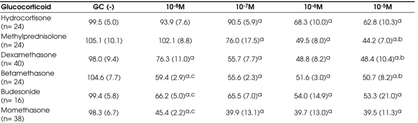
Table 1.Percentage of live cells expressed as mean (SD) after treatment with glucocorticoid for 48 hours.
Glucocorticoid GC (-) 10-8M 10-7M 10-6M 10-5M
Hydrocortisone
(n= 24) 99.5 (5.0) 93.9 (7.6) 90.5 (5.9)a 68.3 (10.0)a 62.8 (10.3)a Methylprednisolone
(n= 24) 105.1 (10.1) 102.1 (8.8) 76.0 (17.5)a 49.5 (8.0)a 44.2 (7.0)a,b Dexamethasone
(n= 40) 98.0 (9.4) 76.3 (11.0)a 55.7 (7.7)a 48.8 (8.2)a 48.4 (10.4)a,b Betamethasone
(n= 24) 104.6 (7.7) 59.4 (2.9)a,c 55.6 (2.3)a 51.6 (3.0)a 50.7 (8.2)a,b Budesonide
(n= 16) 99.4 (5.8) 66.2 (5.0)a,c 65.5 (7.0)a 54.0 (14.9)a 53.3 (21.0)a Momethasone
(n= 38) 98.3 (6.7) 45.4 (2.2)
a,c 39.9 (13.1)a 39.7 (13.0)a 39.5 (11.3)a
* corticosensitive lymphoblasts (c7/14 cell line); GC(-): maximal cell survival without glucocorticoid; M= molar; n= total number of point-experiments performed for each glucocorticoid
a: significant reduction when compared to basal values, p< 0.05 (Anova Repeated Measures)
nisolone, between 10-8M to 10-7M for
dexametha-sone, and smaller than 10-8M for betamethasone,
budesonide and momethasone (figure 1).
DISCUSSION
Glucocorticoids have specific biologic effects in sever-al organ systems, depending on their pharmacokinetic characteristics and inherent actions exerted through their specific nuclear receptors (GR). C hemical changes in cortisol molecule can enhance glucocorti-coid or mineralocortiglucocorti-coid activities, determining
exert thymolytic actions (8), and to inhibit skin fibrob-lat growth rate (9).
There is a considerable variation between these previously described potencies. Additionally, just a small number of synthetic GCs were compared by the same technique, preventing direct comparison of the anti-inflammatory potencies among different gluco-corticoids. Limited information is also available for comparison among recently synthesized novel gluco-corticoids. Glucocorticoid receptor binding capability can be detected by radioreceptor-assay, and this char-acteristic has been correlated to the anti-inflammatory potency of these steroids. U sing this method, anti-inflammatory potency observed for methylpredniso-lone, dexamethasone and betamethasone were conside-red higher than that established by other methods (10). Studies evaluating GC antiproliferative effects are even more scarce. U sing an MTT assay to compare prednisolone and dexamethasone on its relative antileukemic activity, a 16-fold higher potency was observed for dexamethasone (11). Another study eval-uated the relative cytotoxicity of these two glucocorti-coids by flow-cytometric analysis of cells from patients with acute lymphoblastic leukemia, and the authors concluded that dexamethasone had a cytotoxic activi-ty five to six times higher than prednisolone (12). D espite the existing data evaluating and comparing the antiproliferative and apoptotic GC actions, these studies usually compare only two glucocorticoids (dexamethasone and prednisolone).
In this study, the relative potency of eight dif-ferent glucocorticoids were compared regarding their antiproliferative and apoptotic activity, by examining cell survival. We described GC potencies considering the minimal glucocorticoid concentration able to start its effect on cell survival, the concentration at which the maximal reduction was obtained, the concentra-tion at which 50% of the maximal effect was detected (EC50), and the maximal cell reduction observed after 48h of steroid therapy. This is the first report compar-ing multiple glucocorticoids in their effects or cell sur-vival. As a group, hydrocortisone, methylprednisolone and dexamethasone started their anti-proliferative and apoptotic effect at “physiological” concentrations
(10-7M). The same effect was observed with
budes-onide and momethasone but at a 10-times smaller concentration. Momethasone was the GC able to in-duce the grater reduction on cell number and to requi-re the smallest dose to start its effects.
We observed in this study, employing different glucocorticoids, that betamethasone, budesonide and momethasone have their EC50 at similar levels and
under the 10-8 molar concentration, suggesting that
further studies should evaluate even smaller doses of these compounds.
The discrepancy between anti-inflammatory and the cell proliferation and apoptotic potencies observed in this study are potentially related to the unique pathways involved in cell cycle control and apoptosis, different from pathways activated during inflammation. These discrepancies on relative gluco-corticoid potency suggest that, if the regulation of cell number is the major target of therapy, specific dosage and type of glucocorticoid should be titrated for this specific effect. Future studies should determine the rel-ative potency of new synthetic glucocorticoids and establish these effects for even smaller concentrations.
ACKNOWLEDGMENTS
We are deeply grateful to D r. George P. Chrousos for his extensive collaboration and important suggestions for this study. This study was supported by a research grant of FAPESP – Proccess # 98/ 10680-7. We also thank the Editorial assistance offered by the Support Center for Scientific Publication of Santa Casa, SP – Faculty of Medical Sciences – Brazil.
REFERENCES
1. Schimmer BP, Parker KL. Hormônio adrenocorticotrófico; esteróides adrenocorticais e seus análogos sintéticos; inibidores da síntese e das ações dos hormônios adrenocorticais. In: Goodman LS, Gilman A, eds. As bases farmacológicas da terapêutica. 10ª ed. Rio de Janeiro:McGraw-Hill, 2003. p.1241-61.
2. Grossmann C, Scholz T, Rochel M, Bumke-Vogt C, Oelk-ers W, Pfeiffer AF, et al. Transactivation via the human glucocorticoid and mineralocorticoid receptor by ther-apeutically used steroids in CV-1 cells: a comparison of their glucocorticoid and mineralocorticoid properties.
Eur J Endocrinol 2004;151:397-406.
3. Zurier RB, Weissmann G. Antimmunologic and antinflam-matory effects of steroid therapy. Med Clin North Am 1973;57:1295-307.
4. Longui CA, Vottero A, Adamson PC, Cole DE, Kino T, Monte O, et al. Low glucocorticoid receptor alpha/beta ratio in T-cell lymphoblastic leukemia. Horm Metab Res 2000;32:401-6.
5. McGahon AJ, Martin SJ, Bissonnette RP, Mahboubi A, Shi YF, Mogil RJ, et al. The end of the (cell) line: methods for the study of apoptosis in vitro. Methods cell biol 1995;46:153-85.
6. Cantrill HL, Waltman SR, Palmberg PF, Zink HA, Becker B.
In vitrodetermination of relative corticosteroid potency.
7. Maurer M, Trajanoski Z, Frey G, Hiroi N, Galon J, Willen-berg HS, et al. Differential gene expression profile of glu-cocorticoids, testosterone, and dehydroepiandros-terone in human cells. Horm Metab Res 2001;33:691-5.
8. Tonelli G, Thibault L, Ringler I. A bioassay for the con-comitant assesment of the antiphlogistic and thimolytic activities of topically applied corticoids. Endocrinology 1965;77:625-34.
9. Liddle GW. Clinical pharmacology of the anti-inflamma-tory steroids. Clin Pharmacol Ther 1961;2:615-35.
10. Ballard PL, Carter JP, Graham BS, Baxter JD. A radiore-ceptor assay for evaluation of the plasma glucocorti-coid activity of natural and synthetic steroids in man. J Clin Endocrinol Metab 1975;41:290-304.
11. Kaspers GJ, Veerman AJ, Popp-Snijders C, Lomecky M, Van Zantwijk CH, Swinkels LM, et al. Comparison of the antileukemic activity in vitro of dexamethasone and
prednisolone in childhood acute lymphoblastic leukemia. Med Pediatr Oncol 1996;27:114-21.
12. Ito C, Evans WE, McNinch L, Coustan-Smith E, Mahmoud H, Pui CH, et al. Comparative cytotoxicity of dexam-ethasone and prednisolone in childhood acute lym-phoblastic leukemia. J Clin Oncol 1996;14:2370-6.
Endereço para correspondência:
Carlos Alberto Longui
R. Pimenta Bueno 65, apto 102 03060-000 São Paulo, SP Fax: (11) 6618-4480