O
RIGINALA
RTICLE Revista Brasileira de FisioterapiaHigh-voltage electrical stimulation improves
nerve regeneration after sciatic crush injury
Estimulação elétrica de alta voltagem favorece a regeneração nervosa após
compressão do nervo isquiático
Rosana M. Teodori, Andréia M. Silva, Meiricris T. Silva, Larissa S. Oliveira, Maria L. O. Polacow, Elaine C. O. Guirro
Abstract
Background: Peripheral nerve injury causes prolonged functional limitation being a clinical challenge to identify resources that accelerates its recovery. Objectives: To investigate the effect of high-voltage electrical stimulation (HVES) on the morphometric and functional characteristics of the regenerated nerve after crush injury in rats. Methods: Twenty Wistar rats were randomly allocated into 4 groups: Control (CON) - without injury and without HVES; Denervated (D) - sciatic nerve crush only; Denervated + HVES - sciatic nerve crush and HVES; SHAM - without injury but HVES. The HVES and SHAM groups were stimulated (100 Hz; minimum voltage of 100 V, 20 μs, 100 μs interpulse interval) for 30 min/day, 5 days/week. The sciatic functional index (SFI) was evaluated before the injury and at the 7th, 14th and 21st postoperatory (PO) days.
Neural components and the area density of connective tissue, blood vessels and macrophages were analyzed. Results: Axonal diameter was higher on the HVES than on D group, reaching almost 80% above the control values after 21 days (p<0.05). Fiber diameter and myelin sheath thickness were higher on the HVES than on D group (p<0.05) reaching 96.5% and 100% of the control values, respectively. Functional recovery at the 14th PO day was better on group HVES. The macrophages and connective tissue area density was lower on the HVES group,
while blood vessels number did not differ among groups. Conclusions: The HVES accelerated the functional recovery, potentiated the nerve fibers maturation and decreased macrophages and connective tissue area density, suggesting acceleration of neural repair.
Keywords: nerve crush; peripheral nerves; nerve regeneration; electric stimulation; sciatic nerve; physical therapy modalities.
Resumo
Contextualização: Lesões nervosas periféricas provocam limitação funcional prolongada, sendo um desafio para a clínica identificar recursos que acelerem sua recuperação. Objetivos: Investigar a influência da estimulação elétrica de alta voltagem (EEAV) sobre a morfologia e a função do nervo regenerado após esmagamento em ratos. Métodos: Vinte ratos Wistar foram divididos nos grupos: controle (CON) – sem lesão e sem EEAV; desnervado (D) – esmagamento do nervo isquiático; desnervado + EEAV (EEAV) – esmagamento do nervo e EEAV; SHAM – sem lesão, porém submetido à EEAV. Os grupos EEAV e SHAM foram estimulados (100 Hz, tensão mínima de 100 V; 20 µs e 100 µs interpulso) 30 min/dia, 5 dias/semana. O índice funcional do ciático (IFC) foi avaliado antes da lesão, nos 7º, 14º e 21º dias pós-operatório (PO). Componentes neurais, densidade de área de tecido conjuntivo, de vasos sanguíneos e macrófagos foram analisados. Resultados: O diâmetro axonal foi maior no grupo EEAV que no grupo D, atingindo quase 80% dos valores-controle após 21 dias (p<0,05). O diâmetro das fibras e espessura das bainhas de mielina foram maiores no grupo EEAV que no D (p<0,05), alcançando 96,5% e 100% dos valores-controle, respectivamente. A recuperação funcional no 14º dia PO foi melhor no grupo EEAV. A densidade de área de macrófagos e tecido conjuntivo foi menor no grupo EEAV, enquanto o número de vasos sanguíneos não diferiu entre os grupos. Conclusões: A EEAV acelerou a recuperação funcional, potencializou a maturação das fibras nervosas regeneradas e promoveu diminuição da densidade de área de macrófagos e tecido conjuntivo no nervo, sugerindo aceleração do reparo neural.
Palavras-chave: compressão nervosa; nervos periféricos; regeneração nervosa; estimulação elétrica; nervo isquiático; modalidades de fisioterapia.
Received: 08/09/2010 – Revised: 14/03/2011 – Accepted: 10/05/2011
Master’s Program in Physical Therapy, Universidade Metodista de Piracicaba (UNIMEP), Piracicaba, SP, Brazil
Correspondence to: Rosana Macher Teodori, Universidade Metodista de Piracicaba, School of Health Sciences, Master’s Program in Physical Therapy, Neuromuscular, Plasticity Laboratory, Rodovia do Açúcar, Km 156, Taquaral, CEP 13.400-911, Piracicaba, SP, Brazil, e-mail: [email protected]
Introduction
Traumatic injury of peripheral nerves is common and results in signiicant functional deicits. After crush injuries, regeneration occurs spontaneously. In rats, no axons are found in the muscle ten days after injury because the muscle rein-nervation begins two weeks after the injury1,2.
Poly-innervation is observed two days after the neuritis reach the muscle, given that ifteen days after nerve crush 25% of mus-cle ibers are poly-innervated. he increase of neuritis sprouting and growth, as well as the formation of synapses between 14 and 25 days after injury indicate that nerve impulses may be released to the muscle2. Around the 25th day, muscle membrane rest
po-tential, contraction force, sensitivity to acetylcholine, and acetyl-cholinesterase levels have returned to normal. he process that eliminates excessive synaptic contacts starts at this time and, around the 60th day, muscle ibers become mono-innervated.
he remaining nerve terminals are increased in size and after 90 days the postsynaptic cleft is almost completely occupied1.
However, at the end of the regeneration morphological al-terations on regenerated axons remain. After section injuries, the nerve presents reduced axonal caliber and decreased my-elin sheath thickness3. Even in crush injuries, nerve
morpho-metric properties are not fully recovered4.
In humans, the most notable characteristic of denervation is muscular atrophy, which is determined by the reduction in quantity of actin and myosin myoilaments, and results in a decrease of iber diameter and loss of muscle force5.
Lu, Huang and Carlso6 demonstrated the negative efects
caused by an increase of intramuscular connective tissue after denervation, which alters muscle function by delaying nutrient exchange between muscle ibers and the capillary system, as well as interfering with axonal growth during reinnervation. Due to slow progress inherent to nerve recovery7 the structural
condi-tions of a denervated muscle remain widely altered during nerve recovery in humans, which compromises the return to work. Since the maturation of regenerated nerve is essential for the nerve conduction speed and functional recovery, the main clini-cal challenge is to identify mechanisms to accelerate the nerve regeneration.
It is known that animal tissues are endowed with intrinsic electricity that is involved with fundamental physiological pro-cesses, such as nerve conduction and muscle contraction8. he
complete motor function recovery after nerve injury requires the occurrence of morphological and physiological processes that determine the return of motoneurons electrical activity on the involved muscles9.
he use of muscle electrical stimulation after peripheral nerve injury for the prevention of progressive muscle atrophy, although there is weak evidence that, can be beneicial when
applied under favorable conditions10. his fact reinforces the
im-portance of controlled trials for the establishment of new treat-ment strategies to stimulate peripheral nerve regeneration.
High-voltage electrical stimulation (HVES) has gained ac-ceptance in both experimental and clinical research11,12. Most
studies that use HVES investigate its circulatory13 and
regen-erative properties on skin ulcers healing14.
he application of negative electric ields increases regener-ation of the peripheral nerves in mammals15. However, there are
no studies suggesting the efect of HVES on the peripheral nerve regeneration, which justiies the need for need for high quality studies that will permit a greater understanding of HVES and its role on this process, as well as will stimulate discussion of its use on the peripheral nerve injury treatment in humans. hus, the hypothesis of the present study is that HVES can shorten the regeneration and maturation of injured nerves.
herefore, the aim of this study was to investigate the in-luence of high-voltage electrical stimulation on the morpho-metric and functional characteristics of the regenerated nerve after crush injury in rats.
Methods
Twenty male Wistar rats (210.80±10.79 g) were randomly allocated into 4 groups (n=5 each): Control (CON) - animals without injury and without HVES; Denervated (D) - sciatic nerve crush; Denervated + HVES (HVES) - sciatic nerve crush and HVES; SHAM - without injury, but subjected to HVES (SHAM).
his study was approved by the Ethics Committee for Animal Experimentation of the Universidade Federal de São Carlos (CEEA/ UFSCar), São Carlos, SP, Brasil, protocol number 037/2008.
Functional gait analysis: Sciatic Functional Index (SFI)
For the functional gait assessment, a pathway of 8.2x42 cm was covered with white paper and the animals were set to walk on it, with their hind paws marked with ingerprinting ink. his registration was performed prior to the crush injury, and at the 7th, 14th and 21st post operatory (PO) days16. Distances between
the extremity of the third inger and the calcaneous - Print Length (PL); between the irst and ifth toes - Total Spreading (TS); and between the second and fourth toes - Intermediary Toes (ITS) were measured in the experimental and normal paws using a MitutoyoTM digital pachymeter with an accuracy of 0.01 mm,
according to the manufacturer´s manual. he obtained values were used in the equation proposed by the same authors16. he
results were expressed as functional loss, being that the value 0 (zero) representing normal function, and the value of -100 (mi-nus one hundred) representing maximum disability.
Nerve injury
he animals in D and HVES groups were submitted to a crush injury (performed surgically) on the left sciatic nerve. hey received intramuscular anesthesia with Ketamine Chlo-ride (1.16 g/10 mL) and Xylazine ChloChlo-ride (2 g/100 mL) at a 3:2 ratio with doses of 0.09 mL/100 g and 0.06 mL/100 g of their body weight, respectively, additionally a digital trichotomy on their gluteal region was performed, bilaterally. he left sciatic nerve was crushed with a haemostatic forceps, 5 mm proximal to its branching point, by four 20-second clamps with one sec-ond of interval between them17. he same researcher crushed
all nerves.
Animals from the SHAM group had the sciatic nerve ex-posed and maintained intact. All animals were kept under vivarium conditions, subjected to a 12-hour photoperiodic cycle of light/dark, controlled temperatures (23±2 ºC), and had constant access to special food (Labina, PurinaTM) and water.
In the irst two days after surgery, the animals received 4 µL of Sodic Dipirone (500 mg/mL) by oral administration, for anal-gesic efect, every 12 hours.
High Voltage Electrical Stimulation (HVES)
The Neurodyn High Volt - ANVISA 10360310008 - IBRAMEDTM was used in this study. The calibration was
performed with an oscilloscope Tektronix (TDSTM) 210,
with a load of 1000 Ω, while the timer was calibrated us-ing three stopwatches (TechnosTM).
he intervention on HVES and SHAM groups was per-formed under anesthesia (Ketamine Chloride and Xylazine Chloride – 0.045 mL/100 g and 0.03 mL/100 g of body weight, respectively) and was initiated 24 hours after the crush injury, with cathodic stimulation at the motor threshold, for 30 min-utes (100 Hz; minimum voltage of 100 V, 20 μs and 100 μs inter-pulse interval), 5 days a week, during 21 days. A silicon-carbon active electrode (2.0x2.0 cm) was placed over the surgical scar, while the dispersive electrode (4.0x4.0 cm) was positioned par-allel to the active, preserving a distance of 1 cm between them. Sterile gel was used on the skin shaved as a conductor.
Data collection
After 21 days, the animals were anesthetized by the same procedures as described for the nerve injury. he left sciatic nerve was ixed in situ at 4° C during 10 minutes, with modiied Karnovsky ixative containing 1% of paraformaldehyde and 2% of glutaraldehyde in a sodium cacodylate bufer at 0.1 M, pH 7.3. Next, the nerve segment distal to the injury was removed and the animals were euthanized by cervical dislocation.
he sciatic nerve fragments were maintained in the same ixative solution for 24 hours and postixed in osmium tetrox-ide at 1% in sodium cacodylate bufer at 0.1 M, pH 7.3, for two hours, immersed in 5% uranyl during 24 hours for en bloc stain-ing, dehydrated in increasing solutions (30% to 100%) of ac-etone and included in AralditeTM 502 resin. Transversal section
cuts (1 µm) of the distal nerve obtained 5 mm far to the crush were stained with toluidine blue at 1% borax aqueous solution for observation in light microscopy. In the control group, the transversal section cuts were obtained from the same region as the experimental groups.
Quantitative and morphometric analysis
For these analysis the examiner was blinded. he number and diameter of axons and the diameter of nerve ibers were determined3. In this study we used an image analysis system
(Image-Pro Plus 6.2 - Media CyberneticsTM). From these data,
the myelin sheath thickness was calculated.
For the area density analysis of connective tissue, blood ves-sels and macrophages, ive non-serial sections were selected, in which ive ields with a 1000x magniication were randomly photographed using an Ininity Lite (Lumenera CorporationTM)
digital camera attached to an OlympusTM BX 41 light
micro-scope, which was connected to an image analysis system (Im-age-Pro Plus 6.2 - Media CyberneticsTM, with a 100x objective
lens). Following, a planimetry by a point-countingsystem18 was
used, in which a grid with 450 line intersections per ield, total-izing 56.250 intersections per experimental group, was applied in order to obtain the percentage of area density.
Statistical analysis
he Shapiro-Wilk normality test and a One Way ANOVA, followed by the post-hoc Tukey test were used for the num-ber and density of blood vessels, density of macrophages and connective tissue, axon number, nerve morphometry and functional analysis. One Way ANOVA followed by Tamhane’s T2 test (between-group comparisons) was used to calculate axonal diameter. To analyze the SFI between-group diferences, Two Way ANOVA, followed by post hoc LSD test were utilized. he signiicance level of 5 % was considered for all analyses.
Results
Quantitative and morphometric analysis
No diferences on the axons number was observed in all groups. he axonal and iber diameter, as well as the myelin
sheath thickness, were higher on the HVES group than on the D group. hese results are displayed in Table 1.
Histological analysis
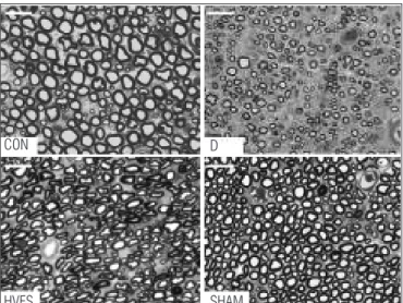
Figure 1 presents the morphological characteristics of the normal sciatic nerve, with axons surrounded by myelin sheaths with thickness proportional to its diameter (CON). In the denervated group (D), the axons, as well as the myelinated nerve ibers, present decreased diameters and myelin sheaths thickness, with an evident increase of neural connective tis-sue. When the injured nerve was subjected to HVES, the re-covery of their morphometric characteristics was observed. As expected, nerves that were not injured but received HVES (SHAM) presented morphological characteristics similar to normal nerves.
Functional gait analysis
As presented in Figure 2, among the four diferent groups, the most efective functional recovery was observed in the rats
allocated to the HVES group at the 14th PO day (p<0.05).
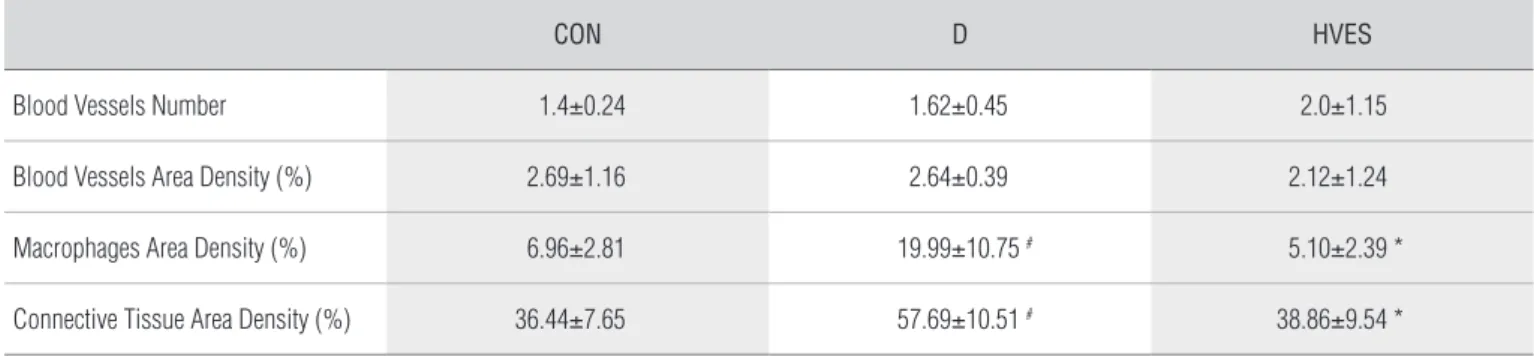
Area density of blood vessels, macrophages and
connective tissue and blood vessels number
Considering that the morphometric analysis revealed no diferences between the CON and SHAM groups, these param-eters, as well as the number of blood vessels, were investigated only on the CON, D and HVES groups.
No between-group diferences in the area density of blood vessels were observed (p>0.05). Macrophages and connective tissue area densities were higher on D than on CON group
Table 1. Mean±SD of quantitative and morphometric analysis on groups: Control (CON); Denervated (D); Denervated and High-voltage Electrical Stimulation (HVES); Without injury but High-voltage Electrical Stimulation (SHAM).
CON D HVES SHAM
Axons Number 9161.60±1394.14 11140.60±1152.53 9780.60±2061.95 10078.00±2261.94
Axons Diameter (µm) 6.73±0.38 3.53±0.43 # 5.38±0.34 # * 6.43±1.16 *
Fibers Diameter (µm) 11.45±0.49 6.66±0.67 # 11.05±0.59 * 11.48±1.21 *
Myelin Sheath Thickness (µm) 1.14±0.11 0.75±0.06 # 1.24±0.11 * 1.21±0.05 *
(#) Differ from CON group; (*) Differ from D group (p<0.05).
Preoperative 7th PO day
D
0
-20
-40
-60
-80
-100
-120
HVES SHAM
14th PO day 21st PO day
Sciatic Funcional Index
Figure 2. Between-Groups comparison of Functional Sciatic Index (FSI) on groups: Denervated (D); Denervated and High Volt Electrical Stimulation (HVES); Without injury but High Volt Electrical Stimulation (SHAM) in different moments.
(*) Differ from the SHAM group; (**) Differ from the D group. Figure 1. Transverse section of sciatic nerve on groups: Control
(CON); Denervated (D); Denervated and High Volt Electrical Stimulation (HVES); and SHAM (SHAM). Note the differences in the axon diameter and myelin sheath thickness, as well as the neural connective tissue area. Toluidine Blue stain. Bar=20 µm.
CON D
HVES SHAM
(p<0.05). he HVES reduced the macrophages and connective tissue area densities after nerve crush injury (p<0.01) with esti-mates being very similar to the control group.
here was no between-group diference (p>0.05) for the number of blood vessels in the nerve (Table 2).
Discussion
In a pilot study, a group of 5 Wistar rats was submitted to the injury method described and had the nerve segment distal collected and histologically analyzed after 6 days. Considering that the myelin sheaths engage in an intense deterioration process 36 to 48 hours after injury19 the observation of either
the presence or the absence of intact nerve ibers would allow for a demonstration of the eicacy of the chosen method. An absence of intact nerve ibers was observed, as well as exten-sive axonal deterioration, making this nervous injury method valid for evaluative studies of diferent resources to be used in the stimulation of peripheral nerve regeneration.
he inluence of low frequency biphasic currents over den-ervated muscle has been established20. Oliveira et al.21 showed
that low frequency electrical stimulation on rat denervated soleus muscle favored axonal sprouting after nerve crush in-jury, since the axon number doubled after 21 days, which was justiied by an increase in endoneural blood low with a con-sequent supply increase of the trophic substances needed for nerve regeneration.
However, despite being a therapeutic resource used in vari-ous clinical treatments, this is the irst report on the efects of the High Voltage Electrical Stimulation (HVES) after periph-eral nerve injury in rats. he clinical advantage of HVES is that the stimulation is confortable and afects sensory, motor and nociceptive nerve ibers. he physical parameters involved in this current allow for several therapeutic efects, such as pain and edema control22, as well as acceleration of tissue repair23,24.
Houghton et al.25 demonstrated the HVES efectiveness
on human’s chronic skin ulcers regeneration. Subjects treated with HVES presented 44.3% reduction in ulcer area after 4 weeks, while those that received conventional treatment im-proved only 16%.HVES can inluence blood low, but this efect depends on the physical parameters used as well as the stimu-lation site23. Mohr, Akers and Wessman26 observed signiicant
increase on blood low velocity in the rat hind limb during and after HVES application with a cathodic stimulation.
In this study we used the cathodic stimulation, however the blood low velocity was not assessed, but the number and area density of blood vessels, where no diference was observed when the HVES was applied. his suggests that this resource only inluence the velocity of blood low, apparently not pro-moting angiogenesis.
his study used HVES at the motor threshold because a rhythmic muscle contraction is required to increase arterial blood low to the stimulated area. However, it seems neces-sary to investigate the characteristics of blood low in the area where stimulation was applied, since the quantitative analysis of blood vessels failed to identify any inluence of HVES. It is possible that the use of diferent parameters may contribute to understanding the efects of HVES on blood low in regener-ated nerves.
After two to four weeks of the sciatic nerve crush in rats, the axon number in the distal segment tends to increase by two-fold7 due to axonal sprouting. In this study, both
den-ervated groups showed number of axons similar to control group, showing that HVES did not inluence the number of regenerated ibers.
he HVEScrosses the skin and produces negligible thermal and electrochemical efect, which allows for a greater current density to reach the target tissues, besides producing efects on vascular system, since the rhythmic muscular contraction/ relaxation increases blood low on the muscle and neighboring tissues27.
Table 2. Mean±SD of neural blood vessels number and area density; macrophages and connective tissue area density on groups: Control (CON); Denervated (D); Denervated and High-voltage Electrical Stimulation (HVES).
CON D HVES
Blood Vessels Number 1.4±0.24 1.62±0.45 2.0±1.15
Blood Vessels Area Density (%) 2.69±1.16 2.64±0.39 2.12±1.24
Macrophages Area Density (%) 6.96±2.81 19.99±10.75 # 5.10±2.39 *
Connective Tissue Area Density (%) 36.44±7.65 57.69±10.51 # 38.86±9.54 *
(#) Differ from CON group; (*) Differ from D group (p<0.05).
It is possible that the cathodic stimulation on motor threshold applied at injured peripheral nerve has promoted a transient efect on blood low to connective tissues surround-ing the nerve, in this sense favorsurround-ing the uptake of nutrients and neurotrophic factors essential to the nerve’s morphological and physiological restoration, without changing the area density and the number of blood vessels, since these morphometric characteristics refer to the nerve analyzed 24 hours after the end of stimulation.
he results of the SHAM group demonstrated that the HVES application on normal nerve does not inluence its quantita-tive and morphometric characteristics, highlighting the efect of HVES on the maturation of regenerated nerve ibers.
In this study, all morphometric parameters were com-pletely recovered on the HVES group, with the exception of the axon diameter, since it did not reach control values after 21 days of injury. Considering that the nerve maturation is only completed when it is reconnected to the muscle and when the synaptic elimination occurs, which happens only around the 60th day after crush injuries1,2 it is possible to suggest that
HVESspeed axonal maturation, once that these results were observed only 21 days after injury.
he HVES increased myelin sheath thickness in comparison to group D, which was proportional to axon diameter and auto-matically relected on the nerve ibers diameter. hese results exceeded the expectations of morphological recovery of injured nerve ibers, because, according to Schröder4, after crush injuries,
the axons diameter can reach control values after 6 months, but the myelin sheath thickness reaches only 79% of normal values after 1 year. Mira28 identiied the presence of regenerated nerve
ibers after rat sciatic nerve crush between the 10th and 15th day,
however, the variation of iber diameters remained the same after two years of injury. In the present study, the diameter of regener-ated axons after HVES treatment reached 79.9% of control values, while the nerve ibers diameter reached 96.5% and the myelin sheath thickness reaches 100% of the control values. hese results demonstrate the beneits of HVESon the maturation of regener-ated nerves after crush, as well as the earliness with which this oc-curred after its application, emphasizing the importance of future studies to demonstrate its role in transection nerve injury.
hese results are reinforced when the morphometric data of other nerve elements are examined, such as the connective tissue. he macrophages, responsible for cell debris phago-cytosis, presented the highest area density in D group at the 21st day. In the HVES group, there was signiicant reduction
of these cells, demonstrating a more advanced stage of re-generated tissue maturation. Probably, HVES stimulated the macrophages migration to the injury site because according
to Orida and Feldman29 the macrophages tend to migrate
toward the anode.
In this study, there was no inluence of HVES on normal nerve function. On the denervated groups, the functional behavior followed the evidence already mentioned by Carmignoto et al.1 and Gorio et al.2, where complete function
loss was observed between the 7th and the 14th day, and
recovery at the 21st PO day, when the muscle is almost
completely re-innervated.
he between-group analysis showed that all groups pre-sented normal function at the preoperatory period, whereas at the 7th POday, denervated groups showed signiicant function
reduction. At the 14th PO day, these values remained close to
-100 in D group, while there was signiicant functional recovery on the HVES group at the same time period. his fact dem-onstrates the HVES efectiveness in accelerating recovery, not only regarding the morphological characteristics of regenerated nerve, but also the restoration of muscles control by inferior motoneurons. At the 21st PO day, the two denervated groups
recovered their function with no diference between HVES and D groups being observed.
hese results suggest that the HVES use accelerated muscle reinnervation. Consequently, the acetylcholine re-lease on the neuromuscular junction would be occurring precociously because, according to Gorio et al.2, after 15 days
of nerve crush in rats, 25% of muscle ibers are reinnervated. It was observed in D group that at the 14th PO day there was
no functional recovery, probably due to the fact that only one a fourth of muscle ibers were reinnervated, which would not relect as improved function. However, on the HVES group, SFI values suggest that a larger number of muscle ibers were reinnervated, resulting in better contraction quality and im-provement of function.
Despite the fact that the SFI results show that at the 21st
POday all groups reached the normality values, once nerve re-generation and functional recovery occur spontaneously after crush injury7 is evident the acceleration of functional recovery
in the HVES group, reinforcing the need to investigate the functional parameters on nerve section injuries.
It should be highlighted the importance of early functional recovery when considering the efects of denervation to the muscle. After denervation, muscle atrophy rapidly occurs, due to the loss of myoibrillar protein components that represent 60% of muscle proteins30, since contractile activity is
impor-tant for the maintenance of the muscle iber’s cross-sectional area10. Denervation reduces the muscle iber cross-sectional
area, increases the area density of the intramuscular connec-tive tissue, undermining the muscle reinnervation, limiting the
References
1. Carmignoto G, Finesso M, Siliprandi R, Gorio A. Muscle reinnervation – I. Restoration of transmitter release mechanisms. Neuroscience. 1983;8(3):392-401.
2. Gorio A, Carmignoto G, Finesso M, Polato P, Nunzi MG. Muscle reinnervation – II. Sprouting, synapse formation and repression. Neuroscience. 1983;8(3):406-16.
3. Santo Neto H, Pertille A, Teodori RM, Somazz MC, Marques MJ. Primary nerve repair by muscle autografts prepared with local anesthetic. Microsurgery. 2004;24(3):188-93.
4. Schröder JM. Altered ratio between axon diameter and myelin sheath thickness in regenerated nerve fibers. Brain Res. 1972;45(1):49-65.
5. Sheffler LR, Chae J. Neuromuscular electrical stimulation in neurorehabilitation. Muscle Nerve. 2007;35(5):562-90.
6. Lu DX, Huang SK, Carlson BM. Electron microscopic study of long-term denervated rat skeletal muscle. Anat Rec. 1997;248(3):355-65.
7. Fawcett JW, Keynes RJ. Peripheral nerve regeneration. Annu Rev Neurosci. 1990;13:43-60.
8. Hess D, El Manira A. Characterization of a high-voltage-activated IA current with a role in spike timing and locomotor pattern generation. Proc Natl Acad Sci U S A. 2001;98(9):5270-81.
9. English AW, Schwartz G, Meador W, Sabatier MJ, Mulligan A. Electrical stimulation promotes peripheral axon regeneration by enhanced neuronal neurotrophin signaling. Dev Neurobiol. 2007;67(2):158-72.
10. Dow DE, Cederna PS, Hassett CA, Kostrominova TY, Faulkner JA, Dennis RG. Number of contractions to maintain mass and force of a denervated rat muscle. Muscle Nerve. 2004;30(1):77-86.
11. Mendel FC, Wylegala JA, Fish DR. Influence of high voltage pulsed current on edema formation following impact injury in rats. Phys Ther. 1992;72(9):668-73.
12. Garcia LB, Guirro ECO. Efeitos da estimulação de alta voltage no linfedema pós-mastectomia. Rev Bras Fisioter. 2005;9(2):243-8.
13. Taylor K, Mendel FC, Fish DR, Hard R, Burton HW. Effect of high-voltage pulsed current and alternating current on macromolecular leakage in hamster cheek pouch microcirculation. Phys Ther. 1997;77(12):1729-40.
14. Davini R, Nunes CV, Guirro ECO, Guirro RRJ. Estimulação elétrica de alta voltage: uma opção de tratamento. Rev Bras Fisioter. 2005;9(3):249-56.
15. Román GC, Strahlendorf HK, Coates PW, Rowley BA. Stimulation of sciatic nerve regeneration in the adult rat by low-intensity electric current. Exp Neurol. 1987;98(2):222-32.
16. Bain JR, Mackinnon SE, Hunter DA. Functional evaluation of complete sciatic peroneal, and posterior tibial nerve lesions in the rat. Plast Reconstr Surg. 1989;83(1):129-38.
17. Lima SC, Caierão QM, Peviani SM, Russo TL, Somazz MC, Salvini TF, et al. Muscle and nerve
responses after different intervals of electrical stimulation sessions on denervated rat muscle. Am J Phys Med Rehabil. 2009;88(2):126-35.
18. Mathieu O, Cruz-Orive LM, Hoppeler H, Weibel ER. Measuring error and sampling variation in stereology: comparison of the efficiency of various methods for planar image analysis. J Microsc. 1981;121(Pt 1):75-88.
19. Dubový P. Schwann cells and endoneurial extracellular matrix molecules as potential cues for sorting of regenerated axons: a review. Anat Sci Int. 2004;79(4):198-208.
20. Caierão QM, Betini J, Teodori RM, Minamoto VB. The effect of time interval between electrical stimulation on the denervated rat muscle. Rev Bras Fisioter. 2008;12(2):143-8.
21. Oliveira LS, Sobral LL, Takeda SYM, Betini J, Guirro RRJ, Somazz MC, et al. Estimulación eléctrica y natación en la fase aguda de la axonotmesis: influencia sobre la regeneración nerviosa y la recuperación funcional. Rev Neurol. 2008;47(1):11-5.
22. Holcomb W, Rubley MD, Girouard TJ. Effect of the simultaneous application of NMES and HVPC on knee extension torque. J Sport Rehabil. 2007;16(4):307-18.
23. Bettany JA, Fish DR, Mendel FC. Influence of high voltage pulsed direct current on edema formation following impact injury. Phys Ther. 1990;70(4):219-24.
24. Baker LL, Chambers R, DeMuth SK, Villar F. Effects of electrical stimulation on wound healing in patients with diabetic ulcers. Diabetes Care. 1997;20(3):405-12.
25. Houghton PE, Kincaid CB, Lovell M, Campbell KE, Keast DH, Woodbury MG, et al. Effect of electrical stimulation on chronic leg ulcer size and appearance. Phys Ther. 2003;83(1):17-28.
26. Mohr T, Akers T, Wessman HC. Effect of high voltage stimulation on blood flow in the rat hind limb. Phys Ther. 1987;67(4):526-33.
27. Griffin JW, Newsome LS, Stralka SW, Wright PE. Reduction of chronic posttraumatic hand edema: a comparison of high voltage pulsed current, intermittent pneumatic compression, and placebo treatments. Phys Ther. 1990;70(5):279-86.
28. Mira JC. Quantitative studies of the regeneration of rat myelinated nerve fibres: variations in the number and size of regenerating fibres after repeated localized freezings. J Anat. 1979;129(Pt1):77-93.
29. Orida N, Feldman JD. Directional protrusive pseudopodial activity and motility in macrophages induced by extracellular electric fields. Cell Motil. 1982;2(3):243-56.
30. Furuno K, Goodman MN, Goldberg AL. Role of different proteolytic systems in the degradation of muscle proteins during denervation atrophy. J Biol Chem. 1990;265(15):8550-7.
31. Carter AJ, Kristmundsdottir F, Gilmour J, Glasby MA. Changes in muscle cytoarchitecture after peripheral nerve injury and repair. A quantitative and qualitative study. J Hand Surg Br. 1998;23(3):365-9.
interaction of nerve terminals with acetylcholine receptors at the neuromuscular junction31.
he HVES application accelerated nerve regeneration, pro-pitiating to the muscle the possibility of an earlier voluntary activity recovery, thereby preventing the development of den-ervation deleterious efects.
Concluding remarks
It is concluded that HVES accelerated the regenerated nerve ibers maturation after crush injury, as well as functional recovery. Our results suggest that the utilization of HVES could contribute to a more favorable regeneration of peripheral nerve
injury by aiding in a premature functional recovery, reducing treatment costs, and in providing a faster reintegration of the subjects to his or her labor activity.
Additional studies may contribute to specify the time of nerve regeneration initiation after HVES application, as well as to promote the use of this resource on human peripheral nerve injury treatment.
Acknowledgments
To the Conselho Nacional de Desenvolvimento Cientíico e Tecnológico (CNPq), for the research productivity grant (Process nº 307041/2008-5).