Article
Printed in Brazil - ©2017 Sociedade Brasileira de Química0103 - 5053 $6.00+0.00*e-mail: [email protected], [email protected]
Tailored Silica Nanoparticles Surface to Increase Drug Load and Enhance
Bactericidal Response
Luciane F. de Oliveira,a Karim Bouchmella,a,b Agustin S. Picco,a,b Larissa B. Capeletti,a,c Kaliandra A. Gonçalves,d João Henrique Z. dos Santos,c Jörg Kobarge and Mateus B. Cardoso*,a,b
aLaboratório Nacional de Luz Síncrotron, LNLS and bLaboratório Nacional de Nanotecnologia,
LNNano, CNPEM, CP 6192, 13083-970 Campinas-SP, Brazil
cUniversidade Federal do Rio Grande do Sul, 91501-970 Porto Alegre-RS, Brazil
dLaboratório Nacional de Biociências, LNBio, CP 6192, 13083-970 Campinas-SP, Brazil
eFaculdade de Ciências Farmacêuticas e Departamento de Bioquímica e Biologia Tecidual,
Instituto de Biologia, Universidade Estadual de Campinas, UNICAMP, CP 6154, 13083-970 Campinas-SP, Brazil
Nanoparticles’ surface properties can be used as triggers to regulate or even enhance biological response and generate tailored structures to substitute conventional antibiotics. Here, silica nanoparticles surface was duly tuned in order to increase the water-insoluble drug load (curcumin) and improve the antibacterial activity. Our main motivation was based on the electrostatic attraction between the positively charged amino groups and the negatively charged curcumin and/or bacteria membrane. In addition, the variation of amino grafting amount on silica nanoparticles indicated that the grafting increase was directly related to the extent of drug entrapped into the nanoparticles as well as to the bactericidal activity. The combination of amino-functionalized silica nanoparticles associated with the presence of curcumin allowed to produce a dual bactericidal system that shows promising perspective for its use in biomedical applications.
Keywords: amino-functionalized silica nanoparticles, curcumin, bactericidal activity, Escherichia coli
Introduction
Pathogenic microorganisms have proved to develop resistance to currently available antibiotics becoming a
worldwide concern.1-5 Thus, it is necessary to find new
systems or drugs presenting alternative mechanisms of action to substitute the existing commercial antibiotics.
Nanoparticles are promising materials for therapeutic applications since they possess unique physical and
chemical properties.6-9 Thus, silica-based nanoparticles
have been widely investigated and used in different fields, including materials science and biomedicine, due to their stability, high hydrophilic surface, biocompatibility
and easy surface functionalization.10-13 In particular,
mesoporous silica nanoparticles are potential candidates as drug carriers since they provide the possibility of encapsulating and delivering large quantities of drugs due
to large surface areas and pore volumes.14-20 Using the
well-known silane chemistry, silica nanoparticles have been functionalized with a variety of different functional
groups such as carboxyl,21 vinyl,22 amino,23 mercapto24
and epoxy.25 The surface functionalization with organic
functional groups allows increasing the storage capacity
of drugs or biomolecules within the silica pores.21,25-28
Moreover, the chemical surface modification of the nanoparticles is an important tool to control the interaction of nanoparticles with biological systems while reducing
toxicity and increasing the therapeutic effects.29-32 Typical
surface modification methods via covalent bonds are
(i) the co-condensation (one-pot synthesis method); (ii) the
postsynthetic grafting (PSG) and (iii) the use of bissilylated
organic precursors that generate periodic mesoporous
organosilicas (PMOs).33-35 The PSG involves modification
kept, the materials can be selectively functionalized with a wide variety of functional groups and the resulting structure exhibits high hydrothermal stability.
Nanostructured silica and curcumin (CCM) complexes
were first reported in 2011 by Jin et al.36 They have shown
the selective immobilization of CCM onto the internal cavity of mesoporous hollow silica particles as well as their drug
release properties. Later, few works37,38 have demonstrated
that bare silica porous systems are able to entrap CCM and are effective against tumor cells. More recently, a
plethora of works39-45 demonstrating that surface-modified
curcumin-loaded silica nanoparticles can be successfully and selectively used against different types of cancer cells were reported. However, to the best of our knowledge, nothing has been done in order to use nanostructured silica and curcumin (CCM) complexes as bactericidal agents.
In our previous works,46-54 we investigated the correlation
between the nanoparticles physical properties and their behavior in biological systems.These properties, that include size, surface charge and hydrophobicity, play an important role for the interaction between nanomaterials and biological systems. In the present study, we go further and show for the first time the bactericidal properties of amino-functionalized curcumin-loaded silica nanoparticles obtained from PSG method. In addition, we studied the influence on the amount of amino groups in the extent of encapsulation of curcumin and the resulting silica nanoparticles bactericidal activity. The bactericidal properties of these materials were tested
against Escherichia coli (E. coli) to evaluate the
amino-functionalized silica as a platform for water-insoluble drug carrier.
Experimental
Chemicals
Tetraethoxysilane (TEOS), (3-aminopropyl) triethoxysilane (APTES), curcumin and ammonium
hydroxide (26 wt.% NH3 in water) were obtained from
Sigma-Aldrich. Ethanol was obtained from J. T. Baker. Peptone, sodium chloride (NaCl), yeast extract and bacteriological agar were purchased from Bio Rad. All chemicals and reagents were used as received without further purification. Water used in all procedures was obtained from a water purification system (Purelab from
ELGA) and had a measured resistivity of 18.2 MΩ cm.
Preparation of bare silica nanoparticles (SiO2)
The bare mesoporous silica nanoparticles were prepared
using the modified Stöber method. SiO2 nanoparticles
were prepared by mixing 380 µL of TEOS and 12 mL of ethanol under continuous stirring for 30 min. Then,
570 µL of NH4OH were added to the mixture and stirred for
24 hours at room temperature. The particles were separated by centrifugation (10000 rpm, 20 minutes, Eppendorf Centrifuge Model 5804), washed with ethanol and dried in air at room temperature for one day.
Amino-functionalized silica nanoparticles (SiO2-NH2)
synthesis
SiO2-NH2 nanoparticles were prepared by mixing
380 µL of TEOS and 12 mL of ethanol under continuous
stirring for 30 min. Then, 570 µL of NH4OH were added to
the mixture and stirred for 24 hours at room temperature. After this time, 200 or 400 µL of APTES were added to the mixture and kept under stirring for 24 hours. The particles were separated by centrifugation (10000 rpm, 20 minutes, Eppendorf Centrifuge Model 5804), washed with ethanol and dried in air at room temperature for one day.
Synthesis of amino-functionalized curcumin-loaded silica nanoparticles (CCM/SiO2-NH2)
Amino-functionalized curcumin-loaded silica
(CCM/SiO2-NH2) nanoparticles were prepared by mixing
380 µL of TEOS, 12 mL of ethanol and 60 µL of curcumin solution (5% m/v) under continuous stirring for 30 min.
Then, 570 µL of NH4OH were added to the mixture and
stirred for 24 hours at room temperature. After this time, 200 or 400 µL of APTES were added to the mixture and kept under stirring for 24 hours. The particles were separated by centrifugation (10000 rpm, 20 minutes, Eppendorf Centrifuge Model 5804), washed with ethanol and dried in air at room temperature for one day. Supernatant was used to determine the encapsulation yield.
Ninhydrin assay
The amino groups on the modified SiO2-NH2 surface
570 nm with an ultraviolet-visible spectrometer (Agilent 8453 equipment).
Characterization of silica nanoparticles
Size and morphology of the nanoparticles were investigated by transmission electron microscopy (TEM). TEM images were taken using JEOL JEM-2010 instrument at the Brazilian Laboratory of Nanosciences (LNNano) operating at an acceleration voltage of 200 kV.
The structural organization of nanoparticles was investigated through the small-angle X-ray scattering (SAXS) technique. The SAXS experiments were carried out on the
D1B beamline at the LNLS using a wavelength λ = 1.488 nm
and the Pilatus 300 k detector placed 1549.8 mm away from the sample. The X-ray beam was monochromatized with a multilayer monochromator and collimated by a set of slits defining a pin-hole geometry. All measurements were performed at room temperature. Silver behenate powder was used as a standard to calibrate the sample-to-detector distance, the detector tilt and the direct beam position. The isotropic scattering patterns were radially averaged. Fitting procedures were carried out using the SASfit software.
The amount of entrapped curcumin was determined by the UV-Vis adsorption (measured at 520 nm) taking into account the remaining curcumin in the supernatant after the reaction has been completed. The UV-Vis absorption spectra were obtained using an Agilent 8453 UV-Vis spectrophotometer (Agilent Technologies).
The nitrogen adsorption-desorption isotherms were obtained at 77 K using a Tristar II 3020 (Micromeretics) instrument. The specific surface area is assessed according to the standard BET (Brunauer-Emmett-Teller) method in
the 0.05-0.30 P/P0 range. The total micropore volume of the
sample was estimated using t-Plot analysis. The samples
were degassed at 80 °C for 2 h under vacuum before the measurements.
The determination of surface charge of the nanoparticles
was performed on a dispersion (40 µg mL-1) of nanoparticles
in a 40 mmol L-1 KCl solution. Complementary dynamic
light scattering (DLS) experiments in biological media were performed to verify the colloidal stability of the synthesized nanoparticles. Both experiments mentioned above were performed in a Zetasizer Nano-ZS ZEN3600 instrument (Malvern Instruments Ltd., UK) operating with a He-Ne (633 nm) laser.
Fourier transform infrared (FTIR) spectra were recorded in a transmission mode on a PerkinElmer FT-IR spectrophotometer (model Spectrum Two) using KBr pellets under ambient conditions. The pellets were
subjected to 32 scans at a resolution of 4 cm-1.
Bactericidal susceptibility tests
E. coli was grown in Agar plates for 16-20 hours at 37 ºC.
A single colony was taken and transferred into 200 mL of PSI medium composed by 3.2, 2, 1, 0.08 and 0.1 g of peptone, yeast extract, sodium chloride, potassium dihydrogen phosphate and disodium hydrogen phosphate, respectively. The mixture was incubated for ca. 3 hours at 37 ºC under vigorous shaking. The bacteria were aseptically transferred to a sterile ice-cold polypropylene tube, stored on ice for 10 minutes and centrifuged at 4000 rpm (Thermo Scientific- Sorvall RC+) for 15 minutes at 4 ºC. The pellet was resuspended in 25 mL of buffer I (6 mL solution of potassium
hydroxide 1 mol L-1, 6 mL solution of acetic acid 1 mol L-1,
10 mL solution of magnesium chloride 1 mol L-1, 2.41 g of
rubidium chloride, 0.294 g of calcium chloride and 30 mL glycerol 90%, pH was adjusted to 5.8 in final volume of 200 mL) which was stored on ice for 15 minutes followed by centrifugation at 4000 rpm (Thermo Scientific-Sorvall RC+) for 10 minutes at 4 ºC. The pellet was resuspended in 8 mL
of buffer II (0.42 g MoPs, 3-(N-morpholino)propanesulfonic
acid, 2.2 g of calcium chloride, 0.24 g of rubidium chloride, 30 mL glycerol 90%, pH was adjusted to 7.0 in a final volume
of 200 mL). 20 µL of a suspension containing E. coli were
added to a flask containing 5 mL of Luria Bertani (LB)
broth (containing 10 g L-1 of peptone and NaCl and 5 g L-1
of yeast extract). The flask was stirred for 5 hours with an orbital shaker and then 1 µL of this solution was diluted with 50 mL of LB broth. At this point, bacteria diluted suspension (50 µL), 1 mL of LB broth and 700 µL of aqueous suspension of silica nanoparticles were mixed in the test tubes and incubated at 37 °C under vigorous shaking (200 rpm). After 5 h of incubation, the bacteria growth was determined by measuring the optical density (OD) and successive dilutions were realized to reach a bacteria concentration in the range
of 2000-4000 CFU mL-1. For the bactericidal susceptibility
tests, 100 µL of the ultimate diluted bacteria-silica solution were dispersed on a standard sized Agar plate to obtain an E. coli plate count in the range of 200-400 CFU. The
Agar plates were incubated at 37 °C overnight. For each material, the experiments were conducted in triplicate. The same procedure described above was also performed using autoclaved water for all controls. The bactericidal activity of the materials was evaluated by counting the number of
colonies formed on E. coli Agar plates.
Results and Discussion
A schematic representation for the formation and encapsulation of curcumin (CCM) into silica nanoparticles
Bare silica nanoparticles, SiO2 (1), were generated
through a slightly modified Stöber method55 with
tetraethyl orthosilicate (TEOS) and ammonia used as
the silica precursor and catalyst, respectively. The SiO2
surface was then amino-functionalized by reacting (1)
with (3-aminopropyl)-triethoxysilane (APTES) as
previously described in the literature.56 Different amounts
of aminopropyl groups were grafted on the SiO2 surface
by changing APTES concentration used during reaction
procedures (200 or 400 µL resulting SiO2-NH2-200 (2a)
or SiO2-NH2-400 (2b), respectively). Amino quantification
on SiO2-NH2 surface was determined by using ninhydrin
assay.57 Ninhydrin reacts with amino groups to produce a
characteristic purple color of the ninhydrin-amino complex. Then, the obtained optical density is proportional to the amount of amino group on the silica surface. The total
amount of amino groups per gram of silica was 1.26 and
2.01 µmol for SiO2-NH2-200 (2a)or SiO2-NH2-400 (2b),
respectively.
The curcumin encapsulation inside silica nanoparticles was also carried out through one-pot synthesis method.
First, curcumin-loaded silica nanoparticles, CCM/SiO2 (3),
were obtained by hydrolysis and condensation of TEOS in the presence of alcoholic curcumin solution. Then,
CCM/SiO2 (3) was amino functionalized using different
amounts of APTES as the silylation agent resulting in
CCM/SiO2-NH2-200 (4a) and CCM/SiO2-NH2-400 (4b)
(200 and 400 µL of APTES, respectively). The amount of curcumin encapsulated by the nanoparticles was determined by UV-Vis, subtracting the remaining amount of curcumin found in the supernatant from the total of curcumin used along the reaction procedure. We found that the curcumin
entrapment yield was 15, 32 and 75% for CCM/SiO2 (3),
CCM/SiO2-NH2-200 (4a)and CCM/SiO2-NH2-400 (4b),
respectively. Thus, amino surface functionalization plays an important role in the curcumin encapsulation yield. We attribute this encapsulation enhancement to the electrostatic attraction/repulsion forces between the main reactants of the system since curcumin and silica present
negative charges while amino groups are considerably positive structures. Due to opposite charges, there will be strong affinity between amino groups and curcumin and the encapsulation will be enhanced. On the other hand, both silica and curcumin have negative charges resulting in low affinity. Thus, the low entrapment yield for
CCM/SiO2 (3) can be attributed to the repulsive forces
between silica and curcumin.
The morphology and size distribution of the synthesized nanoparticles were primarily obtained by transmission electron microscopy (TEM). The bare silica
nanoparticles (1) have a spherical structure with narrow
particle size distribution of 52 ± 13 nm. TEM images (Figure S1, Supplementary Information section) of amino-functionalized silica nanoparticles obtained from different amounts of aminopropyl groups show a slightly difference
in size if compared to bare silica. The SiO2-NH2-200 (2a)
nanoparticle presented a bimodal distribution with average sizes of 56 ± 18 and 73 ± 7 nm. Taking into account the area of each population, the one centered at 73 nm corresponds to about 30% of the total sample population. The average diameter determined through the size distribution for
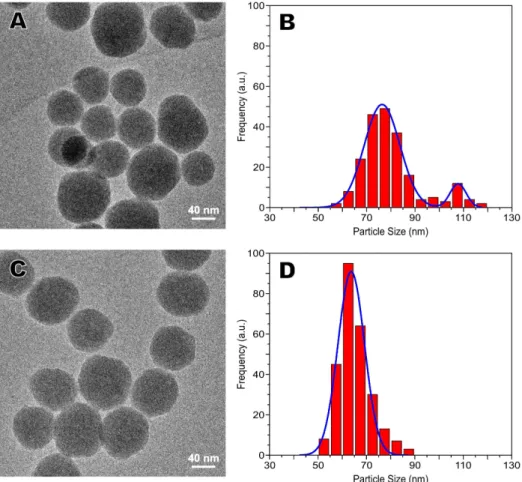
SiO2-NH2-400 (2b) is 62 ± 22 nm. Figures 1A and 1C
present TEM images for CCM/SiO2-NH2-200 (4a) and
CCM/SiO2-NH2-400 (4b), respectively. As expected, there
was a subtle increase in particle size due to the presence of curcumin. The particle morphology was maintained
and (4a) presented a bimodal distribution with average
sizes of 76.4 ± 17 and 107.7 ± 8 nm while 66 ± 13 nm was
observed for (4b) (Figures 1B and 1D). Taking into account
the area of each population in Figure 1B, the one centered at 107.7 nm corresponds to about 10% of the total sample population. The nature of this bimodal distribution when
we have used 200 µL of APTES (samples 2a and 4a) is
out of the scope of this work and has no influence on the biological results.
SAXS curve for CCM/SiO2-NH2-400 (4b) and its best
corresponding fit.
Similar SAXS profiles were observed for all studied
systems (Figure S2,Supplementary Information section).
For all of them, a polydisperse sphere model taking into
account a Gaussian size distribution and subtle interparticle
correlation at low-q was applied. The quality of the SAXS
fits (Figure 2) as well as the TEM images indicated that the nanoparticles are mainly spherical in shape. It was observed that the mean diameter determined from the fit for bare
silica, SiO2-NH2-200, SiO2-NH2-400, CCM/SiO2-NH2-200
and CCM/SiO2-NH2-400 were 53, 56 and 73, 66, 79 and
71 nm, respectively. It is important to highlight that these values are in close agreement with TEM images (Table 1).
The nitrogen adsorption/desorption isotherm for all samples are shown in Figure 3. All studied samples presented similar nitrogen adsorption/desorption isotherms. After functionalization and encapsulation, the isotherms of
SiO2-NH2-200, SiO2-NH2-400, CCM-SiO2-NH2-200 and
CCM-SiO2-NH2-400 kept the same characteristics of SiO2.
There is almost no difference in the shape of the hysteresis loop, suggesting that the pore shape was not significantly changed after grafting with APTES and encapsulation with curcumin. However, the adsorbed nitrogen amount was reduced implying a decrease of pore volume of the particles.
According to IUPAC classification, mesoporous silica nanoparticles clearly show a type II isotherm with an H1 hysteresis loop that is often obtained for materials Figure 1. TEM images of (A) CCM/SiO2-NH2-200 and (C) CCM/SiO2-NH2-400 nanoparticles. Size distribution histograms of (B) CCM/SiO2-NH2-200 and (D) CCM/SiO2-NH2-400.
consisting of agglomerates or compacts of approximately
uniform spheres.58 For all the materials, t-Plot analysis (S
µ)
indicated that most of the specific surface area was located
in mesopores (SBET). This result agrees well with the
literature59 that describes Stöber silica particles containing
microporous and mesoporous compartments in their
structure. In a previous study,44 using high-resolution
transmission electron microscopy, we showed that this type of materials is formed by elementary silica spheres.
The FTIR spectra of the bare silica (1), SiO2-NH2-400 (2b)
and CMC/SiO2-NH2-400 (4b) were obtained to characterize
the chemical bonds and surface organic groups in
mesoporous silica nanoparticles. FTIR of (1) is presented
in Figure 4 (dashed blue line) where absorption bands
due to OH (3446 cm-1), H
2O (1635 cm-1), Si–O–Si
(νas 1097 cm-1, ν
s 796 cm-1) and Si-OH (νs 960 cm-1)
bonds are observed (the whole FTIR spectra is presented in the Supplementary Information section Figure S3). After reaction with APTES, the FTIR spectrum shows new
absorption peaks observed at 2920, 2850 and 1470 cm-1,
which are assigned to C−H stretching and bending
vibrations from the aminopropyl groups, respectively (solid black line). The presence of amino groups was
also confirmed through the weak absorption at 1542 cm-1,
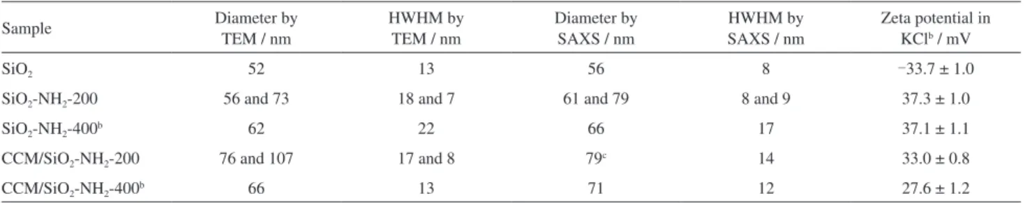
Table 1. Diameters and HWHMa obtained from different techniques and zeta potentials for different nanoparticles
Sample Diameter by
TEM / nm
HWHM by TEM / nm
Diameter by SAXS / nm
HWHM by SAXS / nm
Zeta potential in KClb / mV
SiO2 52 13 56 8 -33.7 ± 1.0
SiO2-NH2-200 56 and 73 18 and 7 61 and 79 8 and 9 37.3 ± 1.0
SiO2-NH2-400b 62 22 66 17 37.1 ± 1.1
CCM/SiO2-NH2-200 76 and 107 17 and 8 79c 14 33.0 ± 0.8
CCM/SiO2-NH2-400b 66 13 71 12 27.6 ± 1.2
aHWHM: half width at half maximum; bthese structures were used as precursors to obtain folate-functionalized silica nanoparticles (reference 44); conly one size distribution was enough to properly fit the scattering data; TEM: transmission electron microscopy; SAXS: small-angle X-ray scattering.
Figure 3. (A) Nitrogen adsorption-desorption isotherms of SiO2, SiO2-NH2-200, SiO2-NH2-400, CCM/SiO2-NH2-200 and CCM/SiO2-NH2-400; (B) surface area determined by BET (SBET) and t-Plot (Sµ) tests for all studied samples.
which is attributed to typical bending vibration of NH2. For
CCM/SiO2-NH2-400 (4b), the absorption peaks of curcumin
could not be observed. This result was expected since the amount of curcumin compared to silica is considerably small and the equipment is not sensitive enough to detect such small amount of CCM.
Zeta potential was also employed to confirm the encapsulation of curcumin seen by UV-Vis. The surface charge measured as zeta potential is presented in Table 1 for all nanoparticles. The zeta potential provides the nanoparticles surface charge and can be also related to the colloidal dispersions stability. A higher zeta potential results in a larger electrostatic repulsion between the particles decreasing the aggregation and providing improved stability to the dispersion. Colloids with low zeta potential tend to undergo aggregation. A value of 30 mV or higher in module (positive or negative) can be taken as a theoretical value to indicate colloidal stability. Samples
with zeta potential between -30 and +30 mV typically tend
to aggregate. The zeta potential of bare silica is negative due to the negatively charged hydroxyl groups on the
silica surface. As expected, SiO2-NH2 (2a and 2b) presents
positive value for zeta potential, indicating that the silanol groups were covalently bonded to the APTES molecules.
For CCM/SiO2-NH2 (4a and 4b), a slight decrease in zeta
potential occurs due to the presence of curcumin in the nanoparticle. This is likely due to the presence of curcumin fraction that might be found on the nanoparticles surface. These results confirm the presence of amino groups and encapsulated curcumin into/onto the mesoporous silica. As one can observe, the absolute values measured indicate that the nanoparticles are within the stable colloidal range. In addition, DLS of these nanoparticles was measured in biological media. These results together with CCM delivery experiments are presented in the Supplementary Information section as Figures S4 and S5.
Nanoparticles bactericidal properties were evaluated
against E. coli as a typical gram-negative bacterium using
the viable cell count method which measures the bacterial
growth. For this study, SiO2, SiO2-NH2-200, SiO2-NH2-400,
CCM/SiO2-NH2-200 and CCM/SiO2-NH2-400 (1, 2a,
2b, 4a and 4b, respectively) were inoculated with
E. coli solution in LB broth. The total mass for each
structure was deduced based on the amount of curcumin encapsulated in each nanoparticle (details are given in the Supplementary Information section). Thus, 32 mg for the
silica structures 2a and 4a were used, while for 2b and 4b,
13 mg were employed. As a point of comparison, we have also chosen to use two amounts of silica (13 and 32 mg,
samples were named SiO2-13 and SiO2-32, respectively)
to evaluate the effect of bare silica amount against E. coli.
On the other hand, curcumin is insoluble in LB broth and,
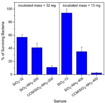
therefore, could not be used as control. Figure 5shows the
percentage of surviving E. coli for all nanoparticles. The
figure is divided considering the total incubated mass per
experiment.
The percentages of surviving bacteria for SiO2-32,
S i O2- N H2- 2 0 0 , C C M / S i O2- N H2- 2 0 0 , S i O2- 1 3 ,
SiO2-NH2-400 and CCM/SiO2-NH2-400 were 57 ± 4,
40 ± 7, 11 ± 3, 94 ± 4, 36 ± 7 and 3 ± 1%, respectively.
For samples of bare silica (SiO2-32 and SiO2-13), the
antimicrobial activity is diminished as the concentration
of the silica is reduced. SiO2-NH2-200 (2a) and SiO2
-NH2-400 (2b) had enhanced biocidal efficacy than their
bare silica counterparts, regardless of the quantities
employed SiO2-13 and SiO2-32. This effective antibacterial
performance of silica nanoparticles without curcumin can be explained taking into account the influence of particle
sizes and surface charges. According to the literature,46
smaller particles exhibit enhanced bactericidal activity than the larger ones. However, our results do not follow this
trend. Although bare silica (1) presents smaller size than
the amine-coated nanoparticles (2a and 2b), SiO2-NH2 was
more effective to prevent bacterial growth. Therefore, it is clear that the subtle difference in particle size (Table 1) is not able to explain these bactericidal efficiency results. Thus, we suggest that the surface charge contribution seems to be more important to the bactericidal activity than the particle size for this particular case (similar nanoparticles sizes). Figure 5. Antimicrobial activity of silica nanoparticles against
The zeta potential measurements for SiO2-NH2 (2a and
2b) exhibit positive values due to the positively charged
amine groups on the silica surface (Table 1). As the bacterial cell surface is negatively charged, it is expected that the electrostatic attraction with the positively charged nanoparticles can be enhanced if compared to the negatively
charged ones.44,60,61 Thereby, SiO
2-NH2 nanoparticules (2a
and 2b) are more electrostatically attracted by negative
bacterial cell surface than SiO2 (1) resulting in superior
bactericidal properties. When comparing the results for
SiO2-NH2-200 and SiO2-NH2-400 (2a and 2b), we observed
that the bactericidal efficacy is very similar. However, it is
important to emphasize that the amount of SiO2-NH2-200
used along the tests is much higher (around three times
larger) than that of SiO2-NH2-400. Hu et al.62 reported
that the cationic amino groups can associate with anions on the bacterial wall, suppress its biosynthesis, disrupt the mass transport across the wall and accelerate the death of the bacteria. Thus, our results are in agreement with their findings and the presence and amount of amino groups were essential in the bacterial growth inhibition.
Nanoparticles containing curcumin (CCM/SiO2-NH2-200
and CCM/SiO2-NH2-400) exhibited superior bactericidal
effects when compared with other nanoparticles. It is important to highlight here that when the synthesis of bare
silica is performed in the presence of curcumin (3), the
total drug load is very low. Due to the small amount of
curcumin, the bactericidal efficiency of CCM/SiO2 (3) is
very close to the one of SiO2 (1). SiO2-NH2 samples (2a
and 2b) and CCM/SiO2-NH2 (4a and 4b) exhibit similar
particle sizes and surface charges, which demonstrate that the bactericidal effect differences can be attributed to the curcumin presence. In addition, the differences in the
results obtained between CCM/SiO2-NH2-200 (4a) and
CCM/SiO2-NH2-400 (4b)cannot be explained by surface
charge effect since all structures present similar zeta potentials. In this case, the nanoparticles present slight difference in particle size and different amounts of amino
groups on the surface. In addition, (4a)present a slightly
larger amount of total CCM if compared to sample (4b).
The CCM/SiO2-NH2-200 exhibits nanoparticles with two
populations and large size distribution. On the order hand,
CCM/SiO2-NH2-400 has a monomodal distribution, larger
curcumin intra-particle load and higher contribution due to the amino groups density on the nanoparticles surface. Our results highlight that the bactericidal properties of curcumin were maintained after encapsulation and that the contribution of the amino groups is essential during bacterial growth inhibition. Thus, a dual bactericidal system was obtained due to the presence of curcumin associated to the amine group on the nanoparticles surface.
Katsu et al.63 reported that the outer membrane of
gram-negative bacteria prevents the penetration of hydrophobic antibiotics into cells. Hence, the cationic compounds are used to enhance the permeability of the outer membrane and thereby increase the sensitivity of gram-negative bacteria to several antibiotics. Thereby, we believe that curcumin had their bactericidal properties enhanced by the amino-functionalized silica. The interaction between the positively charged amino-functionalized silica and the negatively charged bacteria cell wall induces the preferential delivery of curcumin around the bacteria cell wall, leading to an increased local concentration of drug and consequent bacteria death. This strong interaction is also seen with mammalian cells since these nanoparticles present considerable toxicity especially after 48 h of incubation. This poisoning effect was somehow predicted since mammalian cells are highly susceptible to positively
charged structures.44,60 Although we are sure that these
nanoparticles cannot be used as single drug delivery vectors, the overall surface modification strategy as well as the nanoparticles presented here can definitively be incorporated into hybrid materials when cytotoxic effects are mitigated. Thus, we can envisage the use of our approach to produce composite materials with great potential as drug delivery vehicles since it is a versatile and mild method to solubilize and protect water-insoluble drugs while the nanoparticle surface can be tailored depending on the material as well as the final use.
Conclusions
Supplementary Information
Supplementary data including TEM and size distribution images, SAXS profile, FTIR spectra, DLS measurements in biological media, curcumin release and detailed samples description for biological experiments are available free of charge at http://jbcs.sbq.org.br as a PDF file.
Acknowledgments
M. B. C. would like to thank FAPESP (No. 2014/22322-2
and 2015/25406-5)and the productivity research fellowship
granted by CNPq (No. 309107/2014-8). We acknowledge LNLS (CNPEM, Brazil) for SAXS measurements (proposal SAXS1-11058) and LNNano (CNPEM, Brazil) for TEM measurements (proposal TEM-MSC-15277).
References
1. Llor, C.; Bjerrum, L.; Ther. Adv. Drug Saf.2014, 5, 229.
2. Boucher, H. W.; Talbot, G. H.; Benjamin, D. K.; Bradley, J.; Guidos, R. J.; Jones, R. N.; Murray, B. E.; Bonomo, R. A.; Gilbert, D.; Clin. Infect. Dis.2013, 56, 1685.
3. Falagas, M. E.; Rafailidis, P. I.; Matthaiou, D. K.; Virtzili, S.; Nikita, D.; Michalopoulos, A.; Int. J. Antimicrob. Agents2008,
32, 450.
4. Chen, L. F.; Chopra, T.; Kaye, K. S.; Med. Clin. North Am. 2011, 95, 647.
5. Adhikari, N.; J. Inst. Med.2009, 31, 1.
6. Daniel, M. C.; Astruc, D.; Chem. Rev.2004, 104, 293.
7. Cobley, C. M.; Chen, J.; Cho, E. C.; Wang, L. V.; Xia, Y.; Chem. Soc. Rev. 2011, 40, 44.
8. Huang, K.; Ma, H.; Liu, J.; Huo, S.; Kumar, A.; Wei, T.; Zhang, X.; Jin, S.; Gan, Y.; Wang, P. C.; He, S.; Zhang, X.; Liang, X. J.; ACS Nano2012, 6, 4483.
9. Redl, F. X.; Black, C. T.; Papaefthymiou, G. C.; Sandstrom, R. L.; Yin, M.; Zeng, H.; Murray, C. B.; O’Brien, S. P.; J. Am. Chem. Soc.2004, 126, 14583.
10. Lee, J. E.; Lee, N.; Kim, H.; Kim, J. H. J. H.; Choi, S. H.; Kim, T.; Song, I. C.; Park, S. P.; Moon, W. K.; Hyeon, T.; J. Am. Chem. Soc.2010, 132, 552.
11. Tu, H.-L.; Lin, Y.-S.; Lin, H.; Hung, Y.; Lo, L.; Chen, Y.; Mou, C.; Adv. Mater.2009, 21, 172.
12. Kim, J.; Lee, J. E.; Lee, J.; Jang, Y.; Kim, S. W.; An, K.; Yu, J. H.; Hyeon, T.; Angew. Chem., Int. Ed.2006, 45, 4789. 13. Lin, Y. S.; Hung, Y.; Lin, H. Y.; Tseng, Y. H.; Chen, Y. F.; Mou,
C. Y.; Adv. Mater.2007, 19, 577.
14. Vivero-Escoto, J. L.; Slowing, I. I.; Trewyn, B. G.; Lin, V. S.-Y.;
Small2010, 6, 1952.
15. Vallet-Regí, M.; Balas, F.; Arcos, D.; Angew. Chem., Int. Ed. 2007, 46, 7548.
16. Cotí, K. K.; Belowich, M. E.; Liong, M.; Ambrogio, M. W.; Lau, Y.; Khatib, H.; Zink, J. I.; Khashab, N. M.; Stoddart, J. F.;
Nanoscale2009, 1, 16.
17. Trewyn, B. G.; Slowing, I. I.; Giri, S.; Chen, H.-T.; Lin, V. S.-Y.;
Acc. Chem. Res.2007, 40, 846.
18. Saha, S.; Leung, K. C. F.; Nguyen, T. D.; Stoddart, J. F.; Zink, J. I.; Adv. Funct. Mater.2007, 17, 685.
19. Rosenholm, J. M.; Sahlgren, C.; Linden, M.; Nanoscale2010, 2, 1870.
20. Ambrogio, M. W.; Thomas, C. R.; Zhao, Y. L.; Zink, J. I.; Stoddart, J. F.; Acc. Chem. Res.2011, 44, 903.
21. Bagwe, R. P.; Hilliard, L. R.; Tan, W.; Langmuir2006, 22,
4357.
22. Maria Chong, A. S.; Zhao, X. S.; Appl. Surf. Sci.2004, 237,
398.
23. Cauda, V.; Schlossbauer, A.; Kecht, J.; Zürner, A.; Bein, T.; J. Am. Chem. Soc.2009, 131, 11361.
24. Möller, K.; Kobler, J.; Bein, T.; Adv. Funct. Mater.2007, 17,
605.
25. Lü, Y.; Lu, G.; Wang, Y.; Guo, Y.; Guo, Y.; Zhang, Z.; Wang, Y.; Liu, X.; Adv. Funct. Mater.2007, 17, 2160.
26. Sevimli, F.; Yilmaz, A.; Microporous Mesoporous Mater.2012, 158, 281.
27. Chu, L.; Daniels, M. W.; Francis, L. F.; Chem. Mater.1997, 9,
2577.
28. Nieto, A.; Colilla, M.; Balas, F.; Vallet-Regí, M.; Langmuir 2010, 26, 5038.
29. Puddu, V.; Perry, C. C.; Langmuir2014, 30, 227.
30. Kim, S. T.; Saha, K.; Kim, C.; Rotello, V. M.; Acc. Chem. Res. 2013, 46, 681.
31. Mout, R.; Moyano, D. F.; Rana, S.; Rotello, V. M.; Chem. Soc. Rev.2012, 41, 2539.
32. Moyano, D. F.; Saha, K.; Prakash, G.; Yan, B.; Kong, H.; Yazdani, M.; Rotello, V. M.; ACS Nano2014, 8, 6748.
33. Hoffmann, F.; Cornelius, M.; Morell, J.; Fröba, M.; Angew. Chem., Int. Ed.2006, 45, 3216.
34. Tarn, D.; Ashley, C. E.; Xue, M.; Carnes, E. C.; Zink, J. I.; Brinker, C. J.; Acc. Chem. Res.2013, 46, 792.
35. Du, X.; He, J.; Langmuir2011, 27, 2972.
36. Jin, D.; Park, K.-W.; Lee, J. H.; Song, K.; Kim, J.-G.; Seo, M. L.; Jung, J. H.; J. Mater. Chem.2011, 21, 3641.
37. Siddharth Jambhrunkar, Surajit Karmakar, Amirali Popat, M. Y. C. Y.; RSC Adv.2014, 4, 1166.
38. Hamam, F.; Al-Remawi, M.; J. Funct. Foods2014, 8, 87.
39. Jambhrunkar, S.; Qu, Z.; Popat, A.; Yang, J.; Noonan, O.; Acauan, L.; Ahmad Nor, Y.; Yu, C.; Karmakar, S.; Mol. Pharm. 2014, 11, 3642.
40. Singh, S. P.; Sharma, M.; Gupta, P. K.; Int. J. Biol. Macromol. 2015, 74, 162.
42. Wang, J.; Wang, Y.; Liu, Q.; Yang, L.; Zhu, R.; Yu, C.; Wang, S.;
ACS Appl. Mater. Interfaces, in press, DOI: acsami.6b08400.
43. Kotcherlakota, R.; Barui, A. K.; Prashar, S.; Fajardo, M.; Briones, D.; Rodríguez-Diéguez, A.; Patra, C. R.; Gómez-Ruiz, S.; Biomater. Sci.2016, 4, 448.
44. de Oliveira, L. F.; Bouchmella, K.; Gonçalves, K. D. A.; Bettini, J.; Kobarg, J.; Cardoso, M. B.; Langmuir2016, 32, 3217. 45. Datz, S.; Engelke, H.; Schirnding, C. V.; Nguyen, L.; Bein, T.;
Microporous Mesoporous Mater.2016, 225, 371.
46. dal Lago, V.; França de Oliveira, L.; de Almeida Gonçalves, K.; Kobarg, J.; Borba Cardoso, M.; J. Mater. Chem.2011, 21, 12267.
47. França de Oliveira, L.; de Almeida Gonçalves, K.; Boreli, F. H.; Kobarg, J.; Cardoso, M. B.; J. Mater. Chem.2012, 22, 22851.
48. Capeletti, L. B.; de Oliveira, L. F.; Gonçalves, K. D. A.; de Oliveira, J. F. A.; Saito, Â.; Kobarg, J.; dos Santos, J. H. Z.; Cardoso, M. B.; Langmuir2014, 30, 7456.
49. de Oliveira, J. F. A.; Cardoso, M. B.; Langmuir2014, 30, 4879.
50. França de Oliveira, L.; Gonçalves, J. O.; Gonçalves, K. D. A.; Kobarg, J.; Cardoso, M. B.; J. Biomed. Nanotechnol.2013, 9,
1817.
51. de Souza e Silva, J. M.; Pastorello, M.; Kobarg, J.; Cardoso, M. B.; Mazali, I. O.; ChemPhysChem2013, 14, 4075. 52. Silva, H. F. O.; Lima, K. M. G.; Cardoso, M. B.; Oliveira,
J. F. A.; Melo, M. C. N.; Sant’Anna, C.; Eugênio, M.; Gasparotto, L. H. S.; RSC Adv.2015, 5, 66886.
53. Ribeiro de Barros, H.; Cardoso, M. B.; Camargo de Oliveira, C.; Cavichiolo Franco, C. R.; de Lima Belan, D.; Vidotti, M.; Riegel-Vidotti, I. C.; RSC Adv.2016, 6, 9411.
54. de Souza e Silva, J. M.; Hanchuk, T. D. M.; Santos, M. I.; Kobarg, J.; Bajgelman, M. C.; Cardoso, M. B.; ACS Appl. Mater. Interfaces2016, 8, 16564.
55. Stöber, W.; Fink, A.; Bohn, E.; J. Colloid Interface Sci.1968, 26, 62.
56. Mahalingam, V.; Onclin, S.; Péter, M.; Ravoo, B. J.; Huskens, J.; Reinhoudt, D. N.; Langmuir2004, 20, 11756.
57. Taylor, I.; Howard, A. G.; Anal. Chim. Acta1993, 271, 77.
58. Leirose, G. D. S.; Cardoso, M. B.; J. Pharm. Sci.2011, 100, 2826.
59. Szekeres, M.; Dékány, I.; de Keizer, A.; Colloids Surf., A1998,
141, 327.
60. Catherine, M. G.; Cusker, C. D. M.; Tuna, Y.; Rotello, V. M.;
Bioconjugate Chem.2004, 15, 897.
61. Hayden, S. C.; Zhao, G.; Saha, K.; Phillips, R. L.; Li, X.; Miranda, O. R.; Rotello, V. M.; El-Sayed, M. A.; Schmidt-Krey, I.; Bunz, U. H. F.; J. Am. Chem. Soc.2012, 134, 6920. 62. Hu, S. G.; Jou, C. H.; Yang, M. C.; Biomaterials2003, 24, 2685.
63. Katsu, T.; Nakagawa, H.; Yasuda, K.; Antimicrob Agents Chemother. 2002, 46, 1073.
Submitted: September 19, 2016
Published online: February 6, 2017