Internet Journal of Medical Update
Journal home page: http://www.akspublication.com/ijmu
Original Work
15
Pulse Wave Velocity and Electroneurophysiological Evaluation in patients
of Rheumatoid Arthritis
Geetanjali Sharma
*ΨMD, Sushma Sood
**MD, R. Handa
†MD and H. Singh
‡MD
*
Assistant Professor,
**Senior Professor and Head, Department of Physiology
†
Resident,
‡Senior Professor and Head, Department of Rheumatology
University of Health Sciences, Rohtak, Haryana, India
(Received 11 July 2010 and accepted 02 December 2010)
ABSTRACT: Rheumatoid arthritis is a chronic systemic inflammatory disease of undetermined etiology involving the synovial membranes and articular structures of multiple joints and is also associated with carditis, pleuritis, hepatitis, peripheral neuropathy and vasculitis. The present study was undertaken to investigate arterial stiffness using carotid-radial and femoral-dorsalis pedis pulse wave velocity measurements and electrophysiological tests for peripheral nervous system involvement. 25 patients (aged between 20-60 years) with rheumatoid arthritis according to the criteria of the American College of Rheumatology and 25 control subjects of the same age and sex were recruited. In the motor conduction studies, out of 25 patients of Rheumatoid arthritis, 6 had clinical evidence of peripheral neuropathy. 11 patients showed pure sensory neuropathy (44%), 10 showed mixed sensory motor neuropathy (40%) while 4 showed normal motor and sensory conduction velocity. Two patients (8%) showed features of entrapment neuropathy of median nerve i.e. feature of Carpal tunnel syndrome. In the pulse wave velocity evaluation statistically significant increase in pulse wave velocity between femoral-dorsalis pedis and carotid-radial artery segments was observed in Rheumatoid arthritis patients as compared to the control group. Measurement of carotid-radial and femoral-dorsalis pedis PWV may provide a simple and non-invasive technique for identifying patients at increased risk of vascular disease in Rheumatoid arthritis.
KEY WORDS: Peripheral neuropathy; Nerve conduction; Vascular events INTRODUCTIONᴪ
Rheumatoid arthritis is a chronic systemic inflammatory disease of undetermined etiology involving synovial membranes and articular surfaces of multiple joints in a symmetrical manner. Females are affected approximately three times more than men. It begins with pain, stiffness and swelling of specific joints such as proximal interphalangeal, metacarpophalangeal and knee joints. These include rheumatoid nodules, pulmonary manifestations, pericarditis and vasculitis.1,2 Neurological manifestations in Rheumatoid arthritis include peripheral neuropathy,
ᴪCorrespondence at: 24/9-J, Medical Enclave,
Rohtak-124001, Haryana, India; Phone: 91 121 62213165; Mobile: +919416513989; Email:
cervical myelopathy, entrapment neuropathy and vasculitis.3 Golding & Mackenzie were the first to describe a series of patients with neuropathy and rheumatoid disease4. There are three causes of
conduction studies not only confirm the presence of neuropathy and also give indication of the type of neuropathy9; e.g. axonal neuropathy is characterized by a reduction in amplitude of the compound muscle action potential or compound nerve action potential, demyelination neuropathy is characterized by prolonged latency and prolonged slowing of conduction and chronic compressive lesions produce localized slowing of conduction. Rheumatoid arthritis is associated with increase cardio-vascular morbidity and mortality; pulse wave velocity is an index of arterial stiffness and a marker of cardio-vascular events.
The present study is aimed to investigate the arterial stiffness using carotid-radial and femoral-dorsalis pedis pulse wave velocity measurements in patients of Rheumatoid arthritis and electrophysiological tests to find out the incidence of early peripheral nervous system involvement in patients of Rheumatoid arthritis.
METHODOLOGY
The present study was carried out in the Department of Physiology, Pt. B.D. Sharma University of Health Sciences, Rohtak on 50 subjects (25 patients aged between 20-60 years diagnosed as Rheumatoid arthritis clinically as per American criteria of Rheumatology11 and 25 control subjects matched for age and sex). The duration of disease in the patients was ranging from 10.56 ± 6.7 years (mean ± standard deviation), the DAS score12 was 3.1-5.1and all patients attended the Rheumatology clinic with or without clinical evidence of neuropathy. Patients with diabetes mellitus, severe anaemia, hypothyroidism, alcoholism and other metabolic or toxic causes of neuropathy were excluded from the study.
Recording of nerve conduction velocities
The nerve conduction velocity was recorded using RMS EMG EP Mark II machine (Chandigarh). The placement of Electrodes was as per Mishra et al14. The parameters recorded were:
• Motor conduction studies: Median, ulnar,
tibial and peroneal conduction velocities (CV), distal latencies (L) and amplitudes (A).
• Sensory conduction studies: Sensory median
and sural conduction velocity (CV), distal latencies (L) and amplitudes (A) by ring electrodes.
The cut off points for each parameter were considered as below:
Upper limb (motor): CV < 52.12, L > 3.75, and A < 10.04 are indicative of neuropathy.
Lower Limb (motor): CV < 44.2, L > 8.4 and A < 8.93 are indicative of neuropathy.
Lower Limb (sensory): CV < 41.2, L >3.63 and A < 0.22 are indicative of neuropathy.
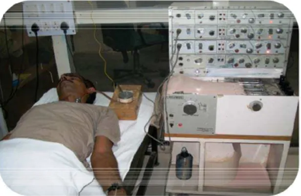
Recording of Pulse Wave Velocity (PWV) The pulse wave velocity was recorded with the help of Polywrite-4 channel machine (INCO) (Figure 1) as from the left carotid in the neck to the left radial artery at the wrist (C-R) and from the left femoral artery just below the inguinal ligament to the left Dorsalis pedis artery in the foot (F-DP). The pulse wave at various points was recorded using pulse transducers having in built photo-detectors and light sources. Recording of arterial pulse was taken on 4 channel polygraph paper (Polywrite INCO) at a paper speed of 25 mm/sec.13
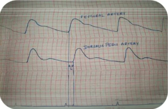
Figure 1: Polywrite for recording PWV For the arterial segment C-R, the pulse wave velocity was obtained by measuring the time delay between the onset of pulse waves in the carotid and radial arteries. Similarly, the time delay between the onset of pulse waves in the femoral and dorsalis pedis aterieswas measured for F-DP segment. The time delay (t) of the pulse wave was measured using these points and was expressed in milliseconds. The length of the arterial segments (L) was measured on the surface of body of the patient with the help of measuring tape. The pulse wave velocity (meters/sec) was calculated by using the formula L/t. Only clearly defined tracings were analyzed (Figure 2 and 3).
17
Figure 3: Tracing for F-DP segment Statistical analysis
The standard statistical procedure unpaired t-test (2 tailed) was used to compare values in both control and Guillain Barre Syndrome groups and ‘p’ value was derived. P<0.001 indicates statistically significant findings. The statistical analysis was done on Microsoft Office Excel 2007.
RESULTS
Out of 25 patients of Rheumatoid arthritis, 23 were females while two were males with mean age 44.4
± 10.8 years. The mean duration of disease was 10.56 ± 6.1 years.
Conduction studies
Out of 25 patients of Rheumatoid arthritis, 6 had clinical evidence of peripheral neuropathy in the form of paraesthesia, loss of deep tendon reflexes and loss of vibration sense while remaining 19 subjects showed no clinical evidence of neuropathy. Eleven patients showed pure sensory neuropathy (44%) i.e. decrease in sensory conduction velocity, increase in latency and slight decrease in amplitude of median and sural nerve.; Ten patients showed mixed sensory motor neuropathy (40%) i.e. decrease in motor and sensory conduction velocity, slight increase in latency and markedly decrease amplitude of median ulnar, tibial and peroneal nerves. The decrease was more pronounced in lower limbs. Four subjects showed normal motor and sensory conduction velocity. Two patients (8%) showed features of entrapment neuropathy of median nerve i.e. feature of Carpal tunnel syndrome.
Table 1 shows Mean and standard deviation of motor & sensory conduction velocities.
Table 1: Mean ± standard deviation of motor & sensory conduction velocities
Motor Sensory
Median Ulnar Tibial Peroneal Median Sural
C RA C RA C RA C RA C RA C RA
C V (m/s) 57.32 ± 5.20 50 ± 5.6 57.2 ± 5.4 55.9 ± 4.3 46.3 ± 2.1 37.5 ± 6.9 43.34 ± 2.1 36.08 ± 3.3 54.82 ± 1.06 46.3 ± 5.8 42.4 ± 1.2 34.2 ± 7.2 L (milli secs) 3.38 ± 0.37 4.3 ± 0.54 2.5 ± 0.4 3.92 ± 0.4 9.6 ± 1.7 11.03 ± 2.9* 7.6 ± 0.8 10.1 ± 1.2 2.5 ± 0.22** 2.9 ± 0.43 3.4 ± 0.23 4.88 ± 0.93 A (milli volts) 11.2 ± 0.98 6.6 ± 0.98 10.8 ± 1 8.8 ± 1.1 11.13 ± 2.2 5.7 ± 3.3 4.9 ± 1.1 2.7 ± 1.3 0.081 ± 0.007* 0.07 ± 0.03 0.33 ± 0.11 0.184 ± 0.17 *= p<0.05; **= p<0.01; others= p<0.001; C= control group; RA= Rheumatoid group
Pulse Wave velocity
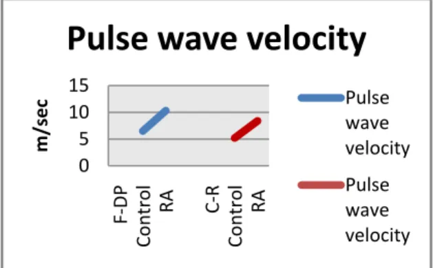
Table 2 shows mean and standard deviation of pulse wave velocity evaluation. There was a significant increase in pulse wave velocity of both Femoral-Dorsalis pedis and Carotid-Radial artery segments in rheumatoid arthritis patients as compared to the control group. (Figure 4)
Table 2: Mean ± standard deviation of pulse wave velocity evaluation
PWV C-R
PWV F-DP
Figure 4: Pulse wave velocity (meter/second) in control and rheumatoid arthritis (RA) groups DISCUSSION
The present study showed that there was an increased incidence of peripheral neuropathy in Rheumatoid arthritis i.e. 21 out of 25 patients (84 %). Although clinical features like paraesthesia, absent distal tendon reflexes and absent vibration sense are good indicators of peripheral neuropathy, the peripheral neuropathy in rheumatoid arthritis is mainly subclinical. Our results indicated that peripheral nervous involvement mainly manifested as mild sensory (44%) and mixed sensory motor neuropathy in median, tibial, peroneal and sural nerves (40%). Entrapment neuropathy was seen in only two patients that too in the median nerve (8%). This nerve involvement was ranging from axonal neuropathy as evident by marked decrease in amplitude as compared to motor conduction velocity to segmental demyelination in some cases. This finding is consistent with the findings of Lanzillo et al15in which 21 out of 26 (80.7 %) of the patients exhibited sensory motor neuropathy and of Yazdchi et al16 where sensory motor neuropathy was observed in 57.1% and pure sensory neuropathy in 35.7% subjects. Agarwal et al17 in 2008 also found sensory motor type of neuropathy to be the most common type in patients of Rheumatoid arthritis. Similar results were also reported by Aneja et al3 where entrapment neuropathy was reported in 9% cases, pure sensory involvement in 12% subjects, 20% showed mixed sensory motor neuropathy while 12% cases showed pure motor neuropathy. Rheumatoid arthritis is associated with increased cardio-vascular mortality. Chronic inflammation may impair vascular function and lead to increased arterial stiffness which is an important determinant of cardio-vascular risk.18Pulse wave velocity is defined as the velocity of the arterial pulse as it moves along the vessel wall. It is an indicator of arterial elasticity.19 A close association between increased PWV and atherosclerosis development has been reported.20 In the present study, PWV was
increase in PWV in F-DP and C-R artery segment. These findings are in consistent with Yildiz10, Kaisa21 and Klocke18. The inflammation in rheumatoid arthritis may impair endothelial function, arterial compliance and arterial elasticity and may act as a contributory factor in the initiation or progression of atherosclerosis.22,23 In the present study, 18 patients were C-reactive protein and rheumatoid antibody positive which is again predictive of atherosclerosis which is again in consistent with the statement made by Ridker24.
CONCLUSION
Thus to summarize, occlusive vascular disease in the vasa nervosum was considered to be the major cause of peripheral neuropathy and inflammation in vessels may impair vascular function and lead to increase in pulse wave velocity. The same reasoning has been theorized by Weller25and Wong et al22. Further, pulse wave velocity and electrodiagnostic tests are non-invasive techniques and should be recommended in patients of Rheumatoid arthritis as a routine for early detection of peripheral neuropathy and for identifying vascular involvement.
ACKNOWLEDGEMENTS
The authors are deeply thankful to the patients and control subjects for their cooperation during this study. They are acknowledge the sincerity and hard work of Mr. Randhir, Senior Laboratory Technician, Department of Physiology, University of Health Sciences, Rohtak for his technical expertise and help rendered during this work.
REFERENCES
1. Lipsky PE. Rheumatoid Arthritis. Harrison’s Principles of Internal Medicine 17th ed. Fauci A et al editors. New York: McGraw Hill; 2008:2083-92.
2. Chang DJ, Paget SA. Neurological complications of rheumatoid arthritis. Rheum Dis Clin North Am. 1993 Nov;19(4):955-73. 3. Aneja R, Singh MB, Shankar S, et al.
Prevalence of peripheral neuropathy in patients with newly diagnosed rheumatoid arthritis. Indian J Rheumatol. 2007 Jun;2(2):47-50. 4. Hart FD, Golding JR. Rheumatoid neuropathy.
Br Med J. 1960 May;1(5186):1594-600. 5. Prasad K, Bhatia R. Rheumatoid neuropathy.
Indian J Rheumatol. 2007 Jun;2(2):45-6.
6. Gorson KC. Vasculitic neuropathies: an update. Neurologist. 2007 Jan;13(1):12-19. 7. Scott DGI, Bacon PA, Tribe CR. Systemic
rheumatoid vasculitis: a clinical and laboratory 0
5 10 15
F
‐
DP
Control
RA
C
‐
R
Control
RA
m/sec
Pulse
wave
velocity
Pulse
wave
velocity
Pulse
wave
19
8. Sirri A, Guler-Uysal F. The
electroneurophysiological findings in rheumatoid arthritis patients. Electromyogr Clin Neurophysiol. 1999 Oct-Dec;39(7):387-91.
9. Daube JR. Nerve conduction studies. In Amnoff MJ editor. Electrodiagnosis in clinical neurology. 4th ed New York: Churchill Livingstone; 1999:190-6.
10. Yildiz M, Soy M, Kurum T, et al. Increased pulse wave velocity and shortened pulse wave propagation time in young patients with rheumatoid arthritis. Can J Cardiol. 2004 Sep;20(11):1097-100.
11. Arnett FC, Edworthy SM, Bloch DA, et al. The American Rheumatism Association 1987 revised criteria for the classification of rheumatoid arthritis. Arthritis Rheum. 1988 Mar;31(3): 315-24.
12. Prevoo ML, van'T Hof MA, Kuper HH, et al. Modified disease activity scores that include twenty-eight-joint counts development and validation in a prospective longitudinal study of patients with rheumatoid arthritis. Arthritis Rheum. 1995 Jan;38(1):44-8.
13. Singh PI, Sood S, Sood AK. Pulse wave velocity in diabetes. Ind Med Gaz. 1987;121(11):359-61.
14. Mishra US, Kalita J. Nerve conduction, electromyography and evoked potentials. Clinical neurophysiology. 2nd ed. Elsevier: New Delhi 2004:31-84.
15. Lanzillo B, Pappone N, Crisci C, et al. Subclinical peripheral nerve involvement in patients with rheumatoid arthritis. Arthritis Rheum. 1998 Jul; 41(7):1196-202.
16. Yazdchi M, Ebrahim AA, Mikaeeli H, et al. The electrophysiological evaluation of 70 Iranian rheumatoid arthritis patients. J Neurol Sci. (Turkish) 2007;24(3):190-6.
17. Agarwal V, Singh R, Wiclaf, et al. A clinical, electrophysiological and pathological, study of
neuropathy in rheumatoid arthritis. Clin Rheumatol. 2008 Jul;27(7):841-4.
18. Klocke R, Cockcroft JR, Taylor GJ, et al. Arterial stiffness and central blood pressure, as determined by pulse wave analysis in, rheumatoid arthritis. Ann Rheum Dis. 2003 May;62(5):414-8.
19. Blacher J, Asmar R, Djane S, et al. Aortic pulse wave velocity as a marker of cardiovascular risk in hypertensive patients. Hypertension. 1999 May;33(5): 1111-7. 20. Wilkinson IB, Webb DJ, Cockcroft JR. Aortic
pulse-wave velocity. Lancet. 1999 Dec;354(9194):1996-7.
21. Mäki-Petäjä KM, Hall FC, Booth AD, et al. Rheumatoid arthritis is associated with increased aortic pulse-wave velocity, which is reduced by anti-tumor necrosis factor-alpha therapy. Circulation. 2006 Sep;114(11):1185-92.
22. Wong M, Toh L, Wilson A, et al. Reduced arterial elasticity in rheumatoid arthritis and the relationship to vascular disease risk factors and inflammation. Arthritis Rheum. 2003 Jan;48(1):81-9.
23. Van Doornum S, McColl G, Jenkins A, et al. Screening for atherosclerosis in patients with rheumatoid arthritis: comparison of two in vivo tests of vascular function. Arthritis Rheum. 2003 Jan;48(1):72-80.
24. Ridker PM. High-sensitivity C-reactive protein: potential adjunct for global risk assessment in the primary prevention of cardiovascular disease. Circulation. 2001 Apr;103(13):1813-8.