Permeability of
E. coli
Miguel Viveiros1, Myrielle Dupont2, Liliana Rodrigues1, Isabel Couto1,3, Anne Davin-Regli2, Marta Martins1, Jean-Marie Page`s2, Leonard Amaral1
*
1Unit of Mycobacteriology, Unidade de Parasitologia e Microbiologia Me´dicas (UPMM), Instituto de Higiene e Medicina Tropical, Universidade Nova de Lisboa, Lisboa, Portugal,2UMR-MD-1, IFR48, Faculte´s de Me´decine et de Pharmacie, Universite´ de la Me´diterrane´e, Marseille, France,3Centro de Recursos Microbiolo´gicos (CREM), Faculdade de Cieˆncias e Tecnologia, Universidade Nova de Lisboa, Lisboa, Portugal
Background. Membrane permeability is the first step involved in resistance of bacteria to an antibiotic. The number and activity of efflux pumps and outer membrane proteins that constitute porins play major roles in the definition of intrinsic resistance in Gram-negative bacteria that is altered under antibiotic exposure.Methodology/Principal Findings.Here we describe the genetic regulation of porins and efflux pumps of Escherichia coli during prolonged exposure to increasing concentrations of tetracycline and demonstrate, with the aid of quantitative real-time reverse transcriptase-polymerase chain reaction methodology and western blot detection, the sequence order of genetic expression of regulatory genes, their relationship to each other, and the ensuing increased activity of genes that code for transporter proteins of efflux pumps and down-regulation of porin expression. Conclusions/Significance. This study demonstrates that, in addition to the transcriptional regulation of genes coding for membrane proteins, the post-translational regulation of proteins involved in the permeability of Gram-negative bacteria also plays a major role in the physiological adaptation to antibiotic exposure. A model is presented that summarizes events during the physiological adaptation ofE. colito tetracycline exposure.
Citation: Viveiros M, Dupont M, Rodrigues L, Couto I, Davin-Regli A, et al (2007) Antibiotic Stress, Genetic Response and Altered Permeability ofE. coli. PLoS ONE 2(4): e365. doi:10.1371/journal.pone.0000365
INTRODUCTION
Intrinsic antibiotic resistance in Gram-negative bacteria (without chromosomal mutation or the acquisition of mobile genetic elements encoding resistance determinants) can be increased by preventing the antibiotic from entering the cell. This can be achieved by the control of the outer membrane permeability and by the effectiveness of the efflux (active pumping out) of antibiotics [1–3]. The effectiveness of the outer membrane of Gram-negative bacteria as a barrier only delays the influx of various antibiotics, detergents and dyes. Intrinsic resistance to antibiotic agents is brought about by efflux pumps, which extrude the drug from the periplasmic space to the environment, enabling bacterium to survive in the presence of these noxious agents [1,4]. Additional resistance is afforded by over-expressed efflux pumps that extrude a wide variety of unrelated antibiotics. Over-expressed efflux pumps of Gram-negative bacteria result in a multidrug resistant (MDR) phenotype known to be a prevalent form of clinical resistance [3].
We have previously demonstrated that it is possible to induce high-level resistance to tetracycline (TET) in susceptibleEscherichia coliK-12 by a gradual, step-wise increase in the exposure to the antibiotic [5]. The induction process takes about 110 days and this resistance can be reversed by either transfer to drug free medium or by the use of Phe-Arg-napthylamide (PAbN), an inhibitor of the AcrAB efflux pump system [3–5]. The nine major inner membrane transporter genes ofE. coliK-12 were over-expressed after prolonged exposure to TET, with the acrBbeing the most expressed transporter gene and a clear connection between the induced activity of the AcrAB system and TET induced resistance was demonstrated [5]. Besides becoming resistant to TET, the induced strain became resistant to a variety of other antibiotics, detergents and dyes that are not substrates of the AcrAB system [3–5]. The development of this MDR phenotype led us to explore and analyse the interplay between the major efflux pump systems present in E. coli and the control of the outer membrane permeability through the regulation of the porin channels.
In E. coli, outer membrane permeability is regulated by the
balance of porin proteins, the diffusion channels that are the major route for passage of small hydrophilic compounds [1,6,7]. The two major outer membrane proteins (OMPs) inE. coliare OmpC and OmpF, consisting of three 16-strandedb-barrels defining a trans-membrane pore in the outer trans-membrane porin [8,9].
Highly expressed under optimal environmental conditions, their level of expression is adjusted when it is necessary to minimize penetration of noxious compounds or maximize access to nutrients [7,10,11]. It has been demonstrated that the level of expression of the porins OmpC and OmpF not only controls the permeability of the outer membrane to glucose and nitrogen uptake under nutrient limitation [10,11], but may also be differentially regulated by the concentration of certain antibiotics in the environment [4,12,13]. The OmpC and OmpF coding genes are transcription-ally regulated by a two-component signal transduction regulatory system consisting of the OmpR and EnvZ proteins [14]. Recently, it has been shown that the over-expression of OmpX, structurally related to the eight-bstrand OmpA (a major OMP involved in the
Academic Editor:Debbie Fox, The Research Institute for Children, United States of America
ReceivedJanuary 31, 2007;AcceptedMarch 8, 2007;PublishedApril 11, 2007
Copyright:ß2007 Viveiros et al. This is an open-access article distributed under the terms of the Creative Commons Attribution License, which permits unrestricted use, distribution, and reproduction in any medium, provided the original author and source are credited.
Funding:This work was supported by grants (i) Luso-French Integrated Actions (CRUP/2006-F-33/06) (ii) EU-FSE/FEDER-POCI/SAU-MMO/59370/2004 and (iii) the Universite´ de la Me´diterrane´e, the Service de Sante´ des Arme´es and from Eloi Collery Prix Acade´mie Nationale de Me´decine (J.-M.P.). L. Rodrigues and M. Martins were supported by grants SFRH/BD/24931/2005 and SFRH/BD/14319/ 2003, respectively, from Fundac¸a˜o para a Cieˆncia e Tecnologia (FCT) of Portugal.
Competing Interests:The authors have declared that no competing interests exist.
stabilization of the bacterial membrane), leads to a decrease in the expression of OmpC and OmpF porins and a decreased susceptibility to beta-lactams and other antibiotics inE. coli[15]. Because mutants with decreased expression of porins show only small increases in the minimum inhibitory concentration (MIC) of relevant antibiotics, the complete shut down of influx of small molecules intoE. colidoes not readily occur [16].
E. coli has been shown to have at least nine distinct
proton-dependent efflux pump systems that bestow resistance to two or more antibiotics (MDR). The genes coding for each of these efflux pumps are emrE [17], acrEF (formerly envCD) [18], emrAB [19], emrD[20],acrAB-tolC [21],mdfABC [22],tehA[23],acrD(anacrB homolog) [24] and yhiUV [25]. They belong to one of three genetically and structurally defined families: the major facilitator superfamily (MFS –emrD, mdfA, emrB), the resistance nodulation-cell division family (RND- acrB, acrF, acrD, yhiV), and the small multidrug resistance family (SMR –emrE, tehA) [3,4]. The tripartite AcrAB-TolC system is the most well-studied MDR pump system consisting of an inner membrane efflux transporter (AcrB) that removes antibiotics from the cytoplasm to the periplasm, where the linker protein (AcrA) directs the inter-membrane transport of the antibiotic through the outer membrane channel (TolC) to the environment [1,3,6]. The major efflux pump systems inE. coliare from the RND family and have broad substrate specificity. Their expression is controlled by systemic transcriptional activators like the MarA, encoded by the multiple antibiotic resistance operon
marRAB[26], and homologs like SoxS and Rob [4,27]. MarA not
only controls the expression of the efflux pump systems, but is also involved in the control of porin expression (by decreasing it) through the activation ofmicF, a small antisense RNA that binds with ompF mRNA preventing its translation, and activates the expression of the porin expression down-regulator OmpX [15,28]. These global activators, when induced by oxidative stress or the presence of noxious compounds in the environment, enhance resistance of enterobactereaceae to a variety of antibiotics, hence an MDR phenotype [4,31,37]. Moreover, they control the degree of intrinsic resistance of enterobactereaceae and increase the level of efflux pump expression. The regulation of porin level and expression of MDR efflux pumps has been suggested to occur by a common pathway and/or a cascade of events [4,29].
Our studies of step-wise induction of TET resistance, by gradual exposure of E. coli K-12 wild-type to TET, may afford an understanding of the genetic regulation of MDR efflux pumps, their interplay, and relationship to the permeability barrier, all of which are involved during the TET resistance induction process [5]. Therefore, with the aid of quantitative real-time reverse transcriptase-polymerase chain reaction (RT-PCR) methodology and western blot detection we have analysed and correlated the activity of regulatory genes that affect the MDR phenotype, of genes that code for transporter proteins of RND efflux pumps, of genes that code OMPs; and the level of OMPs during the process of induced resistance of E. coli K-12 wild-type by prolonged exposure to increasing concentrations of TET.
RESULTS AND DISCUSSION
The results obtained in this study and presented by Figure 1 are discussed in terms of relationships that have been established for regulatory and responding genes.
Genes responding to stress:
soxS and rob After resistance to TET has been established at the highest initial concentration of the antibiotic (i.e.1.5 mg/L), the response of thesoxSgene is 2.8 times more active than that of
the unexposed control. This response is further increased to 3.5 times after resistance to 4 mg/L has been induced. However, by the time the strain has become resistant to 10 mg/L of TET, the response of the gene has been reduced to a level below that initially observed, suggesting that the stress genesoxSperforms its functions quite early under conditions of antibiotic pressure. The activity of the rob gene during the process of TET induced resistance is significantly increased after the bacterium becomes resistant to 4 mg/L of TET. As was the case of the other stress-response gene soxS, the increased activity noted is apparently not required for higher levels of resistance (i.e.10 mg/L). Although rob has been reported to respond to exposure to solvents, detergents and metals [30,31], in the current study an antibiotic response is included. The parallel response of both stress-responding genes noted in our study supports the conclusions of Michanet al.[32].
marA, marB and marR The regulatory product of gene marRis known to down-regulate the activity of genesmarAandmarB by binding to the promoter region of the operatormarO. Because TET is known to bind to the product ofmarR, and this produces an MDR phenotype [33], once the repressor activity is inhibited, the universal regulatormarAwould be expected to increase its activity. This expectation is confirmed by data in Figure 1. In this case, one can see that of all of the regulator genes, it is marA which is increased to the highest level (9.7 fold) at the time that the organism has developed resistance to 10 mg/L of TET.
Although nothing is known of the role of marB during MDR phenotypic expression, our study suggests that its function may pre-cede that ofmarA. At this time, other than noting an increased activity of both of these genes, we do not know their precise relationship during the development of TET resistance that results from prolonged exposure to increasing concentrations of this antibiotic.
micF Function ofmicF has been attributed to down-regulate OMPs [34] and is activated bymarA, robandsox[35]. The increase of activity ofmicFreaches its maximum level when the organism has become resistant to 10 mg/L of TET and parallels the rise of activity ofmarA. This behaviour of micF is consistent with that illustrated by others [28]. The over-production of MicF has been previously reported to decrease the amount ofompFmRNA [35]. We noted a three fold decrease of porin mRNA after resistance to 4 and 10 mg/L TET had been induced (Figure 1). This variation may be caused by the MicF effect onompFmRNA; similarly MicC may have the same effect onompCmRNA stability. In contrast, it is important to note that the level of ompF mRNA and ompC mRNA of TET exposed cells remained, at least, similar to that observed for the untreated control (ratio of 1 at 10 mg/L of TET).
Efflux pump genes
acrABand the other efflux pump transporter genes The
level decreases. This suggests that the stress imposed during the early stages of exposure to TET requires the cooperation of all of the efflux pumps and that, as the level of activity of the two main efflux pumps of the bacterium is increased, there is a reduction in activity of the other pumps. The increased activity ofacrAB-tolC and the increased synthesis of AcrA detected by immunoblot in TET induced cells (data not shown), parallel the increased activity of the regulator marA; a relationship that is consistent with that proposed by Barbosaet al[28].
Stress regulator genes of OMP
The ompR and envZ genes are regulators of OMPs that permit hydrophilic compounds to readily enter the cell. ompR and envZ
belong to the two-component signalling family and modulate gene activities ofompFandompC, the two major OMP genes that code for the tri-barrel porin [7]. WhenE. coliis placed under stress, a cascade of gene activities is initiated, involving several global regulators such as MarA and MicF, which result in the down-regulation of porins [11,27,34]. This down-regulation results in decreased activity of ompF and ompC. As shown in Figure 1, whereas the increase in the expression of theompRandenvZgenes is maintained for the duration of exposure to increasing concentrations of TET, the response of theompA, CandFgenes is transiently increased and subsequently reduced to levels comparable to those of theE. coli cells that were not exposed to TET.
BecausemicFis considered to be a post-transcriptional regulator of porins, the activity of ompF and ompC may be related to the Figure 1. Relative expression of outer membrane proteins, regulators and inner membrane transporter genes. Data from three independent total mRNA extractions ofE. coliAG100 physiologically adapted to increasing concentrations of TET compared to its parental non-induced strain grown in the absence of TET as described in Materials and Methods. A ratio of 1 corresponds to no alterations in expression compared with untreated control cells. Values were corrected for standard deviation range.
expression of this gene. Interestingly, we observed a high increase in themicFexpression in TET induced cells (Figure 1). MicF binds the ompF mRNA generating an RNA duplex that alters the translation and mRNA stability. In the case of a MicF multi-copy producing strain, a putative factor (factor X) is believed to be essential for ompF mRNA destabilization and degradation [37]. Since over-production of MicF is observed in TET induced cells, a decrease ofompFmRNA would be expected as described recently with OmpC [38] (compare TET induced/control cell). In contrast, the porin mRNA level is similar to that produced in untreated cells. We may assume that as previously reported [37], the factor X becomes limited and cannot induce porin mRNA degradation.
The analyses of genes involved in the increased resistance to TET suggest that the up-regulation of efflux pump genes is accompanied by a decrease of OmpF and OmpC synthesis. Evaluation of this suggestion was made by the use of immunoblot analyses of OmpC and OmpF proteins of the strain that is resistant to 10 mg/L of TET. As evident from Figure 2, OmpC is reduced and OmpF is markedly reduced in the strain that has become resistant to 10 mg/L of TET. The observed alterations in porin content were confirmed by the use of the antibody that recognizes the specific internal loop domain of general porins (Figure 2). These results are consistent with the notion that when the bacterium is placed under antibiotic stress, in conditions that permit it to adjust (namely slow exposure to sub-lethal concentrations of the antibiotic and nutrient availability), resistance is increased by the up-regulation of efflux pumps and down-up-regulation of porins.
OmpA and OmpX OmpA is considered to be a structural OMP that contributes to the integrity of the cell envelope as a
tri-barrel structure [39]. It does not appear to have a role in functions normally attributed to porins. In our study, there was a transient increase ofompAexpression when the organism became resistant to 4 mg/L of TET (Figure 1). It may well be that as the exposure to increasing concentrations of TET reduces protein synthesis, the need for structural strengthening of the cell envelope takes place. However, with the increased effectiveness of efflux pumps and down-regulation of porins C and F, fewer molecules of antibiotic would be expected to reach their ribosomal targets even when resistance to TET has increased to 10 mg/L, and the extra need for OmpA is obviated.
TheompXgene codes for the outer membrane protein OmpX and over-production of this protein induces a reduction of the porin level in Enterobacter aerogenes [15]. As is evident from the results presented, the activity ofompXis the highest of all of the genes evaluated (Figure 1). Because the level of OmpX detected is also increased in TET induced strains (Figure 2), we propose that the regulatory role for this OMP involves a direct effect on porin assembly. Two hypotheses may be proposed: 1) OmpX alters the normal synthesis of OMPs, or 2) a component such as a chaperone is required for the construction of nascent porin [39]. Concerning the first hypothesis, no modification of OmpA synthesis was noted in TET induced strains (Figure 2) suggesting a more specific effect of OmpX on the porin expression. In this respect, the over-production of OmpX+ TolC in the TET-10 mg/L strains may induce a saturation of OMP chaperones, such as YaeT and YfiO, that are necessary for the insertion of stably folded proteins into the outer membrane and subsequent construction of the tri-barrel porin [39,40]. The increase of OmpX may then impair the normal assembly of porins. The unstable unfolded porin monomers will then be degraded by Deg proteases, serine-type proteases that play an important role in the proteolysis of misfolded and damaged proteins, to avoid toxic accumulation of abortive membrane protein [13], leading to drastic decrease of porin content as has been observed in the TET induced cells. This hypothesis is supported by recent data showing competition between TolC and porins during assembly [41] and by the role of DegP protease that removes the mis-folded membrane proteins accumulated within the periplasm [13,41]. In addition, the degradation of mis-assembled unfolded forms of porin occurs very rapidly due to their unstable conformation [42]. Interestingly, an increased activity of genes that code for proteases inE. coli[41,43] was noted in our study (Figure 3). The activity ofdegP, clpP, rsePanddegSis increased from two to four fold after the organism has become resistant to 10 mg/L of TET and may account for the large reduction of porins due to the degradation of unfolded forms of OmpC and OmpF (Figure 2).
Living organisms have the capacity to adapt to changing environments without the need to rely on mutations, which are infrequent and thereby slow, to be incorporated into a population in a given environment. In the case of the efflux of toxic compounds, physiological adaptation of a cell to a given substance in a given environment begins with an event that takes place at or within the cell envelope and results in a sensor type of stress response. This eventually results in genetic activity that encodes for additional units of that same efflux pump that extrude a broad range of substrates. The addition of more efflux pumps into the cell envelope increases the survival of the organism. This scenario can be mimicked in the laboratory by the gradual, step-wise increase in the concentration of an antibiotic that permit members of the population to sequentially respond by adding more and more pump units to the cell envelope. In this study,E. coliAG100 becomes increasingly resistant to TET when exposure to TET is gradually increased. The characterisation of these TET induced Figure 2. Immunodetection of the outer membrane proteins.The
detections were carried out using antisera prepared against OmpC porin (A), OmpF porin (B), antigenic peptide located inside the internal loop 3 porin (C), OmpA (D) and OmpX (E) respectively. Immunodetec-tion were carried out with total cell extracts from non-induced AG100 (1, 2) and 10 mg/mL TET-induced (3, 4) strains grown in LB and MH media (odd and even lanes respectively).
doi:10.1371/journal.pone.0000365.g002
strains by our study shows that the increased expression of efflux pumps is not the only mechanism involved in the physiological adaptation processes to TET pressure. There is a well-regulated and coordinated interplay between events at the genetic level and protein folding that decrease permeability of the cell envelope and increase efflux pump activity.
In the presence of initial non-lethal concentrations of TET, the wild-type E. coli reacts through the activation of early stress responses as seen by the immediate increase of the global regulators like MarA, SoxS, Rob and the activation of membrane and periplasmic proteases that release sigma factors in order to regulate the two major outer membrane proteins OmpC and OmpF. Following this initial stress response, a long-term adaptative response becomes noticeable with a sustainable in-crease of MarA that is not followed by the other two global regulators (SoxS and Rob) and, instead, is followed by two specific down-regulators of OmpC and OmpF expression, MicF and OmpX. Concomitantly, the over-expression of MarA leads to the transcriptional activation of AcrAB-TolC expression, the major efflux pump system ofE. colialong with an increased expression of the other efflux systems. This is the basis for the development of an MDR phenotype [4,44,45]. The gradual step-wise physiological adaptation ofE. colito TET forces the cell to answer to a constant stressful environment by the activation of a cascade of long-term events that are summarized in Figure 4.
This is the first report that describes, in addition to the transcriptional regulation of genes coding for membrane proteins, a post-translational regulation of proteins involved in the membrane permeability in Gram-negative antibiotic resistant bacteria. This double control that severely reduces the amount of porins of the outer membrane is directly connected with the production of proteases which eliminates the non-assembled
trimers of porins. Therefore, the reduced permeability of the TET induced resistant strains, in conjunction with the increased expression of the efflux pumps, guarantees not only the survival of these cells in the presence of TET, but also accounts for the MDR phenotype shown by these cells [5].
The physiological adaptation which results in an MDR phenotype should be taken into account when dealing with
MDRE. coliinfections, as these mechanisms of low-level resistance
can be underestimated and ultimately result in high-level, clinically relevant resistance, not only inE. colibut also in other bacteria [45]. Because the process of MDR physiological adaptation is slow, the adjustment of the antibiotic dose to a level which exceeds the capacity of the bacterium to survive, without reaching levels that are toxic for the patient, may yield a positive outcome. This has been experienced by clinicians who claim cures with antibiotics for which resistance has been reported by the laboratory. Therefore, the quantification of efflux activity that renders the bacterium MDR [46] may provide relevant in-formation for therapeutic guidance.
MATERIALS AND METHODS
Materials
Tetracycline (TET) and Phe-Arg-napthylamide (PAbN) were purchased from Sigma Aldrich Quimica SA, Madrid, Spain. TET solutions were prepared in methanol whereas PAbN solutions were prepared in distilled sterile water, and filtered with 0.2mm syringe filters, on the day of the experiment.E. colicultures were grown in solid (1.5% agar) or liquid Luria Bertani (LB) medium, purchased from Difco, Detroit, Mi, USA, which was supplemented when necessary at the given concentrations of the tested compounds. Mueller-Hinton (Oxoid, Hampshire, UK) was Figure 3. Relative quantification of the expression level of the protease genes.Data from three independent total mRNA extractions ofE. coli AG100 physiologically adapted to increasing concentrations of TET compared to its parental non-induced strain grown in absence of TET as described in Materials and Methods. A ratio of 1 corresponds to no alterations in expression compared with untreated control cells. Values were corrected for standard deviation range.
employed for the determination of the TET MIC by the E-test strip (0.016–256 mg/L), purchased from AB Biodisk (VIVA Diagnostica, Huerth, Germany).
Bacterial Strains
Wild-typeE. coliK-12 AG100 strain (argE3 thi-1 rpsL xyl mtldelta
(gal-uvrB) supE44) [47], was kindly offered by Hiroshi Nikaido,
Department of Molecular and Cell Biology and Chemistry, University of California, Berkely, California, USA.E. coliATCC 25922 was used as quality control for MIC determinations.
Growth conditions, determination of the MIC of TET
and inducing TET resistance of
E. coli
AG100
Growth conditions, preparation of inoculum and determination of the MIC by the broth macrodilution method in LB for each compound employed, and TET MIC by the E-test have beenpreviously described [5,47,48]. The process by which the resistance
ofE. coliAG100 to TET was increased from 2.0 to 12.0 mg/L has
been previously described [5]. Briefly, the MICs of TET for the parental AG100 strain was initially determined as 2.0 mg/L [47]. The tubes employed for the determination of susceptibility to TET that would normally be discarded after a maximum of 18 h were retained in the incubator. By the end of additional 24–48 h the tubes corresponding to concentration just above the MIC yielded evidence of growth. These cultures were tested for purity and TET susceptibility by the broth macrodilution method in LB and E-test [5,48]. These new cultures were used to inoculate media containing increasing concentrations of TET that ranged from that from which the inoculae were prepared to higher concentrations and incubated at 37uC until evidence of full growth was present. New inoculae were prepared from the cultures that contained the highest concentration under which the strains grew. This cycle was repeated until significant increase in the resistance of the strain to Figure 4. Tetracycline activation cascade ofE. coliresistance physiological adaptation.Broken arrows indicate the activation in 1 and 2 over-expression of specific gene (direct TET pressure effect), in 3, the regulation by induced regulators (second level of control), in 4 the effect of activated genes coding for membrane proteins (third level of effect). Thick arrows (5) illustrate the effect of over-production of OMPs and proteases. doi:10.1371/journal.pone.0000365.g004
TET was evident and yieldedE. coliAG100 that were capable of growing in LB broth containing a concentration of TET as high as 10 mg/L (MICs of 12 mg/L) [5].
Expression analyses of the membrane efflux
transporter genes of the nine major
E. coli
proton-dependent efflux pump systems, outer
membrane proteins and regulators, by the use of
real-time reverse transcriptase-polymerase chain
reaction (RT-PCR) methodology
The TET sensitive E. coliAG100 parent strain (MIC 2.0 mg/L) was induced to significant levels of resistance to TET by gradual step-wise exposure to the antibiotic. Transcript levels of the inner membrane efflux transporter genes of the nine majorE. coliefflux pump systems proton pump dependent genes (acrB, acrF, acrD,
mdfA, tehA, yhiV, emrB, emrDandemrE), the linker proteins AcrA and
AcrE, the outer membrane channel TolC, the outer membrane proteins OmpC, OmpF, OmpA, the transcriptional regulators encoded by the multiple antibiotic resistance operon (marRAB) and homologs SoxS and Rob, porin transcription regulatorsompRand envZ, the regulatorsmicFandompX, as well as the protease genes
degP, clpP, rsePanddegSwere determined by quantitative real-time
RT-PCR analyses at the end of four stages of the induction process; control culture (no TET added); the initial stage where the cells are first exposed to TET (MIC) and the cultures that grew in presence of 1.5 mg/L of TET (MIC 2 mg/L); half-way of the induction process where they grew at 4.0 mg/L of TET (MIC 6 mg/L) and at the end of the induction process where they grew at 10 mg/L of TET (MIC 12 mg/L). Gene transcript levels were normalized against the E. coli house-keeping gene GAPDH measured in the same sample. The change of the expression levels of these transporter genes, membrane proteins and regulators is presented by Figure 1 as the relative quantification of the expression level in the TET induced resistant AG100 strains relative to wild-type AG100 grown in the absence of TET at each stage of the induction process. Each result represents the average of three independent cultures grown at its respective TET resistance induction level. A ratio of 1.00 corresponds to no change of expression of the transcript levels to the parental strain. To prevent the degradation of extracted RNA after cell lysis that might alter the expression profile of each sample at the time of harvesting, required for assuring reliable gene expression analyses, total RNA was isolated in an RNAse-free environment with the aid of the RNeasy Protect Mini Kit (Qiagen, Hilden, Germany) according to the manufacturer’s instructions. The integrity, purity and concentration of the extracted RNA templates were assessed by spectrophotometry at 260 nm and agarose gel (1.5%). Purified RNA was stored in RNAse-free water in siliconised tubes and maintained at220uC until quantification was performed.
The real-time quantification of the RNA templates by quantitative real-time one-step RT-PCR was performed in a Rotor-Gene 3000 thermocycler (Corbett Research, Sydney, Australia) strictly adhering to manufacturer recommendations of the QuantiTect SYBR Green RT-PCR Kit (Qiagen, Hilden, Germany). Briefly, each 0.2 ml standard microfuge tube contained, in a final volume of 25ml, 12.5ml of the 26QuantiTect SYBR Green RT-PCR master mix, 0.25ml of 106QuantiTect RT mix, 900 nM of each primer and approx. 20 ng of total RNA in RNAase free water.
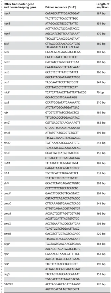
The primers used for real-time RT-PCR quantification of expres-sion of each gene are described in Table 1. These were designed using Primer Express 1.5 Software (Applied Biosystems, CA, USA) based on the sequence entries in the GenBank for E. coli K-12
Table 1.Primers used in this study.
. . . .
Efflux transporter gene
and house-keeping gene Primer sequence (59-39)
Length of amplicon
marA CATAGCATTTTGGACTGGAT 187 bp TACTTTCCTTCAGCTTTTGC
marB ATAGCAGCTGCGCTTATTC 154 bp ACTTATCACTGCCAGTACCC
marR AGCGATCTGTTCAATGAAAT 170 bp TTCAGTTCAACCGGAGTAAT
acrA CTTAGCCCTAACAGGATGTG
TTGAAATTACGCTTCAGGAT 189 bp
acrB CGTACACAGAAAGTGCTCAA 183 bp CGCTTCAACTTTGTTTTCTT
acrD GATTATCTTAGCCGCTTCAA 187 bp CAATGGAGGCTTTAACAAAC
acrE GCCCTCCTTTATTCTGATCT 166 bp GGCTATACGATAAGCATTGG
acrF TAGCAATTTCCTTTGTGGTT 247 bp CCTTTACCCTCTTTCTCCAT
micF TCATCATTAACTTTATTTATTACCG 70 bp GCATCCGGTTGAAATAGG
soxS CCATTGCGATATCAAAAATC 210 bp ATCTTATCGCATGGATTGAC
rob GTCGTCTTTATCCTGACTCG 189 bp TTTGTCACCCTGGAAGATAC
envZ CGTTGAGGTCAACAAAAGTT 185 bp GTCGGTTCTGGATACGAATA
emrB ATTATGTATGCCGTCTGCTT 196 bp TTCGCGTAAAGTTAGAGAGG
emrD TGTTAAACATGGGGATTCTC 243 bp TCAGCATCAGCAAATAACAG
emrE GGATTGCTTATGCTATCTGG 156 bp GTGTGCTTCGTGACAATAAA
mdfA TTTATGCTTTCGGTATTGGT 182 bp GAGATTAAACAGTCCGTTGC
tehA TGCTTCATTCTGGAGTTTCT 232 bp TCATTCTTTGTCCTCTGCTT
yhiV GCACTCTATGAGAGCTGGTC 203 bp CCTTCTTTCTGCATCATCTC
ompF GAACTTCGCTGTTCAGTACC 209 bp CGTACTTCAGACCAGTAGCC
ompC CTTCAAAGGTGAAACTCAGG 241 bp GTTGTCAGAACCGTAGGTGT
ompA ACGACTGGTTAGGTCGTATG 166 bp ACGTTGGATTTAGTGTCTGC
ompX ACCTGAAATACCGCTATGAA 208 bp TCAGTGGTCTGGAATTTACC
ompR GACGTCTTCGTAGTCAGAGC 229 bp TTGAACTTACCGAAAGCAAT
degP TGGTAGTGAACAACGTGAAA 184 bp AACAGGTAGATGGTGCTGTC
clpP CAAAAGGTAAACGTTTTTGC 163 bp AATGATTGACCCGTATGAAG
rseP TTGTTTATTACCTGCCGTTT 190 bp ATTAACAGCACCAGCAGAAT
degS TTCCAGTTAGCAACCAAAAT 153 bp TGACACTTCATTAACCACGA
complete genome (accession number NC_000913). Primer design and PCR experimental conditions were optimised to minimise amplification of contaminating E. coli genomic DNA potentially present in the total RNA sample, as well as for the prevention of non-specific primer annealing. The house-keepingGAPDHgene (coding for D-glyceraldehyde-3-phosphate-dehydrogenase) [49] was chosen as the endogenous reference RNA for relative quantification since it revealed consistent expression levels under the experimental conditions with the primers presented in Table 1. Amplification efficiencies of the target genes and reference gene were determined through the amplification of one-step RT-PCR template dilution series and PCR conditions were optimised until comparable amplification efficiencies were obtained for identical amounts of template RNA (absolute slope values less then 0.1) from calibration curve plots according to Applied Biosystems User Bulletin #2 (Applied Biosystems, CA, USA) recommendations [50].
Amplification ofGAPDHand other genes of interest were then run in separate tubes using the same amount of total RNA retrieved from the same sample. Thermal cycling conditions consisted of an initial reverse transcription step at 50uC during 30 min, an initial PCR activation step at 95uC for 15 min followed by 35 cycles of denaturation (94uC, 60 s), annealing (51uC–53uC for 60 s, depending on optimised conditions for the primers used) and extension (72uC for 60 s).
The relative quantities of the mRNA of each gene of interest were determined by the use of the comparative threshold cycle (CT) method. Taking advantage of the fact that samples with higher initial mRNA template concentration reach the significant threshold level for real-time detection at lower PCR cycle numbers than samples containing lower initial template concentrations, it is possible to obtain a quantitative measure of the expression magnitude (DCT) of each gene of interest, normalized by the house-keeping gene (GAPDH) expression in each sample to correct variation in RNA content and amplification efficiencies between samples. The equation 22DDCTallows the relative quantification of differences of each gene expression level between two samples, the sample of interest (the TET induced AG100 strain) and a calibrator or reference strain (the parental AG100 strain). Briefly, from three independent total mRNA extractions form E. coli AG100 and AG100 TET strains, grown under the described conditions,DCT of the reference and samples for each gene tested was obtained by subtracting the CTvalue of the GAPDHgene from the CTvalue
obtained for each gene.DDCTwas calculated by subtracting the average DCT values of the reference strain (AG100) from the corresponding TET induced DCT for each gene tested. The relative quantifications were then calculated by the equation 22DDCT as the number of fold of mRNA quantity differences relative to the calibrator or reference strain [50]. All data was collected and analysed with the aid of the Rotor-Gene 3000 real-time analysis software and the recommendations of the Applied Biosystems User Bulletin#2.
SDS-PAGE analyses and immunoblotting
Bacteria in exponential growth phase were pelleted and solubilized as previously described [51]. Proteins were analysed on 10% SDS-polyacrylamide gel system for OmpC, OmpF, OmpA and AcrA detection and 12% SDS-PAGE gel for OmpX, [15,51]. Gels were stained with Coomassie Brillant Blue R-250 to standardized protein samples. For western blots, proteins were electrotransfered onto nitrocellulose membranes (Schleicher & Schlull, Keene, NH, USA) in transfer buffer (20 mM Tris, 150 mM glycine, 20% isopropanol, 0.05% SDS). An initial saturating step was performed overnight at 4uC with buffered sodium (TBS: 50 mM Tris-HCl, 150 mM NaCl, pH 8) containing skimmed milk powder (10%). The nitrocellulose membranes were then incubated in TBS containing skimmed milk powder (10%) and Triton X-100 (0.2%) for 2 h at room temperature in the presence of polyclonal antibodies directed against denatured OmpC, OmpF, OmpA and OmpX, or with F4 polyclonal antibody directed against the L3 internal loop of E. coli porins [52]. The detection of antigen-antibody complexes was performed with alkaline phosphatase conjugated AffinitiPure goat anti-rabbit IgG antibodies (Jackson ImmunoResearch, West Grove PA, USA).
ACKNOWLEDGMENTS
We thank JM. Bolla and S. Fanning for helpful advice and discussions.
Author Contributions
Conceived and designed the experiments: JP LA MV MD IC. Performed the experiments: MV MD LR MM AD. Analyzed the data: JP LA MV LR MM AD. Contributed reagents/materials/analysis tools: JP LA MV. Wrote the paper: JP LA MV IC.
REFERENCES
1. Nikaido H (2001) Preventing drug access to targets: cell surface permeability barriers and active efflux in bacteria. Semin Cell Dev Biol 12: 215–223. 2. Gootz TD (2006) The forgotten Gram-negative bacilli: what genetic
determi-nants are telling us about the spread of antibiotic resistance. Biochem Pharmacol 71: 1073–1084.
3. Piddock LJ (2006) Multidrug-resistance efflux pumps - not just for resistance. Nat Rev Microbiol 4: 629–636.
4. Davin-Regli A, Page`s JM (2006) Regulation of efflux pumps in Enterobacter-iaceae: genetic and chemical effectors. In: Zmabiles-Cuevos, ed. Antimicrobial resistance in bacteria, C.F., Horizon Biosciences. pp 55–75.
5. Viveiros M, Jesus A, Brito M, Leandro C, Martins M, et al. (2005) Inducement and reversal of tetracycline resistance inEscherichia coliK-12 and expression of proton gradient-dependent multidrug efflux pump genes. Antimicrob Agents Chemother 49: 3578–82.
6. Pa´ges JM, Masi M, Barbe J (2005) Inhibitors of efflux pumps in Gram-negative bacteria. Trends Mol Med 11: 382–9.
7. Nikaido H (2003) Molecular basis of bacterial outer membrane permeability revisited. Microbiol Mol Biol Rev 67: 593–656.
8. Cowan SW, Schirmer T, Rummel G, Steiert M, Ghosh R, et al. (1992) Crystal structures explain functional properties of twoE. coliporins. Nature 358: 727–733. 9. Basle A, Rummel G, Storici P, Rosenbusch JP, Schirmer T (2006) Crystal Structure of Osmoporin OmpC fromE. coliat 2.0. A J Mol Biol 362: 933–942.
10. Liu X, Ferenci T (2001) An analysis of multifactorial influences on the transcriptional control of ompF and ompC porin expression under nutrient limitation. Microbiology 147: 2981–2989.
11. Ferenci T (2005) Maintaining a healthy SPANC balance through regulatory and mutational adaptation. Mol Microbiol 57: 1–8.
12. Randall LP, Woodward MJ (2002) The multiple antibiotic resistance (mar) locus and its significance. Res Vet Sci 72: 87–93.
13. Castillo-Keller M, Vuong P, Misra R (2006) Novel mechanism ofEscherichia coli
porin regulation. J Bacteriol 188: 576–586.
14. Hall MN, Silhavy TJ (1981) TheompBlocus and the regulation of the major outer membrane porin proteins ofEscherichia coliK12. J Mol Biol 146: 23–43. 15. Dupont M, De E, Chollet R, Chevalier J, Page`s JM (2004)Enterobacter aerogenes
OmpX, a cation-selective channel mar- and osmo-regulated. FEBS Lett 569: 27–30. 16. Ma D, Cook DN, Hearst JE, Nikaido H (1994) Efflux pumps and drug resistance
in gram-negative bacteria. Trends Microbiol 2: 489–493.
17. Purewal AS (1991) Nucleotide sequence of the ethidium efflux gene from
Escherichia coli. FEMS Microbiol Lett 66: 229–231.
18. Kawamura-Sato K, Shibayama K, Horii T, Iimuma Y, Arakawa Y, et al. (1999) Role of multiple efflux pumps inEscherichia coli in indole expulsion. FEMS Microbiol Lett 179: 345–352.
19. Lomovskaya O, Lewis K (1992) emr, anEscherichia colilocus for multidrug resistance. Proc Natl Acad Sci USA 89: 8938–8942.
20. Naroditskaya V, Schlosser MJ, Fang NY, Lewis K (1993) AnE. coligeneemrDis involved in adaptation to low energy shock. Biochem Biophys Res Commun 196: 803–809.
21. Ma D, Cook DN, Alberti M, Pon NG, Nikaido H, et al. (1995) GenesacrAand
acrBencode a stress-induced efflux system ofEscherichia coli. Mol Microbiol 16: 45–55.
22. Edgar R, Bibi E (1997) MdfA, anEscherichia colimultidrug resistance protein with an extraordinarily broad spectrum of drug recognition. J Bacteriol 179: 2274–2280.
23. Turner RJ, Taylor DE, Weiner JH (1997) Expression ofEscherichia coliTehA gives resistance to antiseptics and disinfectants similar to that conferred by multidrug resistance efflux pumps. Antimicrob Agents Chemother 41: 440–444. 24. Rosenberg EY, Ma D, Nikaido H (2000) AcrD of Escherichia coli is an
aminoglycoside efflux pump. J Bacteriol 182: 1754–1756.
25. Nishino K, Yamaguchi A (2001) Analysis of a complete library of putative drug transporter genes inEscherichia coli. J Bacteriol 183: 5803–5812.
26. Cohen SP, Hachler H, Levy SB (1993) Genetic and functional analysis of the multiple antibiotic resistance (mar) locus in Escherichia coli. J Bacteriol 175: 1484–1492.
27. Martin RG, Rosner JL (2001) The AraC transcriptional activators. Curr Opin Microbiol 4: 132–137.
28. Barbosa TM, Levy SB (2000) Differential expression of over 60 chromosomal genes inEscherichia coliby constitutive expression of MarA. J Bacteriol 182: 3467–3474.
29. Li XZ, Nikaido H (2004) Efflux-mediated drug resistance in bacteria. Drugs 64: 159–204.
30. Ariza RR, Li Z, Ringstad N, Demple B (1995) Activation of multiple antibiotic resistance and binding of stress-inducible promoters by Escherichia coliRob protein. J Bacteriol 177: 1655–1661.
31. Nakajima H, Kobayashi K, Kobayashi M, Asako H, Aono R (1995) Overexpression of the robA gene increases organic solvent tolerance and multiple antibiotic and heavy metal ion resistance in Escherichia coli. Appl Environ Microbiol 61: 2302–2307.
32. Michan C, Manchado M, Pueyo C (2002) SoxRS down-regulation of rob
transcription. J Bacteriol 184: 4733–4738.
33. Martin RG, Rosner JL (1995) Binding of purified multiple antibiotic-resistance repressor protein (MarR) tomaroperator sequences. Proc Natl Acad Sci USA 92: 5456–5460.
34. Pratt LA, Silhavy TJ (1996) The response regulator SprE controls the stability of RpoS. Proc Natl Acad Sci USA 93: 2488–2492.
35. Guillier M, Gottesman S, Storz G (2006) Modulating the outer membrane with small RNAs. Genes Dev 20: 2338–2348.
36. Bohnert JA, Schuster S, Fahnrich E, Trittler R, Kern WV (2007) Altered spectrum of multidrug resistance associated with a single point mutation in the
Escherichia coli RND-type MDR efflux pump YhiV (MdtF). J Antimicrob Chemother. In press. (Epub ahead of print Oct24, 2006).
37. Delihas N, Forst S (2001) MicF: an antisense RNA gene involved in response of
Escherichia colito global stress factors. J Mol Biol 313: 1–12.
38. Chen S, Zhang A, Blyn LB, Storz G (2004) MicC, a second small-RNA regulator of Omp protein expression inEscherichia coli.J Bacteriol 186: 6689–97. 39. Ruiz N, Kahne D, Silhavy TJ (2006) Advances in understanding bacterial
outer-membrane biogenesis. Nat Rev Microbiol 4: 57–66.
40. Wu T, Malinverni J, Ruiz N, Kim S, Silhavy TJ, et al. (2005) Identification of a multicomponent complex required for outer membrane biogenesis in
Escherichia coli. Cell 121: 235–245.
41. Charlson ES, Werner JN, Misra R (2006) Differential effects ofyfgLmutation on
Escherichia coliouter membrane proteins and lipopolysaccharide. J Bacteriol 188: 7186–7194.
42. Bolla JM, Lazdunski C, Page`s JM (1988) The assembly of the major outer membrane protein OmpF ofEscherichia colidepends on lipid synthesis. EMBO J 7: 3595–3599.
43. Douchin V, Bohn C, Bouloc P (2006) Down-regulation of porins by a small RNA bypasses the essentiality of the regulated intramembrane proteolysis protease RseP inEscherichia coli. J Biol Chem 281: 12253–12259.
44. Gambino L, Gracheck SJ, Miller PF (1993) Overexpression of the MarA positive regulator is sufficient to confer multiple antibiotic resistance inEscherichia coli.
J Bacteriol 175: 2888–2894.
45. Baquero F (2001) Low-level antibacterial resistance: a gateway to clinical resistance. Drug Resist Updat 4: 93–105.
46. Martins M, Santos B, Martins A, Viveiros M, Couto I, et al. (2006) An instrument-free method for the demonstration of efflux pump activity of bacteria. In Vivo 20: 657–664.
47. Okusu H, Ma D, Nikaido H (1996) AcrAB efflux pump plays a major role in the antibiotic resistance phenotype of Escherichia colimultiple-antibiotic resistant (Mar) mutants. J Bacteriol 178: 306–308.
48. Eliopoulos GM, Moellering RC (1991) Antibiotics in Laboratory Medicine. (Lorian V., Ed.) Williams and Wilkins Co., Baltimore, MD. pp 432–492. 49. Branlant G, Branlant C (1985) Nucleotide sequence of theEscherichia coli gap
gene. Different evolutionary behavior of the NAD+-binding domain and of the catalytic domain of D-glyceraldehyde-3-phosphate dehydrogenase. Eur J Biochem 150: 61–66.
50. Langmann T, Mauerer R, Zahn A, Moehle C, Probst M, et al. (2003) Real-time reverse transcription-PCR expression profiling of the complete human ATP-binding cassette transporter superfamily in various tissues. Clin Chem 49: 230–238.
51. Malle´a M, Chevalier J, Bornet C, Eyraud A, Davin-Regli A, et al. (1998) Porin alteration and active efflux: two in vivo drug resistance strategies used by
Enterobacter aerogenes. Microbiology 14: 3003–3009.