DOI: 10.1590/S0100-736X2016000500002
RESUMO.- [Distribuição de linfócitos, células carrea-doras de imunoglobulinas, macrófagos e células den-dríticas em glândulas sexuais acessórias de carneiros
infectados experimentalmente com Actinobacillus se-minis.] A distribuição das células envolvidas na resposta imune em glândulas sexuais acessórias de carneiros
expe-Distribution of lymphocytes, immunoglobulin-containing cells,
macrophages, and dendritic cells in the accessory sex glands of
rams experimentally infected with
Actinobacillus seminis
1Jorge Acosta-Dibarrat2*, Víctor Tenorio-Gutiérrez3, Edgardo Soriano-Vargas2, MartínTalavera-Rojas2, Luis Cal-Pereyra4, Roberto Montes de Oca Jiménez2,
Valente Velázquez-Ordoñez2 and Jorge Tórtora-Pérez5
ABSTRACT.- Acosta-Dibarrat J., Tenorio-Gutiérrez V., Soriano-Vargas E., Talavera-Rojas M., Cal-Pereyra L., Montes de Oca-Jiménez R., Velázquez-Ordoñez V. & Tórtora-Pérez J. 2016. Distribution of lymphocytes, immunoglobulin-containing cells, macrophages, and dendritic cells in the accessory sex glands of rams experimentally infected with Acti-nobacillus seminis. Pesquisa Veterinária Brasileira 36(5):363-372. Centro de Investigación y Estudios Avanzados en Salud Animal, Facultad de Medicina Veterinaria y Zootecnia, Uni-versidad Autónoma del Estado de México, Km 15.5 Carretera Toluca-Atlacomulco, Toluca, ME 50100, México. E-mail: [email protected]
The distribution of cells involved in the immune response in accessory sex glands of rams experimentally infected with Actinobacillus seminis was studied. Twelve one-year old rams were experimentally infected by intraurethral (IU) (n=4) and intraepididymal (IE) (n=4) route, and four control (CON) animals were used. The animals were slaughtered 35 days post-inoculation, samples were taken from accessory sex glands, and bacteriology and
histo-pathology tests were performed. The presence of CD4, CD8 and TCRγδ (WC1) lymphocytes,
CD45RO cells, macrophages (CD14), dendritic cells (CD1b), IgA-, IgG- and IgM-containing cells (IgCC) was determined. Animals of the IE group developed clinical epididymitis. No lesions were seen in rams of the IU group; two of the intraepididymal inoculated CON deve-loped small lesions in the epididymis. A. seminis isolates were achieved from 6:16 (37.5%) accessory sex glands in the IE group, but not in the IU and CON groups. In the CON group, IgA- and IgM- containing cells predominated in the bulbourethral glands and the
dissemina-ted prostate, and they were scarce or null in the vesicles and ampullae. A significant increase of IgA-, IgG- and IgM- containing cells was confirmed in the seminal vesicles, the ampullae
and the bulbourethral glands in the IE group. In the IE and IU groups, an increase in CD4,
CD8, WC1, CD45RO and CD14 was evidenced in the vesicles and ampullae. CD1b dendritic cells were present in the ampullae and vesicles with inflammatory processes. A. seminis tri-ggered a local immune response in the IE and IU groups. These results indicate a different
pattern of infiltrating immune cells in the accessory sex glands of infected A. seminis rams.
INDEX TERMS:Accessory sex glands, rams, Actinobacillus seminis, lymphocytes, immunoglobulin-containing cells, macrophages.
1 Received on October 30, 2015.
Accepted for publication on February 15, 2016.
2 Centro de Investigación y Estudios Avanzados en Salud Animal, Facul-tad de Medicina Veterinaria y Zootecnia, Universidad Autónoma del Esta-do de México, Km 15,5 Carretera Toluca-Atlacomulco, Toluca, ME 50100, México. *Corresponding author: [email protected]
3 Centro Nacional de Investigación Disciplinaria Microbiología
(CENID--Microbiología) INIFAP, Km 15,5 Carretera Federal México, Toluca, Col. Palo Alto, DF 05110, México.
4 Departamento de Patología, Facultad de Veterinaria, Universidad de la República, Alberto Lasplaces, 1550, Montevideo, MO 11600, Uruguay.
rimentalmente infectados com Actinobacillus seminis foi estudada. Doze carneiros de um ano de idade foram ex-perimentalmente infectados via intrauretral (IU) (n=4) e via intraepididimal (IE) (n=4) e quatro animais controles (CON) foram utilizados. Os animais foram abatidos 35 dias após a inoculação, amostras foram retiradas das glândulas sexuais acessórias e testes bacteriológicos e histopatológi-cos foram realizados. A presença de linfócitos CD4, CD8 e
TCRγδ (WC1), células CD45RO, macrófagos (CD14), células
dendríticas (CD1b) e células contendo IgA, IgG and IgM (IgCC) foi determinada. Os animais do grupo IE desenvolve-ram epididimite clínica. Não fodesenvolve-ram visualizadas lesões nos carneiros do grupo IU, dois dos CON inoculados intraepidi-dimalmente desenvolveram pequenas lesões no epidídimo. Isolados de A. seminis foram obtidos de 6:16 (37,5%) nas glândulas sexuais acessórias no grupo IE mas não nos gru-pos IU e CON. No grupo CON células contendo IgA and IgM predominaram nas glândulas bulbouretrais e na próstata e foram escassas ou ausentes nas vesículas e na ampola. Um
incremento significativo de células contendo IgA, IgG and IgM foi confirmado nas vesículas seminais, na ampola e nas
glândulas bulbouretrais no grupo IE. Nos grupos IE e IU foi
evidenciado um aumento em CD4, CD8, WC1, CD45RO e
CD14 nas vesículas e ampola. As células dendríticas CD1b estavam presentes na ampola e nas vesículas com processo
inflamatório. A. seminis induziu uma resposta imune local nos grupos IE e IU. Estes resultados indicam um padrão
di-ferente de células imunes infiltrantes nas glândulas sexuais
acessórias de carneiros infectados por A. seminis.
TERMOS DE INDEXAÇÃO: Glândulas sexuais acessórias, carnei-ros, Actinobacillus seminis, linfócitos, células carreadoras de imu-noglobulinas, macrófagos.
INTRODUCTION
Sheep epididymitis has been mainly associated with Brucella ovis and Actinobacillus seminis infections (Burgess 1982, Moustacas et al. 2013). The pathogenesis of the disease caused by A. seminis has been explored to a small extent; the presence of pathogenicity factors in A. seminis, such as adhesins (Healey et al. 1991) or RTX toxins (Schaller et al. 2000), is likely to contribute to the development of the pa-thology. However, the susceptibility to infection of the acces-sory sex glands, especially of the seminal vesicles in sheep (Foster et al. 1987, Al-Katib & Dennis 2005, Acosta-Dibarrat et al. 2006), bulls (Bagshaw & Ladds 1974, Cavalieri & Van Camp 1997), and humans (Furuya et al. 2004), leads us to believe that other factors, especially immunological factors, would be implicated in such susceptibility.
The male genital tract has to accomplish functions of de-fense against infections, and it must also tolerate germ cells that present differential antigens. Recently, the study of the immune response mechanisms in the reproductive system
has been intensified, focused on the fight against sexually
transmitted diseases (Anderson & Pudney 1999).
The information on the immune response mechanisms in the male reproductive tract in different species, and par-ticularly, the information on the presence and distribution of immune response cells in the reproductive tract of
she-ep, is limited (Acosta-Dibarrat et al. 2007, 2014, Moustacas et al. 2014). In this work, rams were experimentally infec-ted with A. seminis intraurethral (IU) and intraepididymal (IE) inoculation, and the distribution and amount of CD4,
CD8, and TCRγδ lymphocytes, immunoglobulin-containing
cells, macrophages, CD1b dendritic cells and CD45RO cells in the accessory glands were studied.
MATERIALS AND METHODS
Animals and Actinobacillus seminis inoculation. The ani-mals were kept during the performance of the pre vious examine to slaughter facilities in the FES Cuautitlán UNAM. Inoculation, sampling protocols and slaughter were endorsed by the Subcom-mittee Institutional Animal Care Experimentation (SICUAE) of the Faculty of Veterinary Medicine, UNAM. This protocol was appro-ved by UNAM at 20/06/2003.
Twelve one-year-old male Pelibuey sheep were used, from a
flock without clinical history of the disease, with no clinical al -terations, and with negative semen bacteriology and serology against A. seminis and B. ovis. Three groups of four animals each
were formed. The first group was challenged with A. seminis by intraurethral (IU) route; the same suspension was intraepididy-mally (IE) administered to the second group, directly in the epi-didymal tail. Physiologic saline solution (PSS) was administered by IU intraurethral and in the epididymal tail to the third group, which remained as non-infected control (CON).
A. seminis ATCC 15768 strain was used as challenge bacte-rium; it was seeded in blood agar and cultured in PSS. The chal-lenge inoculum had a bacterial concentration of 2.3x10⁹ CFU/ml.
Inoculation by IU route was carried out with 2 ml of the bac-terial suspension, with previous ablation of the urethral process under sedation, using urethral-probing catheters for cats. Inocu-lation by IE route was carried out following scrotum disinfection and cleaning, with 1.5ml of the bacterial suspension in the left epididymal tail, and 0.5ml were applied in the right tail. 1.5ml of PSS was administered to controls by intraurethral and intraepi-didymal route in the tail of the right epididymis.
Sample management. All animals were slaughtered at 35 days post-inoculation, and the seminal vesicles, the ampullae of the deferent duct, the bulbourethral glands and the pelvic urethra (including the disseminated prostate) were obtained.
Samples of the same regions were taken in all cases to per-form histopathology, immunohistochemistry and bacteriology tests (Acosta-Dibarrat et al. 2007, 2014), using a sagittal section of the bulbourethral glands and the apical portion of the vesicles; the pelvic urethra was cross-sectionally cut at a site equidistant from the bulbourethral glands and the seminal vesicles. A medial section was obtained from the ampullae of the deferent duct at the zone equidistant from the deferent duct and its junction with
the pelvic urethra. The immunohistochemistry and immunofluo -rescence samples were included in OCT-based cryopreservative from the Tissiu-Tek brand (Sakura Finetec, Torrance, USA) and were immediately frozen in liquid nitrogen, after passage over nitrogen vapors for 2 minutes, and were subsequently immersed. Samples were withdrawn from nitrogen and kept at -80°C until sectioning.
Successive 4 µm sections were performed with a HistoSTAT
microtome (AO Scientific Instrument, New York, USA) and were
mounted on plates treated with poly-L-Lysin (Sigma Chemical Co,
St. Louis, USA), they were fixed with acetone for 10 min. Four sli -ces of approximately 1.0cm2 were placed on each plate; one slice
was used as control and the other three were used for the
Immunohistochemistry. Slices were hydrated with PBS for 10 minutes and endogenous peroxidase was blocked with Per-oxo-Block (Lab. Zymed, San Francisco, USA) for 45 seconds. An
unspecific blockade was carried out with 10% caprine serum
overnight. Then, primary monoclonal antibodies for the differ-ent receptors were applied (Table 1), which were incubated for 2 hours at room temperature. The biotin-goat anti mouse IgG com-plex was subsequently applied (Lab. Zymed, San Francisco, USA) for 30 minutes; then, the Streptavidin-Peroxidase complex (Lab. Zymed, San Francisco, USA) was administered for 15 minutes; the DAB substrate (Lab. Zymed, San Francisco, USA) was applied until a reaction was observed (2 to 5 minutes) and hematoxylin contrast staining was carried out. Three washings with PBS were performed between each step. Finally, the slices were dehydrated, cleared and mounted.
In the control slice, the primary antibody was replaced with PBS. Additionally, plates with prepuce sections were used as
posi-tive control in all runs, since it had been previously confirmed that
cells positive to all monoclonal antibodies used in this work were present in this organ.
Immunofluorescence. For the determination of
immunoglo-bulin-containing cells (IgCC), tissue Igs were eliminated first by placing the plates in PBS all night; an unspecific blockade with
1% ASB was subsequently performed for 1 hour at room tempe-rature, and three 5-minute washings with PBS were carried out. Anti-ovine IgA primary antibody (Bethyl Lab., Montgomery, USA) was applied in a 1:40 dilution, as well as anti-ovine IgG primary antibody (Bethyl Lab., Montgomery, USA) in a 1:200 dilution, and anti-ovine IgM primary antibody (Bethyl Lab., Montgomery, USA) in 1:60 in PBS with 1% ASB. Incubation was performed for 1 hour at 37°C. Three washings were performed and the secondary anti-body, TRICT-conjugated anti-rabbit IgG antibody (Sigma-Aldrich, St Louis, USA), was applied in a 1:30 dilution (in 10% goat serum and 1% ASB) for one hour at 37°C. Finally, it was washed again 3 times for 5 minutes per washing, and was mounted in glycerin
and observed under an epifluorescence microscope. The primary
antibody was replaced with PBS in the control slice, and the te-chnique described above was then followed. This control was
in-cluded due to the possibility of unspecific labeling by the anti-rab -bit IgG conjugate. In this case, a prepuce section was also used as positive control, on which positive labeling had been previously
demonstrated for the three immunoglobulins.
Cell count and statistical analysis. Cell count was carried
out in ten 40 X 10x fields with the Image Pro Plus 4.5 software.
The cells present in the projected image were counted and the re-sulting average counts were expressed as cells per mm2. The
pos-sible differences between treatments (groups) for each gland and each monoclonal antibody used were determined by the
Krus-kal-Wallis test, followed by the Mann-Whitney test. The difference was considered significant with p<0.05.
RESULTS
The results of the histopathology and bacteriology studies are shown in Table 2. At the moment of slaughter, only the challen-ge bacterium could be re-isolated in the IE group in 3 of 4 she-ep from the she-epididymal tail, in 3 of 4 sheshe-ep from the ampullae, and in 1 of 4 sheep from the seminal vesicle, the prostate and
the bulbourethral gland. Inflammatory lymphoplasmocytary infiltrates, with different intensity degrees, occurred in the
ampullae and seminal vesicles, and were concurrent with the Table 1. Characteristics of the monoclonal antibodies used in
immunohistochemistry tests
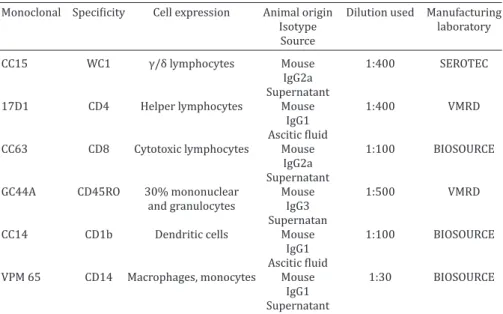
Monoclonal Specificity Cell expression Animal origin Dilution used Manufacturing
Isotype laboratory
Source
CC15 WC1 γ/δ lymphocytes Mouse 1:400 SEROTEC
IgG2a
Supernatant
17D1 CD4 Helper lymphocytes Mouse 1:400 VMRD
IgG1
Ascitic fluid
CC63 CD8 Cytotoxic lymphocytes Mouse 1:100 BIOSOURCE
IgG2a
Supernatant
GC44A CD45RO 30% mononuclear Mouse 1:500 VMRD
and granulocytes IgG3
Supernatan
CC14 CD1b Dendritic cells Mouse 1:100 BIOSOURCE
IgG1
Ascitic fluid
VPM 65 CD14 Macrophages, monocytes Mouse 1:30 BIOSOURCE
IgG1
Supernatant
Table 2. Histopathology and bacteriology of the reproductive tract organs
Group Number Ampulla Seminal Disseminated Bulbourethral
vesicle prostate
IU 1 NC LI+ NC NC
2 NC NC NC NC
3 LI+ LI+ NC NC
4 LI+ NC NC NC
IE 5 LI++ A.s LI+++ LI+ NC
6 LI+ LI+++ LI+ NC
7 LI++ A.s LI +A.s LI +A.s NC A.s
8 LI++ A.s LI+ NC NC
CON 9 NC NC NC NC
10 NC NC NC NC
11 NC NC NC NC
12 NC LI+ NC NC
Fig.1. Number of CD4, CD8, WC1, CD45RO, CD14 and CD1b lymphocytes per mm2 in the accessory sex glands of rams inoculated with A. seminis. IU Intraurethral group, IE Intraepididymal group, CON Control group. a significance compared with the CON group p<0.05, b significance compared with the IE group p<0.05, e compared in the CON group p<0.1, f compared in the IU group p<0.1.
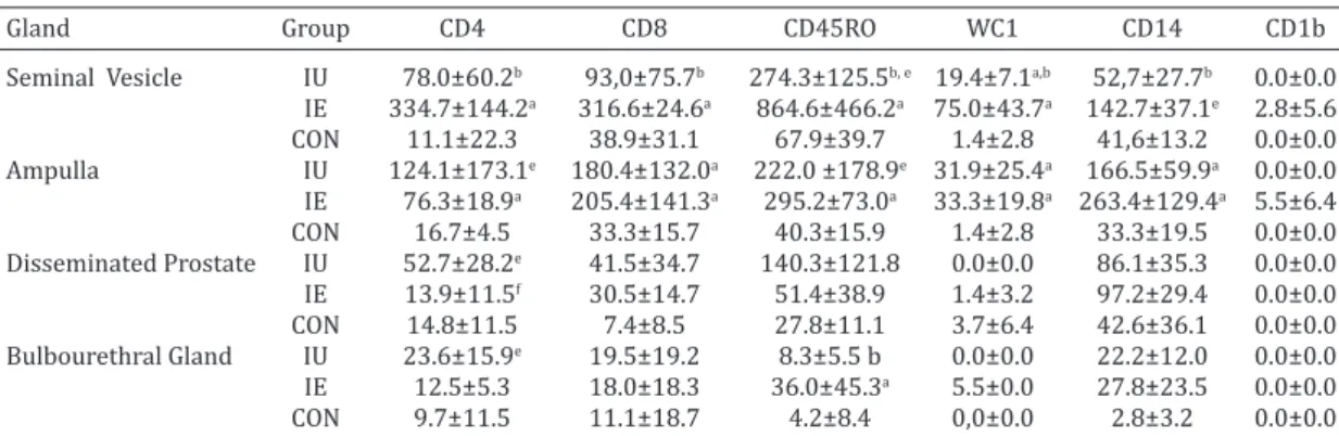
Table 3. CD4, CD8, CD45RO, WC1, CD14 AND CD1b cells per (mm2) in the accessory sex glands of
rams inoculated with Actinobacillus seminis
Gland Group CD4 CD8 CD45RO WC1 CD14 CD1b
Seminal Vesicle IU 78.0±60.2b 93,0±75.7b 274.3±125.5b, e 19.4±7.1a,b 52,7±27.7b 0.0±0.0 IE 334.7±144.2a 316.6±24.6a 864.6±466.2a 75.0±43.7a 142.7±37.1e 2.8±5.6 CON 11.1±22.3 38.9±31.1 67.9±39.7 1.4±2.8 41,6±13.2 0.0±0.0 Ampulla IU 124.1±173.1e 180.4±132.0a 222.0 ±178.9e 31.9±25.4a 166.5±59.9a 0.0±0.0 IE 76.3±18.9a 205.4±141.3a 295.2±73.0a 33.3±19.8a 263.4±129.4a 5.5±6.4 CON 16.7±4.5 33.3±15.7 40.3±15.9 1.4±2.8 33.3±19.5 0.0±0.0 Disseminated Prostate IU 52.7±28.2e 41.5±34.7 140.3±121.8 0.0±0.0 86.1±35.3 0.0±0.0 IE 13.9±11.5f 30.5±14.7 51.4±38.9 1.4±3.2 97.2±29.4 0.0±0.0 CON 14.8±11.5 7.4±8.5 27.8±11.1 3.7±6.4 42.6±36.1 0.0±0.0 Bulbourethral Gland IU 23.6±15.9e 19.5±19.2 8.3±5.5 b 0.0±0.0 22.2±12.0 0.0±0.0 IE 12.5±5.3 18.0±18.3 36.0±45.3a 5.5±0.0 27.8±23.5 0.0±0.0 CON 9.7±11.5 11.1±18.7 4.2±8.4 0,0±0.0 2.8±3.2 0.0±0.0 IE = intraepididymal, IU = intraurethral, CON = control. a Significance compared with the CON group p<0.05, b significance
Actinobacillus seminis isolate in 3:4 animals in the IE group. The average counts obtained for the evaluated cells in each group are summarized in Table 3 and Figure 1. A gre-ater overall cellularity can be observed in the seminal vesi-cles and the ampullae from the IU and IE groups, compared with the control group. The most important differences oc-curred in the ampullae and the seminal vesicles from the animals with concurrent isolation of A. seminis from these structures.
In the ampullae of the deferent duct in the IE group, the-re wethe-re diffethe-rences from the CON group in CD4 (p=0.003),
CD8 (p=0.028), CD45RO (p=0.005), WC1 (P=0.007) and
CD14 (p=0.028) counts, but no differences were noted in CD1b, although the presence of labeled cells was demons-trated (5.5±6.4 mm2). Differences were also found between
the IU and CON groups in CD8 (p=0.028), WC1 (p=0.002)
and CD14 (p=0.003), with a trend in CD45RO (p=0.083).
No significant differences were found between cell popula -tions from the ampullae in the IU and IE groups.
In the seminal vesicles, differences were present
betwe-en the IE and CON groups in CD4, CD8, WC1 and CD45RO
(p=0.029), as well as a tendency in CD14 (p=0.057); these differences were also maintained between the IE and IU
treatments, and were even significant for CD14 (p=0.029). There was a significant difference in the vesicle counts in WC1 between the CON and IU groups (p=0.029), with a
tendency in CD45RO (p=0.057). A scarce number of CD1b (2.8±5.6 mm2) was found in the IE group.
No differences were found in the disseminated prosta-te; a trend was demonstrated only for CD4 between IU, CON and IU, IE (p=0.057).
In the bulbourethral glands, differences were demons-trated between the IE and CON groups, and between the
IE and IU groups in WC1 (p=0.006 and p=0.029, respec -tively) and in CD14 cells between the IU and CON groups (p=0.042).
In the seminal vesicles, lymphoid accumulations were found in the periphery of the acini with presence of CD8 and CD4. CD4 lymphocytes were frequently present in the con-junctival stroma of the glands, and CD8 were present with a closer relationship with the basal membrane of acini (Fig.2). CD45RO were present in both positions and were frequen-tly located within the acini (Fig.3). CD14 labeling occurred in the stroma, on the macrophages and in the endothelia.
Regarding immunoglobulin-containing cells (IgCC), the most frequent labeling in the accessory sex glands corres-ponded to IgA, followed by IgM and IgG. In the CON group, the bulbourethral gland and the disseminated prostate pre-sented IgA- and IgM-containing cells, which were not seen in the ampullae and the vesicle (Table 4, Fig.4)
IgG-containing cells were seen in the IE and IU groups, in a greater proportion in the seminal vesicle and in the def-erent duct ampullae. Coincidently, it was from these glands that A. seminis isolates were obtained more frequently. Statistical differences were found in the ampullae for IgM- and IgG- containing cells between the CON and IE groups (p=0.029), and between the IE and IU groups (p=0.029).
Significant differences were found between IgA-contai -ning cells (p=0.029) in the bulbourethral glands in the CON and IE groups, while IgM-containing cells showed a trend Fig.2. Lymphoid infiltration by an acinus in the seminal vesicle.
Serial sections of seminal vesicle: a) Marking to CD8 (ocher) lymphocytes. IP, obj.20x, b) Marking for CD4 (ocher) lympho-cytes. IP, obj.20x.
Fig.3. CD45RO by an acinus in the seminal vesicle. CD45RO
lym-phocytes infiltrating the stroma and acinar epithelium. IP, obj.
(p=0.059).
Trends were present in the vesicles in the Kruskal Wallis test (p<0.1), but the great variability in the cell counts from
these samples prevents these differences from being evi-dent between the groups (data not shown).
IgG- containing cells were only seen in the IU and IE groups, mainly in the latter, with percentages of 11, 9 and 2 % in the ampulla, the seminal vesicle and the bulbourethral gland, respectively. Conversely, no IgG labeling was seen in
the CON group in any gland.
The greater cellularity observed in the bulbourethral glands and the disseminated prostate stands out in all groups, mainly due to the presence of IgA- and IgM- ning cells (Figure 5). In the bulbourethral gland, IgA- contai-ning cells ranged between 73 and 79%, and IgM- contaicontai-ning cells ranged between 21 and 25%, while in the dissemi-nated prostate, 82 to 90 % were IgA- containing cells and between 10 and 18% were IgM- containing cells , and the
Fig.4. Number of IgA-, IgG- and IgM-containing cells in the accessory sex glands of rams inoculated with A. seminis. IE intraepididymal, IU
positive IgG- containing cells counts were almost null. The histological location of IgCC also showed differen-ces between the glands studied. In the disseminated pros-tate and the bulbourethral gland, IgA- and IgM- containing
cells were observed mainly in the scarce connective tissue surrounding the acini. A clearly positive IgA labeling was observed in the inside of some acini in association with glandular secretion. IgG- containing cells from the seminal vesicles were located in the connective tissue that separa-tes the glandular lobules, while IgA- containing cells were observed in the acini.
DISCUSSION
In this experiment, several facts are confirmed that may
contribute to demonstrating the different susceptibility of the accessory glands to Actiobacillus seminis infection (Al-Katib & Dennis 2005, Acosta-Dibarrat et al. 2006). As previously mentioned regarding A. seminis, the bacterium was isolated more frequently from the ampullae and sem-inal vesicles, suggesting that, in these structures, bacterial establishment and permanence is somehow facilitated, al-though these were the glands that showed the most
nota-ble inflammatory changes behind the epididymides, which
were directly inoculated. In the vesicles and ampullae of the CON group, no positive IgA-, IgM- or IgG-containing cells were found, or they were found in a lower number than in the inoculated groups, a condition that may
facili-tate bacterial establishment. This finding differentiates the
seminal vesicles and the ampullae from the bulbourethral gland and the disseminated prostate, which, conversely, demonstrated an important number of these cells in the aforementioned group.
The distribution of IgCC in control and infected sheep
glands observed in this work is consistent with the findings
demonstrated in bulls, where IgCC are also not found or are present in a very limited number in the seminal vesicles of normal animals (Campero et al. 1989, 1990), while in the
presence of an inflammatory response in the genital tract,
IgCC appear in the ampullae and the seminal vesicles (Cam-pero et al. 1989). It has been noted that the production of IgA and IgG in the seminal vesicles in the bull is an essential component of the local immune response (Bier et al. 1977). However, in other studies, no IgCC are found, or these are found in a very limited number in the seminal vesicles of normal bulls; therefore, it is unlikely that this is the origin of the immunoglobulins present in the semen (Campero et Table 4. IgA-, IgG- and IgM-containing cells in the accessory sex glands per (mm2)
Gland Group IgA-containing cells IgG-containing cells IgM-containing cells
Seminal Vesicle IU 97.4±194.9* 1.4±2.8* 7.0±14.0*
IE 319.9±233.9* 52.8±78.5* 110.6±75.2*
CON 0.0±0.0* 0.0±0.0* 0.0±0.0*
Ampulla IU 12.4±24.9 0.0±0.0 1.4±2.8
IE 49.1±76.4 16.7±15.8 a,b 123.5±128.1 a,b
CON 11.2±15.9 0.0±0.0 0.0±0.0
Disseminated Prostate IU 280.3±106.1 1.4±2.8 32.5±64.9
IE 243.7±94.9 0.0±0.0 48.1±21.4
CON 143.6±167.2 0.0±0.0 31.8±25.3
Bulbourethral IU 84.5±44.8 0.0±0.0 22.2±7.8
IE 237.4±40.6 a,b 5.5±6.4 81.2±53.1 a,e
CON 58.3±58.5 0.0±0.0 15.3±17.8
IE = intraepididymal, IU = intraurethral, CON = control. a Significance compared with the CON group
p<0.05, b significance compared with the IU group p<0.05, e significance compared with the IU
group p<0.1, * p<0.1 (Kruskal Wallis test) very heterogeneous reactions within the group.
al. 1989, 1990).
Foster et al. (1988), using 5 one-year-old sheep and 12 sheep older than 4 years, reported the presence of IgA- and IgG-containing cells, mainly in the bulbourethral gland and the prostate, and only occasionally in the seminal vesicles and the ampullae; these results are consistent with the re-sults of this work. However, in contrast with the rere-sults of this work, these authors found high percentages of IgG-con-taining-cells, a condition that only occurred in a small num-ber in the IE group in the seminal vesicles and ampullae
with active inflammatory processes. This difference may
be attributed to the fact that the animals used by Foster et al. (1988), had subclinical alterations of the reproductive system with local infection, since no bacteriological exami-nations of the semen were carried out and they only
veri-fied that the animals were free of B. ovis by serology; they also do not distinguish between young animals and adult animals, and variations are likely to occur in the amount and distribution of IgCC due to age, as has been reported in bulls (Campero et al. 1989).
In humans, positive IgA- and IgM-containing cells are described in the prostate, the bulbourethral gland and the penile urethra, where only a limited number of IgG-con-taining cells are found. However, little is known about the immunobiology of seminal vesicles, where no plasma cells are seen, but an occasional expression of pIgR is found (An-derson & Pundey 1999).
The information available on the presence and distri-bution of the cell types studied in the male reproductive apparatus, especially in the accessory sex glands, is limited. In this study, no differences were found in the distribution
of CD4, CD8, WC1 and macrophages in the disseminated
prostate, the bulbourethral gland, the seminal vesicle and the ampulla of the deferent duct in sheep of the CON group, in contrast to the observations for IgCC. The disseminated prostate is closely related to the prostatic urethra and its
submucosa, strongly infiltrated by CD4 and CD8 lympho -cytes and macrophages, a condition that may favor anti-genic recognition and presentation and may promote the proliferation and differentiation of B lymphocytes at this zone. In the case of bulbourethral glands, this relationship is not clear, therefore, it may be possible that this gland be-haves as an effector site for the immune response, to which cells activated in other parts of the reproductive appara-tus or other organs may arrive (Russell & Mestecky 2002). There is no information available on the presence of endo-thelial VCAM-1, ICAM-1 or MAdCAM-1 receptors in the bul-bourethral glands.
A. seminis triggered an inflammatory response in the accessory glands, mainly in the seminal vesicles and the
ampullae, with a significant increase in CD8, CD4, TCRγδ
and CD14 lymphocytes and CD45RO cells in both the IU and IE groups at 35 days post-inoculation, although in the IU group no A. seminis isolations were achieved, and only two animals demonstrated a serologic response (Acosta-Dibar-rat et al. 2007). These facts suggest an immune response for controlling the establishment and development of the infection induced by the inoculation of the bacterium by IU route, which is considered as the route that more closely
resembles the natural acquisition of A. seminis infection (Jansen 1980, 1983). In the case of IE-challenged animals, the damage caused to the epididymis and the constant presence of bacteria at this site may have prevented the local response of the ampullae and vesicles from eradicat-ing the infection. These facts support the possibility that A. seminis behaves as an opportunistic bacterium, that is only capable of causing disease in the presence of predisposing
factors (Jansen 1983, Walker & Leamaster 1986).
No conclusions can be drawn from this experiment re-garding the type of response induced by the experimental infection with A. seminis. However, in the inflammatory foci
of both the vesicles and the ampullae, there was a signifi
-cant increase of labeled cells for TCR γ/δ (WC1), CD4 and
CD8 lymphocytes, macrophages, and even dendritic cells, particularly in IE animals. The changes in IgCC in these same glands suggest an interaction of type Th1 and Th2 responses in the response to the bacterium and its poten-tial eradication, considering the results for the IU group. It has been reported that lymphocytes from the spleen of mice previously inoculated with A. seminis and challenged in vitro with the whole bacterium, produced IL4, IL-2 and
IFN-γ, suggesting that A. seminis induces both responses (Patlani et al. 2006). The changes observed in cell
popula-tions in this work may have been influenced by the deve -lopment of autoimmune responses (Paolicchi et al. 2000), particularly in the case of IE inoculated animals. However, controls intraepididymally inoculated with SSF demonstra-ted a reduced number of immune response cells compared to treated animals.
A certain proportion of TCR γδ lymphocytes express the WC1 marker on their surface. These cells are abundant
in the peripheral blood and jejunal mucosa of ruminants, especially in young animals, and may provide an immedi-ate mechanism of Th1 cytokine production (Pollock and
Welsh, 2002, McClure 2009). The increase in WC1 in the seminal vesicles and ampullae was significant relative to
the CON group, but its absolute number was lower than
that of CD4 and CD8 lymphocytes. The increase in TCR γδ lymphocytes has been reported in various inflammatory
or infectious diseases (Baldwin et al. 2000). These cells produce keratinocyte growth factor, and may be impli-cated in epithelial repairing processes (Van der Broek et al. 2005).
The common leukocytic antigen CD45 is a modulator of T-lymphocyte activation signal transduction; the CD45RO isoform is present in memory CD4 and CD8 T-lymphocyte subclasses (Bembridge et al., 1993). This isoform is also ex-pressed in monocytes, granulocytes and mononuclear cells
presenting WC1, TCRγδ, CD4 and CD8, but is not expressed
by B lymphocytes (Bembridge et al. 1995). This isoform is particularly abundant in mucosa-resident lymphocytes, especially in the intestinal lamina propria, where it may represent 93%, in contrast with 30% in peripheral blood (Stephen & Hiroshi 1999). Its increase in the vesicle and ampulla of the infected animals appears to be an additional indicator of immune response induction by the presence of the bacterium.
dendritic cells, through which it is capable of presenting lipid and glycolipid antigens to T cells (Porcelli et al. 1998, Rhind 2001, McClure 2009). Positive cells are considered
excellent antigen presenters for TCR γδ lymphocytes, and
are the main cells responsible for the production of an effective response against these intracellular pathogens (Rhind 2001). In this work, only a limited number of posi-tive cells were present in the ampullae and the seminal ves-icles, mainly in the IE group, where they appeared
concur-rently with a significant increase in TCR γδ lymphocytes,
suggesting that dendritic cells would not be relevant in the antigenic presentation in the accessory sex glands of rams. In contrast, macrophage increase in the ampulla and the vesicle in the IU and IE groups would facilitate antigen presentation in the glands in A. seminis infection. CD14, that has been reported to be capable of recognizing LPS present in Gram- bacteria, is found in the macrophages’ surface,
alone or associated with proteins (Wright et al. 1990). The observation of endothelial labeling with CD14 in the
am-pullae and seminal vesicles with inflammatory processes
may be explained, considering that the LPS of Gram-nega-tive bacteria has been reported to stimulate the expression
of this marker in the endothelia, with TNF-α and IL-6 pro -duction (Dai et al. 2002).
CONCLUSIONS
Inoculation with Actinobacillus seminis caused patholo-gic alterations, not only at the site of inoculation in the epi-didymal tails, but also in the accessory glands of the male reproductive system, mainly in the ampullae of the defe-rent duct and the seminal vesicles.
Differences could be found in the distribution and num-ber of IgCC among glands in the CON group. The ampullae and vesicles from this group presented a limited number of cells or no cells at all, while these were abundant in the bul-bourethral gland and the prostate, mainly of the IgA- and IgM- containing cells. In IU- and IE-treated groups, positive IgG-containing cells were present in those organs with a
marked inflammatory response, in the ampullae and vesi -cles and in the inoculated epididymal tails.
The CD4/CD8 ratio did not show a predominance of any of these subtypes in the tissues where A. seminis was iso-lated.
CD1b dendritic cells were scarce in the accessory glands of the reproductive system, where the antigenic presenta-tion funcpresenta-tions in A. seminis infection seem to be performed by CD14 macrophages.
The immunobiology of the accessory glands may be the reason why there are differences in their susceptibility to A. seminis infection, and why they eventually act as a site (refuge) of bacterial permanence.
Acknowledgments.- This research was supported by PAPITT-UNAM, IN206101 and PROMEP/103.5/12/9805. We are grateful to Márcia Monks Jantzen of the Department of Veterinary Preventive Medicine, Fe-deral University of Rio Grande do Sul for the translation of the abstract to the Portuguese.
Conflict of interest stamen. The authors have no competing interest.
REFERENCES
Acosta-Dibarrat J., Buendía-J.A., Soriano-Vargas E., Oca-Jiménez R.M. & Tórtora-Pérez J. 2014. Distribuição de células da resposta imune na ure-tra pélvica e em prepúcio de carneiros. Pesq. Vet. Bras. 34:270-276 Acosta-Dibarrat J., Díaz-Aparicio E., Arellano-Reynoso B., Tenorio-Gutiérrez V.
& Tórtora-Pérez J. 2006. Experimental induction of epididymitis in sheep, by intra-urethral inoculation of Actinobacillus seminis: a bacteriological, se-rological and histopathological study. Téc. Pecu. Méx. 44:257-267. Acosta-Dibarrat J., Díaz-Aparicio E., Tenorio-Gutiérrez V.R., Suárez-Güemes
F. & Tórtora-Pérez J. 2007. Determination of Pathological Changes in the Reproductive Tract, IgG, IgM and IgA Antibodies in Blood, Seminal Plasma and Smegma of Rams Inoculated with Actinobacillus seminis. J. Anim. Vet. Adv. 6:105-113.
Al-Katib W.A. & Dennis S.M. 2005. Experimental transmission of Actinoba-cillus seminis infection to rams. Vet. Rec. 157:143-147.
Anderson D.J. & Pudney J. 1999. Human Male Genital Tract Immunity and Experimental Models, p.1411-1422. In: Ogra P.L., Mestecky J., Lamm M.E., Strober W., Bienenstock J. & McGhee J.R. (Eds), Mucosal Immunolo-gy. 2nd ed. Academic Press, San Diego.
Bagshaw P.A. & Ladds P.W. 1974. A study of the accessory sex glands of bulls in abattoirs in Northern Australia. Aust. Vet. J. 50:489-495. Baldwin C.L., Sathiyaseelan T., Rocchi M. & McKeever M. 2000. Rapid
changes occur in the percentage of circulating bovine WC1+ γδTh1 cells. Res. Vet. Sci. 69:176-180.
Bembridge G.P., McHugh N.D., McKeever D., Awino E., Sopp P., Collins R.A., Gelder K.I. & Howard C.J. 1995. CD45RO expression on bovine T cells: relation to biological function. Immunology 86:537-544.
Bembridge G.P., Parsons K.R., Sopp P., Mac Hugh N.D. & Howard C.J. 1993. Comparison of monoclonal antibodies with potential specificity for re -stricted isoforms of the leukocyte common antigen (CD45R). Vet. Immu-nol. Immunopathol. 39:129-136.
Bier P.J., Hall C.E., Duncan J.R. & Winter A.J. 1977. Measurement of immu -noglobulins in reproductive tract fluids of bulls. Vet. Microbiol. 2:1-11. Burgess G.W. 1982. Ovine contagious epididymitis: a review. Vet. Micro
-biol. 7:551-575.
Campero C.M., Ladds P.W., Hoffmann D. & De’ath G. 1989. Immunoglobu -lin containing cells in normal and inflamed accessory sex glands of bull. Aust. Vet. J. 66:137-140.
Campero C.M., Ladds P.W., Hoffmann D., Duffield B., Watson D. & Fordyce G. 1990. Immunopathology of experimental Brucella abortus strain 19 infec-tion of the genitalia of bulls. Vet. Immunol. Immunopathol. 24:235-246. Cavalieri J. & Van Camp S.D. 1997. Bovine seminal vesiculitis. A review and
update. Vet. Clin. North Am. Food Anim. Pract. 1:233-241.
Dai L., Gong J., Luo Y. & Liu C. 2002. Expression of CD (14) protein in liver sinusoidal endothelial cells during endotoxemia. Zhonghua Gan Zang Bing Za Zhi.10:93-95.
Foster R.A., Ladds P.W., Briggs G.D. & Hoffmann D. 1987. Pathology of the access-ory sex glands of rams infected with Brucella ovis. Aust. Vet. J. 64:248-250. Foster R.A., Ladds P.W., Hoffmann D. & Husband A.J. 1998. Immuno
-globulins and immunoglobulin-containing cells in the reproductive tract of normal rams. Aust. Vet. J. 65:16-20.
Furuya R., Takahashi S., Furuya S., Kunishima Y., Takeyama K. & Tsuka -moto T. 2004. Is seminal vesiculitis a discrete disease entity?. Clinical and microbiological study of seminal vesiculitis in patients with acute epididymitis. J. Urol. 171:1550-1153.
Healey M.C., Hwang H.H., Elsner Y.Y. & Johnston A.V. 1991. A model for demonstrating the adhesion of Actinobacillus seminis to epithelial cells. Can. J. Vet. Res. 55:121-127.
Jansen B.C. 1980. The aetiology of ram epididymitis. Onderstepoort J. Vet. Res. 47:101-107.
Jansen B.C. 1983. The epidemiology of bacterial infection of the genitalia in rams. Onderstepoort J. Vet. Res. 50:275-282.
Moustacas V.S., Silva T.M.A., Costa L.F., Xavier M.N., Carvalho C.A., Costa E.A., Paixao T.A. & Santos R.L. 2013. Species-specific multiplex PCR for the diagnosis of Brucella ovis, Actinobacillus seminis, and Histophilus somni infection in rams. BMC Vet. Res 9:51.
Moustacas V.S., Silva T.M.A., Costa L.F., Carvalho C.A., Santos R.L. & Paixao T.A. 2014. Clinical and Pathological Changes in Rams Experimentally In-fected with Actinobacillus seminis and Histophilus somni. Scient. World
J. 2014:241452.
Paolicchi F.A., Casaro P.A., Gimeno E.J., Kortebani L.G. & Mazzolli A.B. 2000. Antiesperm response in rams experimentally infected with Brucella ovis. Small Rum. Res. 32:7-15.
Patlani M., Núñez A., Salas E., Díaz E., Tenorio V. & Suárez F. 2006. Determi-nación de la respuesta inmune inducida por Actinobacillus seminis en el modelo murino. Memorias (Proceedings) del XX Congreso Panamerica-no de Ciencias Veterinarias, Santiago de Chile, Chile.
Pollock J.M. & Welsh M.D. 2002. The WC1+ δγ T cell population in cattle: a possible role in resistance to intracellular infection. Vet. Immunol. Im-munopathol. 89:105-114.
Porcelli S.A., Segelke B.W., Sugita M., Wilson I.A. & Brenner M.B. 1998. The CD1 family of lipid antigen-presenting molecules. Immunol. Today 19: 362-368.
Rhind S.M. 2001. CD1-The pathology Perspective. Vet. Pathol. 38:611-619. Russell M.W. & Mestecky J. 2002. Humoral immune responses to microbial
infections in the genital tract. Microbes Infect. 4:667-677.
Schaller A., Kuhnert P., De la Puente-Redondo V.A., Nicolet J. & Frey J. 2000. Axp toxins in Pasteurella ceae species from animals. Vet. Microbiol. 74:365-376.
Stephen P.J. & Hiroshi K. 1999. Gastrointestinal Lamina Propia T Cells, p.381-393. In: Ogra P.L., Mestecky J., Lamm M.E., Strober W., Bienenstock J. & McGhee J.R. (Eds), Mucosal Immunology. 2nd ed. Academic Press, San Diego.
Van der Broek A.H.M., Huntley J.F., Mackellar A., Machell J., Taylor M.A. & Miller H.R.P. 2005. Characterization of lesional infiltrates of dendritic cells and T cell subtypes during primary infestation of sheep whit Psoroptes ovis, the sheep scab mite. Vet. Immunol. Immunophatol. 105:141-150. Walker R.L. & Leamaster B.R. 1986. Prevalence of Histophilus ovis and
Actinobacillus seminis in the tract of sheep. Am. J. Vet. Res. 47:1928-1930. Wright S.D., Ramos R.A., Tobias P.S., Ulevitch R.J. & Mathison J.C. 1990.