http://dx.doi.org/10.1590/0037-8682-0019-2015
Major Article
INTRODUCTION
Clinical conditions associated with
intestinal strongyloidiasis in Rio de Janeiro, Brazil
Anna Caryna Cabral
[1],[2], Alena Mayo Iñiguez
[3], Taiza Moreno
[1],[2],
Marcio Neves Bóia
[1],[4]and Filipe Anibal Carvalho-Costa
[2],[5][1]. Disciplina de Doenças Infecciosas e Parasitárias, Hospital Universitário Pedro Ernesto, Universidade do Estado do Rio de Janeiro, Rio de Janeiro, Brasil. [2]. Laboratório de Epidemiologia e Sistemática Molecular, Instituto Oswaldo Cruz, Fundação Oswaldo Cruz, Rio de Janeiro, Brasil. [3]. Laboratório de Biologia de Tripanosomatídeos, Instituto Oswaldo Cruz, Fundação Oswaldo Cruz, Rio de Janeiro, Rio de Janeiro, Brasil. [4]. Laboratório de Biologia e Parasitologia de Mamíferos Silvestres Reservatórios, Instituto Oswaldo Cruz, Fundação Oswaldo Cruz, Rio de Janeiro, Rio de Janeiro, Brasil. [5]. Escritório Regional, Fundação Oswaldo Cruz, Teresina, Piauí, Brasil.
ABSTRACT
Introduction: Strongyloidesstercoralis is a soil-transmitted helminth that produces an infection that can persist for decades. The relationships between certain clinical conditions and strongyloidiasis remains controversial. This study aims to identify the clinical conditions associated with intestinal strongyloidiasis at a reference center for infectious diseases in Rio de Janeiro, Brazil. Methods: The clinical conditions that were assessed included HIV/AIDS, HTLV infection, cardiovascular diseases, diabetes, obstructive respiratory diseases, viral hepatitis, tuberculosis, cancer, chronic renal disease, nutritional/metabolic disorders, psychiatric conditions, rheumatic diseases and dermatologic diseases. We compared 167 S. stercoralis-positive and 133 S. stercoralis-negative patients. Results: After controlling for sex (male/female OR = 2.29; 95% (CI): (1.42 - 3.70),
rheumatic diseases remained signifi cantly associated with intestinal strongyloidiasis (OR: 4.96; 95% CI: 1.34-18.37) in a multiple logistic regression model. With respect to leukocyte counts, patients with strongyloidiasis presented with signifi cantly higher relative eosinophil (10.32% ± 7.2 vs. 4.23% ± 2.92) and monocyte (8.49% ± 7.25 vs. 5.39% ± 4.31) counts and lower segmented neutrophil (52.85% ± 15.31 vs. 61.32% ± 11.4) and lymphocyte counts (28.11% ± 9.72 vs. 30.90% ± 9.51) than
S. stercoralis-negative patients. Conclusions: Strongyloidiasis should be routinely investigated in hospitalized patients with complex conditions facilitate the treatment of patients who will undergo immunosuppressive therapy. Diagnoses should be determined through the use of appropriate parasitological methods, such as the Baermann-Moraes technique.
Keywords: Strongyloidesstercoralis. Co-infection. Risk factors.
Corresponding author: Dra. Anna Caryna Cabral. DIP/HUPE/UERJ.
Av. 28 de Setembro 77/2º Andar, Vila Isabel, 20551-030 Rio de Janeiro, Brasil.
Phone: 55 21 9-8859-0210 e-mail: [email protected]
Received 30 January 2015
Accepted 24 April 2015
Strongyloidesstercoralis is a soil-transmitted helminth that produces an infection that can persist for decades. Infected patients may be asymptomatic or may present with clinical manifestations ranging from symptoms similar to peptic disease to a severe disseminated form of strongyloidiasis with a high fatality rate. Neulmann et al. reported strongyloidiasis prevalence rates of up to 50% in Latin America and Africa(1). The prevalence rates of strongyloidiasis vary across different Brazilian regions; in particular, the Rio Doce basin is an endemic region(2). In Amazonian Brazil, frequencies of S. stercoralis infection range from 4.2% to 5.6%(3) (4).
Humans are the main reservoir and source of S.stercoralis infection. There are three forms of S. stercoralis transmission:
heteroinfection, external autoinfection and internal autoinfection. Internal autoinfection can lead to the development of severe cases of strongyloidiasis, hyperinfection and the spread of the parasite(5) (6) (7). Importantly, disseminated strongyloidiasis,
which produces a fatality rate of up to 87%, may result in sepsis
caused by Gram-negative bacteria that cross the intestinal barrier during the course of the parasite’s life cycle(8).
Because most cases of severe disease are associated with the presence of other conditions that induce changes in the immune response, severe forms of strongyloidiasis may be linked to the dysregulation of cellular immunity. Researchers have observed that patients with severe strongyloidiasis have generally been subjected to corticotherapy. This treatment could cause changes in helminth biology and generate a stimulus for parasite reproduction, increasing a patient’s parasite burden(8). Patients infected with human T-lymphotropic virus (HTLV) exhibit increased susceptibility to infection with S. stercoralis and progression to severe forms of strongyloidiasis. This phenomenon is likely to be associated with elevated interferon
gamma (IFN-γ) levels caused by the deviation of the immune
Cabral AC et al. - Conditions associated strongyloidiasis
RESULTS
DISCUSSION METHODS
infections in patients with pulmonary tuberculosis suggests that helminth-induced immunomodulation may facilitate Mycobacteriumtuberculosis infection and disease progression. The association of parasitism by S. stercoralis with chronic alcoholism has been demonstrated by Zago-Gomes et al.(9). No
clear association between human immunodefi ciency virus (HIV)
infection and strongyloidiasis has been demonstrated.
The relationships between certain clinical conditions and strongyloidiasis remain controversial. A better understanding of these relationships could benefit patients with cellular
immunodefi ciency and patients who are undergoing treatment
regimens that result in severe immunosuppression, particularly in regions where strongyloidiasis is endemic. This study, which was conducted at a reference center for infectious diseases in Rio de Janeiro, Brazil, aims to identify clinical conditions associated with intestinal strongyloidiasis.
The study included patients at the Pedro Ernesto University Hospital of the State University of Rio de Janeiro (UERJ). The study site was the hospital’s Infectious Diseases Unit.
Cases of strongyloidiasis (n = 167) were identifi ed through
the examination of stool samples by the Baermann-Moraes
technique. Briefl y, fresh stool samples were initially processed
in glass funnels containing 20ml of water that had been heated to 45°C. These funnels were connected to 15ml polypropylene tubes. Approximately 10 g of feces wrapped in gauze was placed in the heated water in each funnel. After 60 minutes, the water was collected, centrifuged (500 × g, 1 minute) and examined by light microscopy. Patients were regarded as
positive for strongyloidiasis if rhabditoid and/or fi larioid larvae
morphologically consistent with S. stercoralis were detected by this examination.
Patients who were not infected with S. stercoralis (n = 133) were selected from the log book of the parasitology laboratory; in particular, the uninfected subjects assessed in this study consisted of S. stercoralis-negative patients who immediately followed an S. stercoralis-positive patient. Each S. stercoralis -negative patient was age-matched to a S. stercoralis-positive patient. The baseline clinical conditions of S. stercoralis -negative and S. stercoralis-positive patients were obtained from their medical records. The medians and interquartile ranges for the ages of S. stercoralis-positive and S. stercoralis-negative
patients were 49.3 (38.5-60.4) years and 50.7 (37.8-66.4) years,
respectively. Blood counts were performed with a Pentra DX 120/DF120 (Villanova, Philadelphia, USA).
Frequencies of co-infection with HTLV, HIV and tuberculosis were assessed in S. stercoralis-positive and S. stercoralis -negative patients. In addition, both groups were assessed for the presence of various clinical conditions, such as cardiovascular diseases, diabetes, obstructive respiratory diseases, viral hepatitis, neoplasms, chronic renal disease, nutritional/ metabolic disorders, psychiatric conditions, rheumatic diseases and dermatologic diseases. Odds ratios (ORs) and their
corresponding 95% confi dence intervals (CIs) were calculated.
Because only one specifi c clinical condition was present in each
patient, we did not perform multivariate analysis with all of the explanatory variables. Thus, a multiple logistic regression model was applied that only controlled for sex. We also used Student’s t test to compare the mean leukocyte counts in the two groups
the statistical signifi cance level was 5%.
Table 1 shows that univariate analysis indicated that rheumatic diseases were associated with intestinal strongyloidiasis (OR: 3.65; 95% CI: 1.02-13.11; p-value = 0.03). In the multiple logistic regression, which controlled for sex (male/female
OR: 2.58; 95% CI: 1.57-4.24; p-value < 0.01), rheumatic diseases continued to be signifi cantly associated with intestinal strongyloidiasis (OR: 4.96; 95% CI: 1.34-18.37; p-value = 0.02).
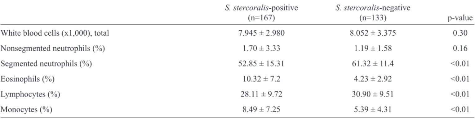
With respect to white blood cell counts, strongyloidiasis patients exhibited higher proportions of eosinophils (10.32%
± 7.2 vs. 4.23% ± 2.92; p-value < 0.01) and monocytes (8.49% ± 7.25 vs. 5.39% ± 4.31, p-value < 0.01) than
S. stercoralis-negative patients. In addition, S. stercoralis -positive patients presented with fewer segmented neutrophils
(52.85% ± 15.31 vs. 61.32% ± 11.4; p-value < 0.01) and lymphocytes (28.11% ± 9.72 versus 30.90% ± 9.51; p-value < 0.01) than S. stercoralis-negative patients (Table 2).
Severe outcomes due to hyperinfection with and the consequent dissemination of S. stercoralis have been associated with clinical conditions that require immunosuppressive therapy(10) (11) (12). In the present study, which did not include cases of disseminated strongyloidiasis, we compared the frequencies of S. stercoralis larvae elimination in patients with distinct clinical conditions. In this context, demonstrations of the presence of intestinal strongyloidiasis reinforce the need to perform parasitological examinations using appropriate techniques on patients who could potentially suffer from immunosuppressive conditions.
In this study, a signifi cant relationship between intestinal
TABLE 1 - Medical conditions associated with intestinal strongyloidiasis in Rio de Janeiro, Brazil.
OR Sex-adjusted
n % n % (95% CI) p-value OR (95% CI) p-value
Sex (male) 119 63.3 48 42.9 2.29 (1.42 - 3.70) <0.01 - -HIV/AIDS 40 24 25 18.8 1.36 (0.77 - 2.38) 0.17 1.19 (0.67 - 2.13) 0.53 HTLV infection 14 8.4 12 9.0 0.92 (0.41 - 2.06) 0.50 0.92 (0.41 -2.06) 0.84
Cardiovascular diseasesa 27 16.2 46 34.6 0.36 (0.21 - 0.62) <0.01 0.40 (0.23 - 0.70) <0.01
Diabetes 8 4.8 4 3.0 1.62 (0.47 - 5.51) 0.31 1.74 (0.42 - 5.12) 0.54
Obstructive respiratory diseasesb 11 6.6 8 6.0 1.10 (0.43- 2.82) 0.52 1.10 (0.42 -2.87) 0.84 Viral hepatitisc 11 6.6 3 2.3 3.05 (0.83 - 11.18) 0.06 3.36 (0.89-12.59) 0.07 Tuberculosisd 10 6.0 4 3.0 2.05 (0.62 - 6.70) 0.17 1.70 (0.51 - 5.65) 0.38 Cancere 17 9.9 10 5.2 1.39 (0.61 - 3.15) 0.28 1.22 (0.53 - 2.81) 0.63
Chronic renal disease 5 3.0 0 0.0 - NA - NA
Nutritional/metabolic disordersf 3 1.8 8 6.0 0.28 (0.07 - 1.09) 0.05 0.27 (0.06- 1.06) 0.06 Psychiatric conditionsg 3 1.8 6 4.5 0.38 (0.09 - 1.57) 0.15 0.52 (0.12- 2.22) 0.38 Rheumatic diseasesh 13 7.8 3 2.3 3.65 (1.02 - 13.11) 0.03 4.96 (1.34 - 18.37) 0.02 Dermatologic diseasesi 5 3.0 4 3.0 0.99 (0.26 - 3.78) 0.62 0.96 (0.24 - 3.74) 0.95
S.: Strongyloides; OR: odds ratio; 95%CI: 95% confi dence interval; HIV/AIDS: human immunodefi ciency virus/acquired immunodefi ciency syndrome; HTLV: human T lymphotropic virus type; NA: not applicable. a61 patients with arterial hypertension, 7 patients with angina pectoris, 4 patients with congestive heart failure, and 1 patient with myocardial infarction; b8 patients with asthma and 11 patients with chronic obstructive pulmonary disease; c6 patients with
hepatitis B and 8 patients with hepatitis C; d12 patients with pulmonary tuberculosis, 1 patient with bone tuberculosis, and 1 patient with lymph node tuberculosis; e5 patients with lymphoma, 1 patient with multiple myeloma, 1 patient with uterine cancer, 1 patient with colon cancer, 1 patient with esophageal cancer,
1 patient with stomach cancer, 2 patients with laryngeal cancer, 1 patient with pancreatic cancer, 3 patients with prostate cancer, and 11 patients with lung cancer; f5 patients with dyslipidemia, 3 patients with hypothyroidism, and 3 patients with obesity; g6 patients with depression, 2 patients with schizophrenia, and 1 patient with anxiety; h6 patients with systemic lupus erythematosus, 3 patients with rheumatoid arthritis, 3 patients with gout, 2 patients with scleroderma, 1 patient with Behcet disease, and 1 patient with Sjogren syndrome; i5 patients with psoriasis and 4 patients with eczema.
S. stercoralis- S. stercoralis -positive (n=167) negative (n=133)
TABLE 2 - White blood cells counts in patients with and without intestinal strongyloidiasis in Rio de Janeiro, Brazil.
S. stercoralis-positive S. stercoralis-negative
(n=167) (n=133) p-value
White blood cells (x1,000), total 7.945 ± 2.980 8.052 ± 3.375 0.30 Nonsegmented neutrophils (%) 1.70 ± 3.33 1.19 ± 1.58 0.16 Segmented neutrophils (%) 52.85 ± 15.31 61.32 ± 11.4 <0.01 Eosinophils (%) 10.32 ± 7.2 4.23 ± 2.92 <0.01 Lymphocytes (%) 28.11 ± 9.72 30.90 ± 9.51 <0.01 Monocytes (%) 8.49 ± 7.25 5.39 ± 4.31 <0.01
S.:Strongyloides.
the sensitivity of the strongyloidiasis detection technique. This phenomenon demonstrates the need to diagnose intestinal strongyloidiasis prior to the start of immunosuppressive and/ or corticoid therapy to avoid the dissemination of the parasite. In this study, we observed no association between intestinal strongyloidiasis and HTLV 1-2 infection. This association has been reported in Brazil by Furtado et al.(17). The synergistic effects of co-infection with HTLV and S. stercoralis, which have been
demonstrated, suggest that co-infected patients exhibit elevated parasite burdens and HTLV-1 proviral loads(18) (19) (20). With respect to the interaction between strongyloidiasis and acquired
immunodefi ciency syndrome (AIDS), our study indicated that
the frequency of HIV infection tended to be higher among S. stercoralis-positive patients than among S. stercoralis -negative patients but that this difference was not statistically
that HIV is a predictor of strongyloidiasis(21). S. stercoralis was the most frequently detected helminth among AIDS patients in Laos(22). In Brazil, a prior investigation found an elevated prevalence of strongyloidiasis during the pre- and post-highly active antiretroviral therapy eras and a greater frequency of strongyloidiasis among male patients than among female patients(23). Similarly, our study found that males were more frequently infected with S. stercoralis than females. More frequent exposure to infective forms of S. stercoralis in the environment has been postulated as the reason for the higher prevalence of strongyloidiasis among males than among females.
Although no association between infection by S. stercoralis and viral hepatitis has been detected, reports have indicated that antiviral therapy with ribavirin could trigger disseminated strongyloidiasis in parasitized patients(24). Our study found that the frequency of infection with hepatitis B and C viruses was slightly higher among patients with strongyloidiasis than in the S. stercoralis-negative group.
Strongyloides stercoralis infection is extremely signifi cant in patients with neoplasms because immunosuppressive chemotherapy can lead to severe forms of this parasitosis(25). Strongyloidiasis in patients who are undergoing chemotherapy should be appropriately treated with effective drugs. In this study, distinct malignancies were present in almost 10% of patients with strongyloidiasis; this rate was 5.2% among S. stercoralis-negative patients. These data reinforce the need to conduct appropriate stool examinations to detect S. stercoralis larvae as part of the routine care of cancer patients. In patients with chronic renal disease, the presence of strongyloidiasis is important due to the possible initiation of immunosuppressive therapy after renal transplantation. Thus, transplant patients should also be evaluated for infection by S. stercoralis(26). Although patients with chronic respiratory diseases typically receive steroids, we observed no association between this group of diseases and strongyloidiasis. Importantly, strongyloidiasis itself can be a cause of obstructive pulmonary symptoms(27).
In this study, we observed that mean relative eosinophil
counts were signifi cantly higher in the group of strongyloidiasis
patients than in the S. stercoralis-negative group. Given this
fi nding and the presence of elevated monocyte counts in the
strongyloidiasis group, it is unsurprising that lower relative neutrophil counts were detected in the strongyloidiasis group than in the S. stercoralis-negative group. The association between strongyloidiasis and eosinophilia has been well established, although the absence of this correlation has also been reported in the literature(28) (29).
We conclude that strongyloidiasis should be routinely investigated in patients hospitalized with complex conditions, irrespective of whether these patients require immunosuppressive therapy. This disease should be diagnosed using appropriate parasitological methods, such as the Baermann-Moraes technique.
REFERENCES
The authors declare that there is no confl ict of interest.
CONFLICT OF INTEREST
1. Neumann I, Ritter R, Mounsey A. Strongyloidesspp as a Cause of Fever of Unknown Origin. J Am Board Fam Med 2012; 25:390-393.
2. Paula FM, Castro E, Gonçalves-Pires MRF, Marçal MG, Campos DM, Costa-Cruz JM. Parasitological and immunological diagnoses of strongyloidiasis in immunocompromised and non-immunocompromised children at Uberlândia, State of Minas Gerais, Brazil. Rev Inst Med Trop Sao Paulo 2000; 42:51-55.
3. Valverde JG, Gomes-Silva A, De Carvalho Moreira CJ, De Souza DL, Jaeger LH, Martins P, et al. Prevalence and epidemiology of intestinal parasitism, as revealed by three distinct techniques in an endemic area in the Brazilian Amazon. Ann Trop Med Parasitol 2011; 105:413-424.
4. Carvalho-Costa FA, Gonçalves AQ, Lassance SL, Silva Neto LM, Salmazo CAA, Boia MN. Giardia lamblia and other intestinal parasitic infections and their relationships with nutritional status in children in brazilian amazon. Rev Inst Med Trop Sao Paulo 2007; 49:147-153.
5. De Bona S, Basso RMC. Hyperinfection by Strongyloides stercoralis associated with chronic use of corticosteroid. Rev Bras Anal Clin 2008; 40:247-250.
6. Andrade EC, Leite ICG, Rodrigues VO, Cesca MG. Parasitoses intestinais: uma revisão sobre os aspectos sociais, epidemiológicos, clínicos e terapêuticos. APS 2010; 13:231-240.
7. Ribeiro LC, Rodrigues-Junior ENA, Silva MD, Takiuchi A, Fontes CJF. Púrpura em paciente com estrongiloidíase disseminada.
Rev Soc Bras Med Trop 2005; 38:255-257.
8. Shorman M, Al-Tawfi q JA. Strongyloides stercoralis
hyperinfection presenting as acute respiratory failure and Gram-negative sepsis in a patient with astroytoma. International J Infect
Dis 2009; 13:288-291.
9. Zago-Gomes MP, Perazzio SF, Gonçalves CS, Pereira FEL. Prevalence of intestinal nematodes in alcooholic patients. Rev Soc Bras Med Trop 2002; 35:571-574.
10. Schär F, Trostdorf U, Giardina F, Khieu V, Muth S, Marti H, et al. Strongyloides stercoralis. Global Distribution and Risk
Factors. PLoS Negl Trop Dis 2013; 7:2288.
11. Paula FM, Costa-Cruz JM. Epidemiological aspects of
strongyloidiasis in Brazil. Parasitology 2011; 138:1331-1340.
12. Gonzalez-Ibarra F, Chevli P, Schachter L, Kaur M, Eivaz-Mohammadi S, Tashtoush B, et al. Strongyloidiasis and diffuse alveolar hemorrhage in a patient with systemic lupus erythematosus.
Case Rep Med 2014; ID 278390.
13. Rajadhyaksha A, Mehra S, Kawale J. Disseminated Strongyloides
in systemic lupus erythematosus and antiphospholipid antibody syndrome: a case report. Int J Rheum Dis 2012; 15:159-161.
14. Caramaschi P, Marocco S, Gobbo M, La Verde V, Volpe A, Bambara L, et al. Systemic lupus erythematosus and strongyloidiasis:
a multifaceted connection. Lupus 2010; 19:872-874.
15. Setoyama M, Fukumaru S, Takasaki T, Yoshida H, Kanzaki T. SLE com a morte por hemorragia pulmonar maciça aguda causada por estrongiloidíase disseminada. Scandinavian J Rheumatol
1997; 26:389-391.
16. Yung EE, Lee CM, Boys J, Chandrasoma PT. Strongyloidiasis hyperinfection in a patient with a history of systemic lupus
erythematosus. Am J Trop Med Hyg 2014; 91:806-809.
17. Furtado KC, Costa CA, Ferreira LS, Martins LC, Linhares AC, Ishikawa EA, et al. Occurrence of strongyloidiasis among patients with HTLV-1/2 seen at the outpatient clinic of the Núcleo de Medicina Tropical, Belém, State of Pará, Brazil. Rev Soc Bras Med Trop 2013; 46:241-243.
18. Satoh M, Toma H, Sato Y, Takara M, Shiroma Y, Kiyuna S,
et al. Reduced effi cacy of treatment of strongyloidiasis in HTLV-I
carriers related to enhanced expression of IFN-gamma and TGF-beta1. Clin Exp Immunol 2002; 127: 354-359.
19. Hirata T, Uchima N, Kishimoto K, Zaha O, Kinjo N, Hokama A, et al. Impairment of host immune response against strongyloides stercoralis by human T cell lymphotropic virus type 1 infection. Am J Trop Med Hyg 2006; 74:246-249.
20. Montes M, Sanchez C, Verdonck K, Lake JE, Gonzalez E, Lopez G, et al. Regulatory T cell expansion in HTLV-1 and strongyloidiasis co-infection is associated with reduced IL-5 responses to Strongyloides stercoralis antigen. PLoS Negl Trop Dis 2009; 3:456.
21. Jongwutiwes U, Waywa D, Silpasakorn S, Wanachiwanawin D, Suputtamongkol Y. Prevalence and risk factors of acquiring
Strongyloides stercoralis infection among patients attending
a tertiary hospital in Thailand. Pathog Glob Health 2014; 108:
137-140.
22. Paboriboune P, Phoumindr N, Borel E, Sourinphoumy K, Phaxayaseng S, Luangkhot E, et al. Intestinal parasitic infections in HIV-infected patients, Lao People's Democratic Republic. PLoS One 2014; 9:24.
23. Bachur TP, Vale JM, Coelho IC, Queiroz TR, Chaves CS. Enteric parasitic infections in HIV/AIDS patients before and after the
highly active antiretroviral therapy. Braz J Infect Dis 2008;
12:115-122.
24. Paraná R, Portugal M, Vitvitski L, Cotrim H, Lyra L, Trepo C.Severe strongyloidiasis during interferon plus ribavirin therapy for chronic HCV infection. Eur J Gastroenterol Hepatol 2000; 12:245-246. 25. Norsarwany M, Abdelrahman Z, Rahmah N, Ariffi n N, Norsyahida
A, Madihah B, et al. Symptomatic chronic strongyloidiasis in children following treatment for solid organ malignancies: case
reports and literature review. Trop Biomed 2012; 29:479-488.
26. Sadjadi SA, Damodaran C, Sharif M. Strongyloides stercoralis
infection in transplanted patients. Am J Case Rep 2013; 14: 205-209.
27. Khan WA, Santhanakrishnan K. Hypereosinophilic syndrome secondary to strongyloides infection: a case of recurrent asthma
exacerbations. BMJ Case Reports 2013; pii: bcr2013009587. 28. Salim N, Schindler T, Abdul U, Rothen J, Genton B, Lweno O, et al.
Enterobiasis and strongyloidiasis and associated co-infections and morbidity markers in infants, preschool-and school-aged children from rural coastal Tanzania: a cross-sectional study. BMC Infect Dis 2014; 14:644.