INTRODUCTION
Corresponding author: Ping Huang. Heart Center/Guangzhou Women and Children’s Medical Center, Guangzhou Medical University. Middle Ren Min Road 318, Yuexiu District, 510000 Guangzhou, China.
Phone: 86 20 8133-0675 e-mail: [email protected] Received 18 March 2015 Accepted 18 May 2015
Infl uenza infection and Kawasaki disease
Xijing Huang
[1], Ping Huang
[1], Li Zhang
[1], Xiaofei Xie
[1], Shuliang Xia
[1],
Fang Gong
[1],
Jia Yuan
[1]and Liling Jin
[1][1]. Heart Center, Guangzhou Women and Children's Medical Center, Guangzhou Medical University, Guangzhou, China.
ABSTRACT
Introduction: The objective of this study was to investigate the possible link between infl uenza (Flu) infection and Kawasaki
disease (KD). Methods: We examined the medical records of 1,053 KD cases and 4,669 infl uenza infection cases hospitalized at
our institute from January 1, 2011 to December 31, 2013. Cases of KD with concomitant infl uenza infection formed the KD + Flu group. Each KD + Flu case was matched with 2 KD cases and 2 infl uenza infection cases, and these cases were assigned to the KD group and Flu group, respectively. The differences in the principal clinical manifestations, course of disease, incomplete KD rate, intravenous immunoglobulin (IVIG) resistance rate, and echocardiographic detection results between the KD + Flu group and KD group were compared. The fever durations and laboratory test results of these three groups were compared. Results:
1) The seasonal variations of the KD + Flu group, KD group and Flu group were similar. 2) The morbidity rate of incomplete KD was higher in the KD + Flu group compared with the KD group. 3) Patients in the KD + Flu group exhibited a longer time to KD diagnosis compared with patients in the KD group. 4) The KD + Flu group exhibited the longest fever duration among the three groups. 5) The CRP and ESR values in the KD + Flu group were higher those in the Flu or KD groups. Conclusions:
Concomitant infl uenza infection affects the clinical manifestations of KD and can impact the laboratory test results and the diagnosis and treatment of the disease. However, it remains unclear whether infl uenza contributes to KD etiology.
Keywords: Kawasaki disease. Infl uenza. Infl uenza virus.
Kawasaki disease (KD) is a systemic vasculitis that predominantly affects infants and young children that are 6 months to 5 years old and has become the leading cause of acquired heart disease among children(1). Although many studies have been performed since the fi rst description of KD in 1967, the etiology of the disease remains unclear, and there is currently no specifi c laboratory diagnostic test for KD(2). Some researchers
have reported that the incidence of KD has increased in recent years(3) (4). A large number of epidemiological and clinical observations have indicated that infectious microorganisms might contribute to KD. Human adenovirus, mycoplasma, streptococci, retroviruses, Epstein-Barr virus, and parainfl uenza type 3 have been implicated in the etiology of KD(5) (6) (7) (8) (9) (10). In this present
study, we explored whether the infl uenza virus represents a candidate KD pathogen through a retrospective study.
METHODS
The medical records of 1,053 KD patients and 4,669 infl uenza infection patients that were hospitalized from January 1, 2011 to December 31, 2013 at our institute (Guangzhou Women and Children's Medical Center, Guangzhou, China) were collected. The diagnosis of KD was made according to the 2004 American Heart Association (AHA) guidelines. Patients with 2 to 3 principal clinical features of KD and unexplained fever for 5 days as well as abnormal laboratory values were classifi ed as exhibiting incomplete KD(11). Patients
with persistent or recrudescent fever in the 48 hours following intravenous immunoglobulin (IVIG) treatments were defi ned as IVIG-resistant cases(12).It should be noted that patients with
infl uenza only were not treated with IVIG.
Routine and biochemical detection
Kawasaki disease patients underwent blood tests for the following parameters on the fi rst day of hospitalization before receiving treatment: white blood cell (WBC), polymorphonuclear leukocytes (PMN), C-reactive protein (CRP), erythrocyte sedimentation rate (ESR), albumin (ALB), alanine aminotransferase (ALT), aspartate aminotransferase (AST), and creatine kinase isoenzyme (CK-MB).
Pathogen detection
RESULTS DISCUSSION
infl uenza A and B, adenovirus, respiratory syncytial virus, and Epstein Barr virus) by immunofl uorescence assay (IFA) and pharyngeal swab tests for the detection of infl uenza virus, adenovirus, respiratory syncytial virus and enteric viruses by fl uorescent polymerase chain reaction technology. Patients with diarrhea were tested for rotavirus by reverse transcription polymerase chain reaction (RT-PCR) and for Shigella using a coagglutination assay.
Grouping and observation index
KD + Flu group: KD patients positive for infl uenza A or/
and B virus but no other detectable pathogens comprised the KD + Flu group. Fifteen patients were classifi ed as KD + Flu cases, including 9 boys and 6 girls aged 2 months to 4 years with a mean age of 2 years.
KD group: each KD + Flu case was matched with 2
pathogen-negative KD patients with respect to age, gender, and date of hospitalization (± 3 weeks). The match cases included 18 boys and 12 girls aged 4 months to 4 years with a mean age of 1.9 years. These patients comprised the KD group.
Flu group: each KD + Flu case was matched with 2 patients
positive for infl uenza A or/and B virus but no other detectable pathogens with respect to age, gender, and date of hospitalization (± 3 weeks). The matched cases included 18 boys and 12 girls aged 5 months to 4 years with a mean age of 2 years. These patients comprised the Flu group.
The above three groups were included in our independent sample design. The age, sex and hospitalization time of two latter groups were matched with the KD + Flu group for comparability. Differences in the clinical manifestations, course of disease, incomplete KD rate, IVIG resistance rate, and coronary artery complications rate between the KD + Flu group and KD group as well as the fever duration and laboratory test results of these three groups were compared using statistical methods.
Descriptive analysis was performed using frequency distributions. Means, standard deviations, medians and inter-quartile ranges (IQ range) were used to summarize the patients’ demographic and baseline characteristics. Fisher’s exact test was used for qualitative variables as appropriate. The Kruskal-Wallis analysis was used for continuous va riables; when the null hypothesis was rejected, we use Bonferroni for multiple comparisons.
A 2-tailed p value ≤ 0.05 was considered signifi cant for comparisons among the three groups. A 2-tailed p value ≤ 0.0167% (Z > 2.93) was considered signifi cant for comparisons between any two of the three groups. The Statistical Package for the Social Sciences 19.0 (IBM SPSS 19.0) was used for data analysis.
Of the 1,053 total KD cases, 705 patients underwent tests for common respiratory pathogens. Of these, 34 cases were positive for infl uenza virus, including 23 infl uenza A infections and 11 infl uenza B infections. The rate of KD with concomitant
influenza was 4.8% (34/705). Among these 34 patients, 11 patients were only infected with infl uenza A virus, 4 patients were only infected with infl uenza B virus, and 19 patients were also infected other pathogens, including mycoplasma, chlamydia, adenovirus, respiratory syncytial virus and Epstein Barr virus. The seasonal variations of the three groups are presented in Figure 1, Figure 2 and Figure 3. As the fi gures
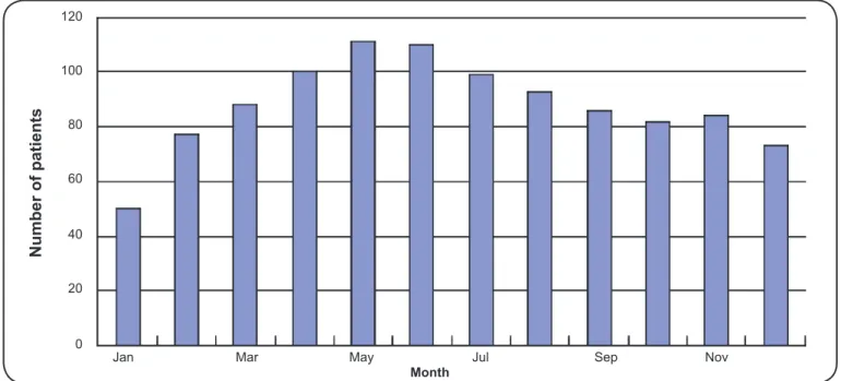
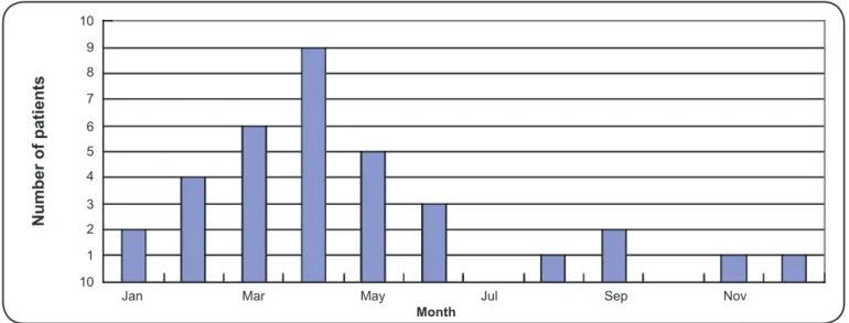
demonstrate, the following common trends were observed: 1) The number of patients steadily increased from January to April and then declined. 2) The number of patients observed in the spring and summer accounted for a larger proportion of the total cases. Moreover, when analyzing the number of patients each month using Pearson’s correlation analysis, we observed that the correlation between the KD group and the KD + Flu group was signifi cant (r = 0.92, p value < 0.01), which partly supports their relationships with respect to seasonal variation.
The clinical manifestations of infl uenza patients in the Flu group included fever, runny nose and stuffy nose. The principal clinical manifestations of the KD patients included fever, bilateral conjunctival injection, mucosal changes, polymorphous rash, changes in the extremities and cervical lymphadenopathy. Therefore, we were only able to compare the duration of fever between the three groups. The mean fever durations (SD) of the KD + Flu group, KD group, and Flu group were 9.13h (4.98), 6.4h (1.76) and 3.2h (1.21), respectively. The fever durations of the KD + Flu group and KD group were longer than that of the Flu group (p value < 0.01, p value < 0.01).
In addition to the infl uenza symptoms, patients in the KD + Flu group exhibited other changes in KD symptoms. We summarized and compared the clinical presentation, clinical course and outcomes of the KD + Flu group and KD group in
Table 1. The morbidity rate of incomplete KD was higher in the
KD + Flu group compared with the KD group (40% vs. 10%, p value = 0.04). Bilateral conjunctival injection was documented more frequently in the KD group than in the KD + Flu group (100% vs. 80%, p value = 0.03). Furthermore, the time required to diagnose patients in the KD + Flu group was longer than the time required to diagnose patients with KD alone (6 vs. 5d, p value =0.03). In summary, atypical symptoms led to an increased rate of incomplete KD, which resulted in a greater time to diagnosis. No statistically difference was observed regarding the occurrence of cardiovascular complications between these two groups.
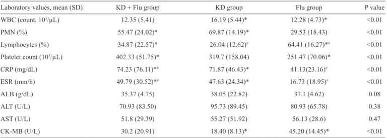
In addition to CK-MB, the majority of the laboratory values in the three groups summarized in Table 2 were abnormal. The numbers of cases with abnormal CK-MB values were 3, 0 and 12 in the KD + Flu, KD and Flu groups, respectively, indicating that CK-MB abnormalities were more common in the KD + Flu group and Flu group than in the KD group (p value < 0.05, p value <0.05, respectively).
120
100
80
60
40
20
0
Number of patients
FIGURE 1 -Monthly variation in KD incidence. From January to May, the number of patients with Kawasaki disease gradually increased; the incidence of Kawasaki disease declined slowly from June to December. The number of KD patients presenting in the spring and summer accounted more than 58% (610/1,053) of the patients seen year-long. KD: Kawasaki disease.
1,000
Jan Feb Mar Apr May Jun Jul Aug Sep Oct Nov Dec
Number of patients
800
600
400
200
0
FIGURE 2 -Monthly variation in infl uenza incidence. From January to May, the number of infl uenza cases gradually increased. The incidence of infl uenza declined signifi cantly from June to December and exhibited a small bump in September; the number of infl uenza patients presenting in spring and summer accounted for more than 65% (3,074/4,667) of the total cases throughout the year.
concerning infl uenza with concomitant KD, but the details about the impact of infl uenza on KD are limited(13). In the present
study, we used a case-controlled study design with the goal of providing more details regarding the role of infl uenza in KD.
The rationale for an infectious etiology is largely based on the epidemiologic features of KD(14). The seasonal variation
of KD patients indicates that an infectious pathogen might be relevant to the etiology of KD. In this study, we observed that the epidemiological features of KD and infl uenza were similar.
First, both KD and infl uenza affected children aged 6 months to 5 years. Second, like most infectious diseases, these diseases are self-limiting and exhibit similar clinical courses. Third, cases of KD with concomitant infl uenza tend to cluster temporally and have a predilection for spring and summer. As Figure 1, Figure 2 and Figure 3 demonstrate, the seasonal variation of the KD + Flu, KD and Flu groups are similar, suggesting that there might be a close relationship between KD and infl uenza.
Jan Mar May Jul Sep Nov
Month
10 9 8 7 6 5 4 3 2 1 10
Number of patients
Jan Mar May Jul Sep Nov
TABLE 1 -Demographics, clinical characteristics, and outcomes of the KD + Flu group and KD group.
KD + Flu group (n=15) KD group (n=30)
Variable n % n % P value
Clinical symptoms
bilateral conjunctival injection 12 80.0 30 100.0 0.03#
mucosal changes 13 86.7 29 96.7 0.25#
polymorphous rash 11 73.3 28 93.3 0.16#
changes of the extremities 9 60.0 22 73.3 0.50#
cervical lymphadenopathy 5 33.3 17 56.7 0.21#
Nonspecifi c symptoms
fl ush or desquamation around the anal skin 7 46.7 10 33.3 0.53#
erythema or induration in the bcg vaccination site 2 13.3 2 6.7 0.60#
Cardiovascular complications
coronary lesion 8 53.3 12 40.0 0.53#
pericardial effusion 0 0.0 5 16.7 0.15#
valvular regurgitation 0 0.0 2 6.7 0.55#
other non-cardiovascular complications 3 20.0 4 13.3 0.67#
Days required for diagnosis
median (IQ range) 6 (4.5-8) 5 (4-6) 0.03*
incomplete KD (n, %) 6 40.0 3 10.0 0.04#
IVIG resistance (n, %) 2 13.3 5 16.6 1.00#
KD: Kawasaki disease; Flu: infl uenza; IVIG: immunoglobulin; IQ range: inter-quartile range; *T- test; # Fisher’s exact test.
FIGURE 3 -Monthly variations in the incidence of concomitant KD and infl uenza. From January to April, the number of KD cases with concomitant infl uenza gradually increased, and the incidence declined signifi cantly from May to December. The number of KD cases with concurrent infl uenza that presented in the spring and summer accounted for more than 70.6% (24/34) of the cases throughout the year. KD: Kawasaki disease.
TABLE 2 - Laboratory values upon admission of the three groups.
Laboratory values, mean (SD) KD + Flu group KD group Flu group P value
WBC (count, 103/µL) 12.35 (5.41) 16.19 (5.44)* 12.28 (4.73)* <0.01
PMN (%) 55.47 (24.02)* 69.87 (14.19)* 29.53 (18.43) <0.01
Lymphocytes (%) 34.87 (22.57)* 26.04 (12.62)# 64.41 (16.27)*# <0.01
Platelet count (103/µL) 402.33 (51.75)* 319.7 (158.04) 251.47 (70.06)* <0.01
CRP (mg/dL) 74.23 (76.11)*# 71.87 (46.43)* 41.13(23.16)# <0.01
ESR (mm/h) 49.79 (30.52)*# 47.63 (24.34)* 16.73 (18.95)# <0.01
ALB (g/dL) 35.37 (4.75) 38.05 (22.82) 37.1 (4.62) 0.08
ALT (U/L) 70.93 (83.50) 95.73 (89.45) 80.93 (65.78) 0.38
AST (U/L) 51.8 (29.39) 55.27 (51.92) 56.13 (28.6) 0.47
CK-MB (U/L) 30.2 (20.91) 18.40 (8.13)* 45.20 (14.45)* <0.01
KD: Kawasaki disease; SD: standard deviation; Flu: infl uenza; WBC: white blood cell; PMN: polymorphonuclear leukocytes; CRP: C-reactive protein; ESR: erythrocyte sedimentation rate; ALB: albumin; ALT: alanine aminotransferase; AST: aspartate aminotransferase; CK-MB: creatine kinase isoenzyme; U/L: units per liter. The Kruskal-Wallis analysis was used for continuous variables; when the null hypothesis was rejected, we used the Bonferroni method for multiple comparisons. *or #indicate statistically signifi cant differences between the two groups (p ≤ 0.0167).
The infl uenza virus is a representative of the Orthomyxoviridae family and includes infl uenza viruses A, B and C. Mild and avirulent viruses infect only the throat and lungs because their hemagglutinin is only cleaved by proteases localized to the throat and lungs. Hemagglutinin can be cleaved by a wide variety of proteases in highly virulent strains, such as H5N1, allowing the virus to spread throughout the body. Infl uenza-infected cells produce large amounts of proinfl ammatory cytokines and chemokines, such as interferon and tumor necrosis factor(15). This massive immune response can cause a life-threatening cytokine storm. Others have suggested that tumor necrosis factor can contribute to the formation of coronary lesions and coronary aneurysms(16). These cytokines or infl ammatory mediators can damage the vascular endothelium, which aggravates or causes KD coronary artery complications. In our study, the CRP and ESR values in the KD + Flu group were higher than those in the Flu group and KD group, which demonstrated that the manifestations of KD with concomitant infl uenza were not entirely due to the KD infl ammatory response and that the infl uenza virus may play an important role in the total infl ammatory response. Furthermore, the fever duration in the KD + Flu group was longer than other groups, indicating that more serious infl ammation occurred in KD with concomitant infl uenza infection. Because of these factors, clinicians should be aware of the possibility of KD with concomitant infl uenza when a longer fever duration is observed in infl uenza or KD patients. Moreover, because patients in the KD + Flu group exhibited fewer principal clinical manifestations of KD, the high CRP and ESR may help diagnose incomplete KD.
Infl uenza can exacerbate a number of diseases, including cardiovascular disease and diabetes, and can lead to viral pneumonia, secondary bacterial pneumonia or other viral/ bacterial infections. Infl uenza is a more common cause of mortality in cardiovascular disease patients than in patients with other chronic diseases(17). Our results demonstrate that the CK-MB
values in the Flu group were higher than in the KD group, and CK-MB abnormalities were observed more frequently in the KD + Flu group and Flu group than in the KD group. The coronary lesion rate was higher in the KD + Flu group than in the KD group (53.3% vs. 40%). Based on the above evidence, we conclude that the infl uenza virus promotes cardiovascular damage in KD patients.
Previous studies have suggested that delayed therapy for KD or incomplete KD is associated with an increased risk of
coronary artery aneurysms(18) (19). In the present study, because
KD patients with concomitant infl uenza infection too longer to diagnose, the fever duration before IVIG was longer for patients in the KD + Flu group than in the KD group. Patients often exhibited insuffi cient criteria for KD diagnosis, such as bilateral conjunctival injection, which was documented less frequently in KD patients with infl uenza, and this lack of criteria may have delayed the diagnosis. Additionally, this lack of criteria may explain why incomplete KD was observed more often in the KD + Flu group. In our study, although the coronary lesion rate was higher in the fi rst group, we could not detect a signifi cant difference between the KD + Flu group and the KD group. These potential differences may require additional study to confi rm.
It is necessary to note that Reye’s syndrome can occur in children who take salicylates while they are experiencing active infection with varicella or infl uenza; this condition has been reported in patients taking high-dose aspirin for a prolonged
period of time after KD(20). Kawasaki disease patients in this
study were treated with aspirin, and the rate of Reye’s syndrome was zero, but vigilance is still necessary. Other antiplatelet drugs, such as clopidogrel or dipyridamole, avoid such risk, but their effects should be studied more thoroughly.
ACKNOWLEDGMENTS
REFERENCES
The authors declare that there is no confl ict of interest.
CONFLICT OF INTEREST
We would like to thank Guangzhou Medical College and Guangzhou Women and Children's Medical Center for providing support and guidance. The above opinions are those of the authors alone.
1. Bayers S, Shulman ST, Paller AS. Kawasaki disease: Part II. Complications and treatment. J Am Acad Dermatol 2013; 69:513. e1-513.e8.
2. Kawasaki T. Acute febrile mucocutaneous syndrome with
lymphoid involvement with specifi c desquamation of the fi ngers
and toes in children. Arerugi 1967; 16:178-222.
3. Park YW, Han JW, Park IS, Kim CH, Yun YS, Cha SH, et al.
Epidemiologic picture of Kawasaki disease in Korea, 2000-2002. Pediatr Int 2005; 47:382-387.
4. Du ZD, Zhao D, Du J, Zhang YL, Lin Y, Liu C, et al. Epidemiologic study on Kawasaki disease in Beijing from 2000 through 2004. Pediatr Infect Dis J 2007; 26:449-451.
5. Jaggi P, Kajon AE, Mejias A, Ramilo O, Leber A. Human
adenovirus infection in Kawasaki disease: a confounding bystander? Clin Infect Dis 2013; 56:58-64.
6. Leen C, Ling S. Mycoplasma infection and Kawasaki disease. Arch Dis Child 1996; 75:266-267.
7. Matsubara K, Fukaya T. The role of superantigens of group A
Streptococcus and Staphylococcus aureus in Kawasaki disease.
Curr Opin Infect Dis 2007; 20:298-303.
8. Nigro G, Midulla M. Retrovirus and Kawasaki disease. Lancet
1986; 2:1045.
9. Kikuta H, Matsumoto S, Osato T. Kawasaki Disease and
Epstein-Barr Virus. Acta Paediatr Jpn 1991; 33:765-770.
10. Moreira A, Leite I, Baptista A, Osório Ferreira E. Kawasaki
disease associated with parainfl uenza type 3 virus infection. Acta
Dermatovenerol Croat 2010; 18:120-123.
11. Newburger JW, Takahashi M, Gerber MA, Gewitz MH, Tani LY,
Burns JC, et al. Diagnosis, treatment, and long-term management
of Kawasaki disease: a statement for health professionals from the Committee on Rheumatic Fever, Endocarditis, and Kawasaki
Disease, Council on Cardiovascular Disease in the Young,
American Heart Association. Pediatrics 2004; 114:1708-1733.
12. Han RK, Sinclair B, Newman A, Silverman ED, Taylor GW, Walsh
P, et al. Recognition and management of Kawasaki disease. CMAJ
2000; 162:807-812.
13. Joshi AV, Jones KD, Buckley AM, Coren ME, Kampmann B.
Kawasaki disease coincident with infl uenza A H1N1/09 infection. Pediatr Int 2011; 53:e1-e2.
14. Bayers S, Shulman ST, Paller AS. Kawasaki disease: Part I.
Diagnosis, clinical features, and pathogenesis. J Am Acad Dermatol 2013; 69:501.e1-501.e11.
15. Schmitz N, Kurrer M, Bachmann MF, Kopf M. Interleukin-1 is responsible for acute lung immunopathology but increases survival
of respiratory infl uenza virus infection. J Virol 2005; 79:6441-6448.
16. Hui-Yuen JS, Duong TT, Yeung RS. TNF-α is necessary for
induction of coronary artery infl ammation and aneurysm formation in an animal model of Kawasaki disease. J Immunol 2006; 176:6294-6301.
17. Glezen WP, Decker M, Perrotta DM. Survey of underlying conditions of persons hospitalized with acute respiratory disease
during infl uenza epidemics in Houston, 1978-1981. Am Rev Respir Dis 1987; 136:550-555.
18. Tse SM, Silverman ED, McCrindle BW, Yeung RS. Early treatment
with intravenous immunoglobulin in patients with Kawasaki disease. J Pediatr 2002; 140:450-455.
19. Berdej-Szczot E, Firek-Pędras M, Szydłowski L,
Krzystolik-Ładzińska J, Klimek K, Małecka-Tendera E. Analysis of risk
factors and prospective evaluation of cardiovascular complications
of Kawasaki disease in children: a single center study. Kardiol Pol
2013; 71:1279-1286.
20. Takahashi M, Mason W, Thomas D, Sinatra F. Reye syndrome
following Kawasaki syndrome confi rmed by liver histopathology in Kawasaki Disease. Paper presented at: Proceedings of the
5th International Kawasaki Disease Symposium; May 22-25;