INTRODUCTION
Corresponding author: Dr. Henrique Silveira Costa. Rua dos Guajajaras 1172/407, Centro, 30180-100 Belo Horizonte, Minas Gerais, Brasil.
Phone: 55 31 3275-4205
e-mail: [email protected]
Received 3 December 2014
Accepted 1 April 2015
Exercise-induced ventricular arrhythmias and
vagal dysfunction in Chagas disease patients
with no apparent cardiac involvement
Henrique Silveira Costa
[1], Maria Carmo Pereira Nunes
[1], Aline Cristina de Souza
[1],
Marcia Maria Oliveira Lima
[2],
Renata Bicalho Carneiro
[1],
Giovane Rodrigo de Sousa
[1]and Manoel Otávio da Costa Rocha
[1][1]. Curso de Pós-Graduação em Ciências da Saúde: Infectologia e Medicina Tropical, Faculdade de Medicina, Universidade Federal de Minas Gerais, Belo Horizonte, Minas Gerais, Brasil. [2]. Departamento de Fisioterapia, Faculdade de Ciências da Saúde, Universidade Federal dos Vales do Jequitinhonha e Mucuri, Diamantina, Minas Gerais, Brasil.
ABSTRACT
Introduction: Exercise-induced ventricular arrhythmia (EIVA) and autonomic imbalance are considered as early markers of
heart disease in Chagas disease (ChD) patients. The objective of the present study was to verify the differences in the occurrence of EIVA and autonomic maneuver indexes between healthy individuals and ChD patients with no apparent cardiac involvement.
Methods: A total of 75 ChD patients with no apparent cardiac involvement, aged 44.7 (8.5) years, and 38 healthy individuals, aged
44.0 (9.2) years, were evaluated using echocardiography, symptom-limited treadmill exercise testing and autonomic function tests. Results: The occurrence of EIVA was higher in the chagasic group (48%) than in the control group (23.7%) during both the effort and the recovery phases. Frequent ventricular contractions occurred only in the patient group. Additionally, the respiratory sinus arrhythmia index was signifi cantly lower in the chagasic individuals compared with the control group. Conclusions: ChD patients with no apparent cardiac involvement had a higher frequency of EIVA as well as more vagal dysfunction by respiratory sinus arrhythmia. These results suggest that even when asymptomatic, ChD patients possess important arrhythmogenic substrates and subclinical disease.
Keywords: Chagas disease. Exercise-induced ventricular arrhythmias. Autonomic function. Treadmill exercise testing.
Chagas disease (ChD) is classifi ed as a neglected tropical disease(1) and continues to be an important cause of heart failure
in many countries in Latin America(2). Beyond the endemic
areas, ChD also represents a worldwide public health problem due to migration of infected people to developed countries, and mainly North America and Europe(3).
In the early stages, ChD patients have no symptoms or electrocardiographic and radiologic abnormalities(4). The
classifi cation with no apparent cardiac involvement has been used for asymptomatic, serologically Trypanosoma cruzi-positive patients with a normal electrocardiogram (ECG) and chest X-ray after a full or partial radiological study of the digestive tract. Because radiological studies of the digestive system of these patients have not been performed, these individuals cannot be classifi ed when they have the indeterminate form of the disease(5).
In any case, the medical literature has indicated an excellent 10-year prognosis for ChD patients without heart involvement(6),
although progressive myocardial damage may occur during the early stages(2) (7). Furthermore, sudden death, mainly resulting
from complications of advanced cardiac involvement, may be the fi rst manifestation of this disease(8), although rare structural
cardiac abnormalities, ventricular arrhythmias and autonomic disturbances may also play an important role(9).
Exercise testing is frequently used as a noninvasive assessment of myocardial ischemia that, along with patient history and physical examination, helps to characterize cardiovascular risk(10). In addition, exercise testing can be used
to identify cardiac arrhythmias, and particularly those triggered by exercise(11). Premature ventricular contractions (PVCs) are
the most frequent arrhythmias observed during exercise(12) and
may increase the risk of death through multiple mechanisms(13),
mainly because they may be markers of ischemic processes(14)
or arrhythmogenic substrates(15). In the setting of ChD,
RESULTS METHODS
Study design
This cross-sectional study was conducted at the Chagas Disease Outpatient Clinic and the Cardiology Service of the Hospital of the Federal University of Minas Gerais, Brazil, a tertiary ChD referral center.
The sample was stratified into two groups according to serology: a chagasic group and a control group. The criteria for inclusion in the chagasic group were the presence of two or more positive serological tests for T. cruzi, the absence of signifi cant
clinical symptoms suggestive of functional impairment due to ChD, a chest X-ray with a normal cardiac silhouette and conventional ECG within the normal limits. The control group included healthy individuals with similar age and gender distributions as the chagasic group. The criteria for exclusion for both groups included the presence of any condition that affected the individual’s ability to perform exercise testing and autonomic maneuvers.
A convenience sample was selected from the Chagas Disease Outpatient Clinic and the community. The selected individuals underwent clinical evaluation, echocardiography, and treadmill exercise and autonomic function tests.
Echocardiographic evaluation
A standard transthoracic two-dimensional (2D) echocardiogram was performed according to the recommendations of the American Society of Echocardiography(16) using a commercially available
echocardiograph (GE Vivid 7, Horten, Norway). Diastolic function was assessed as previously described(17). The left ventricular
ejection fraction (LVEF) was determined using Simpson’s rule.
Treadmill exercise test
All subjects performed a symptom-limited exercise test on a treadmill (Digistress Pulsar, Micromed, Brasilia, Brazil) using the standard Bruce protocol(12). Twelve-lead ECG was
continuously monitored, with data recorded every minute. The heart rate (HR) was determined from the ECG recording. Oxygen uptake (VO2) was estimated using a specifi c formula (VO2max (mL/kg/min) = 2.33 (time in min) + 9.48) to evaluate functional capacity. Chronotropic incompetence (CI) was defi ned as the failure to attain 80% of the HR reserve (HR reserve = age-predicted maximal HR - resting HR)(18).
At the recovery phase, after achieving the maximal workload, all patients spent 1 min in a cool-down period at a speed of 2.4km per hour and a grade of 2.5%(19). After 1 min,
all patients completed the recovery phase in the supine position. For the analysis of EIVA, the prevalence and the total number of PVCs during the exercise and recovery phases were considered. Frequent ventricular ectopy was defi ned as the presence of 7 or more ventricular premature beats per minute during any stage of the exercise test or at recovery(20).
Autonomic function tests
Autonomic function was assessed based on Valsalva maneuvers and respiratory sinus arrhythmia. The Valsalva maneuver was performed according to Oliveira et al.(21). In brief,
in the sitting position, the patient performed a valid maneuver by blowing, with the glottis closed, into a mouthpiece connected to an aneroid manometer by tubing in order to maintain an intraoral pressure of 40mmHg for 15s after a habitual inspiration. The maneuver was considered effective when facial plethora, neck vein distension and abdominal muscle contraction were observed.
The respiratory sinus arrhythmia test was performed with the patient in a sitting position while connected to a Hewlett-Packard electrocardiograph (1504 model) and wearing a nasal clip to avoid nasal respiratory loss(22). The ratio between the
longest expiratory RR interval and the shortest inspiratory RR interval (E:I ratio) was calculated for each respiratory cycle, as was the mean value for 6 cycles. The mean cardiac interval and the mean HR immediately before initiating the maneuver were obtained for the 10 RR intervals immediately preceding the beginning of the maneuver. All calculations were based on the original maximum and minimum cardiac intervals.
Statistical analysis
The sample size used was implemented to detect the prevalence of EIVA in ChD patients based on a previous study(23).
Considering an alpha error of 0.05, a statistical power of 95% was obtained for the sample of 78 chagasic patients.
The data were analyzed using Statistical Package for the Social Sciences (SPSS) version 17.0 (SPSS Inc., Chicago, IL, USA). The normal distribution of the data was verifi ed using the Kolmogorov-Smirnov test. A descriptive analysis yielded the mean and 95% confi dence interval (CI). Categorical variables are presented as an absolute number (percentage). A parametric unpaired t-test, Pearson’s correlation test, the nonparametric Mann-Whitney test and the Spearman rank correlation test were performed for data analysis, with the signifi cance level set at 0.05.
Ethical considerations
ThisstudywasapprovedbytheEthicsCommitteeof the FederalUniversityofMinasGerais,andallpatientsprovided writteninformedconsentbeforeparticipatinginthisstudy.
Characteristics of the sample
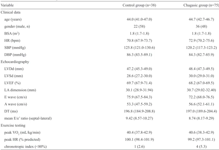
Clinical and demographic characteristics, echocardiographic parameters, functional capacity and chronotropic incompetence were similar between the two groups (Table 1).
In the analysis of myocardial contractility using 2D echocardiography, segmental changes in contractility were detected in fi ve patients. Apical akinesia (apical lesion) was found in two subjects, and hypokinesia of the basal segment of the inferior wall was identifi ed in three patients, one of whom also showed defi cits in contractility in the inferolateral wall. However, the overall contractility was preserved in all patients, with healthy individuals and ChD patients with no apparent cardiac
DISCUSSION
TABLE 1 -Characteristics of the patients.
Variable Control group (n=38) Chagasic group (n=75)
Clinical data
age (years) 44.0 (41.0-47.0) 44.7 (42.7-46.7)
gender (male, n) 22 (58) 36 (48)
BSA (m2) 1.8 (1.7-1.8) 1.8 (1.7-1.8)
HR (bpm) 70.8 (67.9-73.7) 72.9 (70.2-75.6)
SBP (mmHg) 125.8 (121.0-130.6) 120.2 (117.3-123.2)
DBP (mmHg) 86.3 (83.5-89.1) 84.3 (82.7-85.9)
Echocardiography
LVDd (mm) 47.2 (45.3-49.0) 48.4 (47.3-49.5)
LVSd (mm) 28.6 (27.2-30.0) 30.0 (29.0-31.0)
LVEF (%) 69.7 (67.9-71.4) 68.2 (67.0-69.5)
LA dimension (mm) 30.1 (28.9-31.94) 30.7 (29.02-32.40)
E wave (cm/s) 75.9 (67.5-84.3) 72.2 (68.0-76.5)
A wave (cm/s) 53.3 (47.5-59.2) 56.6 (52.1-61.1)
DT (ms) 196.8 (184.9-208.8) 197.0 (189.6-204.4)
mean E/e’ ratio (septal-lateral) 9.42 (8.57-10.27) 8.74 (8.17-9.29)
Exercise testing
peak VO2 (mL/kg/min) 40.4 (37.8-42.9) 40.6 (38.3-42.9) peak HR (% predicted) 100.1 (98.4-101.9) 99.2 (97.3-101.1)
chronotropic index (<80%) 1 (2.6) 4 (5.3)
The data are presented as the mean and 95% confi dence interval (95% CI) or number (percentage). BSA: body surface area; HR: heart rate; bpm: beats per minute; SBP: systolic blood pressure; DBP: diastolic blood pressure, LVDd: left ventricular end-diastolic diameter; LVSd: left ventricular end-systolic diameter; LVEF: left ventricular ejection fraction; LA: left atrium; E: early diastolic transmitral fl ow velocity; A: late transmitral fl ow velocity; DT: deceleration time; E/e’: ratio of the early diastolic transmitral fl ow velocity to the early diastolic mitral annular velocity; peak VO2: peak oxygen uptake; peak HR (% predicted):
percentage of the maximum heart rate achieved.
normal systolic function. Table 1 shows that there was no correlation between segmental changes in contractility and the number of PVCs during exercise testing (r=0.060; p-value=0.539).
Exercise-induced ventricular arrhythmias
The occurrence of PVCs was higher in the chagasic group: 36 (48%) patients, than in the control group: nine (23.7%) individuals, during both the effort and the recovery phases (p-value=0.010 for both). The total numbers of PVCs observed during treadmill exercise testing and at the recovery phase are shown in Figures 1 and 2. Frequent PVCs were observed only in the chagasic group, both during exercise testing: seven (9.3%) patients, and at the recovery phase: three (4%) patients. In contrast, there was no difference in the presence of ventricular tachycardia (VT) between the groups (p-value=0.587).
Assessment of autonomic function
In the assessment of autonomic function, there was no difference in the Valsalva ratio between the groups (p-value=0.855). Moreover, no correlation was found between
the Valsalva ratio and the number of PVCs during exercise or at the recovery phase (r=0.199; p-value=0.102 and r=0.085; p-value=0.487, respectively).
For respiratory sinus arrhythmia, the average ratio between the largest inspiratory and the smallest expiratory cardiac intervals was lower in ChD patients with no apparent cardiac involvement compared with the control group (p-value<0.001). No correlation was found between the respiratory sinus arrhythmia index and the number of PVCs during exercise or at the recovery phase (r=-0.107; p-value=0.381 and r=-0.085; p-value=0.400, respectively).
in ChD patients compared with healthy individuals and frequent ventricular arrhythmias observed only in ChD patients and 2) lower autonomic function, as assessed based on the respiratory sinus arrhythmia index, in patients compared with healthy individuals. These fi ndings support the concept that electrical abnormalities of the heart and autonomic imbalance may precede clinical manifestations in ChD patients(3) (24). We also suggest
that the higher occurrence of EIVA and vagal dysfunction may indicate the presence of subclinical cardiopathy in patients who are considered to be asymptomatic and to have a good prognosis.
59
T
otal number of PVCs during the effort
42 28 19 15 14 11 10 8 7 6 5 3 2 1 0
Control group (n=38) Chagasic group (n=75)
FIGURE 1 - Premature ventricular contractions during exercise testing in the control and chagasic patient groups.
PVCs:premature ventricular contractions.
26
17 10
8 7
6
5
4 3
2
1
0
T
otal number of PVCs at the recovery phas
e
Control group (n=38) Chagasic group (n=75)
FIGURE 2 -Premature ventricular contractions at the recovery phase of exercise testing in the control and chagasic patient groups. PVCs: premature ventricular contractions.
Exercise-induced ventricular arrhythmias in Chagas disease patients
Exercise testing is an important noninvasive prognostic tool that is easily performed, without expensive resources, and the results provide a wealth of information on the state of the autonomic nervous system(15). Moreover, a focused
review(11) showed that the majority of studies suggest that
EIVA is associated with increased cardiovascular morbidity or mortality, and the occurrence of EIVA during the recovery phase of exercise testing has proven to be an even stronger predictor. However, information concerning the prevalence and prognostic implications of EIVA in ChD patients is limited(24).
In a study(25) of 5,754 apparently healthy male subjects, aged
63.6 (10.2) years and without atrial fi brillation and digoxin treatment, the presence of EIVA was significantly higher in individuals with cardiovascular death than in survivors (p-value<0.001) after follow-up, suggesting that EIVA is an independent predictor of cardiovascular death (HR 1.6; 95% CI: 1.1-2.1; p-value=0.002). Furthermore, previous longitudinal studies(20) (26) have demonstrated the prognostic value of
frequent PVCs during exercise. Prakash et al.(26) specifi cally
found signifi cant differences in the percentage of patients with frequent PVCs or ventricular tachycardia between survivors and non-survivors (p-value<0.001). Similarly, Frolkis et al.(20) reported that frequent PVCs during exercise predicted a
higher likelihood of death (fi ve-year death rate, 9% vs 5% for patients without frequent PVCs during exercise; HR 1.8; 95% CI: 1.5-2.1; p-value<0.001).
De Paola et al.(27) attempted to verify the prevalence and
prognostic value of EIVA in 69 ChD patients, aged 46 (12) years, with chagasic cardiomyopathy. Ten (14%) patients had isolated PVCs, but VT was the only variable that signifi cantly infl uenced the occurrence of sudden cardiac death after a follow-up of 24 (15) years. Another study, which examined a prospective cohort(24) of 130 Chagas heart disease patients,
aged 50.7 (10.3) years, also verifi ed the prognostic value of EIVA during a follow-up of 9.9 years (132 days to 17 years). The prevalence of EIVA was 43.1% (95% CI: 34.5-51.7), and in patients with cardiomegaly, the hazard of dying was four times greater in the presence of EIVA (p-value=0.05). However, in the absence of cardiomegaly or low LVEF, the authors suggested that other methods should be considered. The present study encompassed patients with no apparent cardiac involvement, and the differences in the clinical presentation of the patients did not permit additional comparisons.
these patients, even when asymptomatic, possess important arrhythmogenic substrates and subclinical cardiopathy.
At the recovery phase of exercise testing, attenuated vagal reactivation might be associated with ventricular ectopy that is not suppressed(20). Recently, it was demonstrated that
noninvasive parameters are useful for detecting autonomic abnormalities in mild forms of ChD at the recovery phase of exercise testing(28). The present study highlighted a signifi cant
difference in the presence of EIVA both during exercise testing and at the recovery phase in ChD patients, with a higher frequency of ventricular arrhythmias than in healthy individuals.
Autonomic balance in Chagas disease patients
Abnormalities in autonomic function in ChD patients have previously been studied and associated with the destruction of parasympathetic ganglions in the heart, resulting from progressive infl ammation(29). Autonomic balance assessment using the Valsalva
maneuver has been widely used in chagasic patients(21) (30) (31) and
showed no difference between the groups examined in the present study. Similarly, a recent meta-analysis(32) demonstrated that only
one of the seven studies analyzed found a signifi cant difference in the Valsalva ratio between healthy individuals and chagasic patients without cardiopathy. The authors also reported that in certain studies that did not detect any signifi cant difference, other autonomic indexes (such as respiratory sinus arrhythmia) were able to recognize reduced heart vagal modulation in ChD patients without cardiopathy.
For respiratory sinus arrhythmia, our results showed a signifi cant difference between the groups, indicating that vagal dysfunction may occur in the absence of overt heart disease and before the development of left ventricular dysfunction(8). Thus,
left ventricular impairment might not necessarily be the cause of vagal dysfunction(33). Indeed, the reduced respiratory sinus
arrhythmia index found in ChD patients with or without left ventricular dysfunction may indicate a loss of synchronization between the HR and respiratory cycles, which may be related to impaired vagal modulation of the heart(34).
Previous studies(22) (34) have also shown reduced respiratory
sinus arrhythmia indexes in ChD patients compared with healthy individuals (p-value<0.001 and p-value=0.011, respectively), and there were no differences between groups with normal (>50%) and reduced (≤50%) LVEFs(34). In contrast, in the present
study, the main fi nding was the coexistence of vagal dysfunction and EIVA in ChD patients without apparent cardiac involvement. The presence of these early cardiac abnormalities suggests that there is not a clear-cut point that precisely separates individuals without apparent cardiac involvement/indeterminate disease from those with cardiac forms of ChD(21).
The respiratory sinus arrhythmia index is inversely associated with cardiovascular disease and seems to be a strong predictor of sudden cardiac death in cardiac patients (HR 7.4; 95% CI: 3.6-15.1; p-value=0.0001). However, the prognostic signifi cance of a reduced respiratory sinus arrhythmia index in chagasic patients without cardiac involvement needs to be further evaluated in longitudinal multicenter studies with a higher number of patients.
The authors declare that there is no confl ict of interest. CONFLICT OF INTEREST
1. World Health Organization ( WHO). Control of Chagas disease: Second report of the WHO Expert Committee. World Health
Organization 2002; 95:i109.
2. Nunes MC, Carmo AA, Rocha MOC, Ribeiro AL. Mortality prediction in Chagas heart disease. Expert Rev Cardiovasc Ther
2012; 10:1173-1184.
3. Ribeiro AL, Nunes MP, Teixeira MM, Rocha MOC. Diagnosis and management of Chagas disease and cardiomyopathy. Nat Rev
Cardiol 2012; 9:576-589.
4. Ribeiro AL, Rocha MOC. Indeterminate form of Chagas disease: considerations about diagnosis and prognosis. Rev Soc Bras Med
Trop 1998; 31:301-314.
5. Participantes da I Reunião de Pesquisa Aplicada em Doença de Chagas. Comunicação. Validade do conceito de forma indeterminada de doença de Chagas , Araxá, MG. Rev Soc Bras
Med Trop 1985; 18:46.
6. Ribeiro ALP. Disfunção autonômica e arritmia ventricular em chagásicos sem cardiopatia aparente. Rev Soc Bras Med Trop
1997; 30:257-258.
7. Nunes MC, Dones W, Morillo CA, Encina JJ, Ribeiro AL. Chagas disease: an overview of clinical and epidemiological aspects. J Am
Coll Cardiol 2013; 62:767-776.
8. Ribeiro AL, Moraes RS, Ribeiro JP, Ferlin EL, Torres RM, Oliveira E, et al. Parasympathetic dysautonomia precedes left ventricular systolic dysfunction in Chagas disease. Am Heart J
2001; 141:260-265.
9. Rossi MA, Bestetti RB. The challenge of chagasic cardiomyopathy. The pathologic roles of autonomic abnormalities, autoimmune mechanisms and microvascular changes, and therapeutic
implications. Cardiology 1995; 86:1-7.
10. Arena R, Myers J, Williams MA, Gulati M, Kligfi eld P, Balady GJ, et al. Assessment of functional capacity in clinical and
research settings: a scientifi c statement from the American
Heart Association Committee on Exercise, Rehabilitation, and Prevention of the Council on Clinical Cardiology and the Council
on Cardiovascular Nursing. Circulation 2007; 116:329-343.
11. Beckerman J, Froelicher VF. Exercise-test-induced arrhythmias:
a focused review. ACC Curr J Rev 2005; 14:41-44.
12. Fletcher GF, Balady GJ, Amsterdam EA, Chaitman B, Eckel R, Fleg J, et al. Exercise standards for testing and training: a statement for healthcare professionals from the American Heart Association.
Circulation 2001; 104:1694-1740.
13. Morshedi-Meibodi A, Evans JC, Levy D, Larson MG, Vasan RS.
Clinical correlates and prognostic signifi cance of exercise-induced
ventricular premature beats in the community: the Framingham
Heart Study. Circulation 2004; 109:2417-2422.
14. Elhendy A, Chandrasekaran K, Gersh BJ, Mahoney D, Burger KN,
Pellikka PA. Functional and prognostic signifi cance of
exercise-induced ventricular arrhythmias in patients with suspected
coronary artery disease. Am J Cardiol 2002; 90:95-100.
REFERENCES
15. Jouven X, Ducimetiere P. Exercise testing: do frequent premature ventricular depolarizations represent a new criterion of positivity?
Eur Heart J 2001; 22:1759-1761.
16. Lang RM, Bierig M, Devereux RB, Flachskampf FA, Foster E,
Pellikka PA, et al. Recommendations for chamber quantifi cation: a
report from the American Society of Echocardiography's Guidelines
and Standards Committee and the Chamber Quantifi cation Writing
Group, developed in conjunction with the European Association of Echocardiography, a branch of the European Society of Cardiology.
J Am Soc Echocardiogr 2005; 18:1440-1463.
17. Nagueh SF, Appleton CP, Gillebert TC, Marino PN, Oh JK, Smiseth OA, et al. Recommendations for the evaluation of left ventricular
diastolic function by echocardiography. Eur J Echocardiogr 2009;
10:165-193.
18. Azarbal B, Hayes SW, Lewin HC, Hachamovitch R, Cohen I, Berman DS. The incremental prognostic value of percentage of heart rate reserve achieved over myocardial perfusion single-photon emission computed tomography in the prediction of cardiac death and all-cause mortality: superiority over 85% of maximal
age-predicted heart rate. J Am Coll Cardiol 2004; 44:423-430.
19. Cole CR, Blackstone EH, Pashkow FJ, Snader CE, Lauer MS. Heart-rate recovery immediately after exercise as a predictor of
mortality. N Engl J Med 1999; 341:1351-1357.
20. Frolkis JP, Pothier CE, Blackstone EH, Lauer MS. Frequent ventricular ectopy after exercise as a predictor of death. N Engl J
Med 2003; 348:781-790.
21. Oliveira E, Ribeiro AL, Assis Silva F, Torres RM, Rocha MOC. The Valsalva maneuver in Chagas disease patients without
cardiopathy. Int J Cardiol 2002; 82:49-54.
22. Ribeiro AL, Ferreira LM, Oliveira E, Cruzeiro PC, Torres RM, Rocha MOC. Active orthostatic stress and respiratory sinus arrhythmia in patients with Chagas' disease with preserved left ventricular global
systolic function. Arq Bras Cardiol 2004; 83:40-44.
23. Pereira MH, Brito FS, Ambrose JA, Pereira CB, Levi GC, Neto VA, et al. Exercise testing in the latent phase of Chagas' disease.
Clin Cardiol 1984; 7:261-265.
24. Pedrosa RC, Salles JH, Magnanini MM, Bezerra DC, Bloch KV. Prognostic value of exercise-induced ventricular arrhythmia in
Chagas' heart disease. Pacing Clin Electrophysiol 2011; 34:1492-1497.
25. Beckerman J, Mathur A, Stahr S, Myers J, Chun S, Froelicher V. Exercise-induced ventricular arrhythmias and cardiovascular death. Ann Noninvasive Electrocardiol 2005; 10:47-52.
26. Prakash M, Myers J, Froelicher VF, Marcus R, Do D, Kalisetti D, et al. Clinical and exercise test predictors of all-cause mortality: results from > 6,000 consecutive referred male patients. Chest
2001; 120:1003-1013.
27. de Paola AA, Gomes JA, Terzian AB, Miyamoto MH, Martinez Fo EE. Ventricular tachycardia during exercise testing as a predictor of sudden death in patients with chronic chagasic cardiomyopathy
and ventricular arrhythmias. Br Heart J 1995; 74:293-295.
28. Alencar MC, Rocha MOC, Lima MM, Costa HS, Sousa GR, Carneiro RC, et al. Heart rate recovery in asymptomatic patients
with chagas disease. PLoS One 2014; 9:e100753.
29. Junqueira Junior LF, Beraldo PS, Chapadeiro E, Jesus PC. Cardiac autonomic dysfunction and neuroganglionitis in a rat model of
chronic Chagas' disease. Cardiovasc Res 1992; 26:324-329.
30. Odreman RO, Davila DF, Donis JH, Torres A, Ferrer J, Inglessis I. Valsalva maneuver in chagasic patients with documented past
medical history of acute chagasic myocarditis. Int J Cardiol 2004;
93:163-167.
31. Villar JC, Leon H, Morillo CA. Cardiovascular autonomic function testing in asymptomatic T. cruzi carriers: a sensitive method to
identify subclinical Chagas' disease. Int J Cardiol 2004; 93:189-195.
32. Ribeiro AL, Campos MS, Baptista LM, Sousa MR. The Valsalva maneuver in Chagas disease patients without cardiopathy.
Clin Auton Res 2010; 20:79-83.
33. Raadschilders L, Rocha MOC, Sousa L, Nouwen J, Ribeiro AL. Is autonomic function associated with left ventricular systolic function in Chagas heart disease patients undergoing treatment
for heart failure? Rev Soc Bras Med Trop 2014; 47:239-242.
34. Neves VR, Peltola M, Huikuri H, Rocha MOC, Ribeiro AL. Respiratory sinus arrhythmia in Chagas disease. Auton Neurosci