Epilepsy Research (2008)78, 240—243
j o u r n a l h o m e p a g e : w w w . e l s e v i e r . c o m / l o c a t e / e p i l e p s y r e s
SHORT COMMUNICATION
Ictal SPECT in Sturge-Weber syndrome
¨
Ozg¨
ur Bilgin
a,∗, Christian Vollmar
a, Aurelia Peraud
b,
Christian la Fougere
c, Pedro Beleza
a, Soheyl Noachtar
aaUniversity of Munich, Klinikum Grosshadern; Department of Neurology, Germany bUniversity of Munich, Klinikum Grosshadern; Department of Neurosurgery, Germany cUniversity of Munich, Klinikum Grosshadern; Department of Nuclear Medicine, Germany
Received 24 September 2007; received in revised form 31 October 2007; accepted 7 December 2007
KEYWORDS
Sturge-Weber syndrome; Functional hemispherectomy; Ictal SPECT
Summary We report on a patient with right-sided Sturge-Weber syndrome (SWS), in whom earlier functional hemispherectomy failed. Subtraction of ictal and interictal single-photon-emission-computed-tomography (SPECT) superimposed on individual MRI showed a right fronto-orbital hyperperfusion, with a left-sided EEG seizure pattern. Ictal SPECT supported our assumption that right frontal originated seizure pattern propagated to left hemisphere via the remaining right frontal bridge. Right orbito-frontal resection and disconnection from corpus callosum resulted in seizure freedom.
© 2007 Elsevier B.V. All rights reserved.
Sturge-Weber syndrome (SWS) is a sporadically occurring phakomatosis (Juhasz et al., 2007), which frequently leads to epilepsy, hemiparesis, and learning disability in associ-ation with a pial angioma (Aylett et al., 1999). Epilepsy affects most of these patients who may be good can-didates for hemispherectomy if anti-epileptic medication does not control the seizures (Kossoff et al., 2002). Their presurgical evaluation includes non-invasive EEG-video mon-itoring and imaging studies. Ictal perfusion measured by single-photon-emission-computed-tomography (SPECT), particularly if subtracted from interictal SPECT and super-imposed on MRI, can provide additional information in presurgical evaluation of patients with refractory extratem-poral SWS (O’Brien et al., 2004).
∗Corresponding author at: Epilepsy Center, Department of Neu-rology, University of Munich, Marchioninistr. 15, D-81377 Munich, Germany. Tel.: +49 89 7095 5680; fax: +49 89 7095 5686.
E-mail address:[email protected](¨O. Bilgin).
Case report
A 6-year-old boy had frequent daily bilateral asymmetric tonic seizures since he was 8 months old. He had been diag-nosed to have right-sided SWS. He had flaccid hemiparesis on left side more than on the right, and was not able to stand or walk. He was mentally retarded and unable to speak. As he was resistant to multiple anti-epileptic drugs (AED), epilepsy surgery was performed in 2003. Temporal and central lobectomy and frontal and occipital hemi-spherotomy were performed on the right side. He was then seizure-free for 2 years, but the seizures started again in 2005.
When the patient was admitted to our Epilepsy Monitor-ing Unit of the University of Munich Epilepsy Center in 2006 he had multiple daily motor seizures for 6 months. Phenobar-bital treatment failed to control the seizures which occurred approximately 3—5 times a day. The seizures started with vocalization followed by left tonic and bilateral hypermotor seizures.
Ictal SPECT in Sturge-Weber syndrome 241
Figure 1 The interictal EEG shows right fronto-central and parietal sharp waves, continuous slowing, and a reduced background activity in the right hemisphere.
High-resolution magnetic resonance imaging (MRI; 3.0T, GE signa HDx,USA) showed the previous partial hemispherec-tomy and an incomplete hemispherohemispherec-tomy leaving a right frontal bridge to the corpus callosum. A 64-channel-EEG video evaluation with scalp electrodes (10—10 system) was performed. Interictal EEG showed right fronto-central and parietal sharp waves, and continuous slowing, and reduced background activity in the right hemisphere (Fig. 1). Ictal EEG, however, consistently demonstrated left hemisphere seizure patterns which occurred typically about 15 s after the clinical seizure onset (Fig. 2). For further evaluation ictal and interictal brain perfusion single photon emission tomography with a technetium-99m-labeled ethylcysteinate dimer (99mTc-ECD, Neurolite; BMS Pharma, Brussels,
Bel-gium) was performed on a triple-head gamma camera
(Multispect 3; Siemens, Erlangen, Germany) equipped with high-resolution collimators. For ictal SPECT, the patient was injected with radioligand 16 s after the clinical seizure onset. The projection images were reconstructed by filtered backprojection and filtered by a 3D Butterworth low fil-ter. For uniform attenuation correction, Chang’s first-order method was used. For further evaluation, the SPECT data were transferred to a workstation running BRASS software (Hermes/Nuclear Diagnostics, Sweden), which allows reg-istration of a patient’s study on a 3D MRI-based reference template of anatomical ROIs and an identical coregistration of the consecutive interictal study. Subtraction of ictal and interictal SPECT superimposed on the patient’s MRI demon-strated hyperperfusion in the right fronto-orbital cortex (Fig. 3). As it is assumed that the ictal EEG pattern reflects
242 O. Bilgin et al.¨
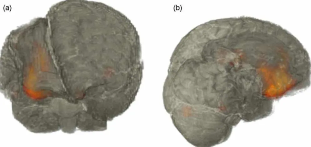
Figure 3 (a and b) Superimposed subtraction of ictal and interictal ECD-SPECT perfusion on the 3D reconstructed MRI showing right orbito-frontal and lateral frontal ictal hyperperfusion.
a seizure that has spread to the normal left hemisphere via the right frontal lobe bridge, we performed a partial right orbito-frontal resection, thus disconnecting the frontal lobe from the corpus callosum. Following this second surgery, the patient has been seizure-free for 14 months now. He has developed both physically and neuropsychologically, and is able to stand and speak a few single words.
Discussion
Our patient demonstrates that even if previous epilepsy surgery, for example, combined hemispherectomy and hemi-spherotomy has failed in SWS patients, they may benefit from repeated surgery if the seizure onset zone can be local-ized. Epilepsy surgery should be considered in these patients if high seizure frequency and developmental deterioration do not respond to AED treatment. Anatomical hemispherec-tomy has an excellent outcome but was formerly reported to be associated with chronic complications such as super-ficial cerebral hemosiderosis (SCH) (Falconer and Wilson, 1969). Therefore, surgical techniques have been introduced to reduce complications, leading to partial hemispherec-tomy and hemispherohemispherec-tomy (Schramm and Behrens, 1997). Functional hemispherectomy has been developed as a less invasive technique (cerebral hemispherotomy), in which cortical resection is minimized and the rest of the affected hemisphere is functionally isolated by transecting its projec-tion and commissural fibers at the corpus callosum (Morino et al., 2007). The purpose of surgery is to disconnect fibers between seizure foci over wide areas of one cerebral hemi-sphere and thus prevent propagation of epileptic discharges to the non-affected hemisphere (Morino et al., 2007).
Although partial hemispherectomy and hemispherotomy are established treatments for medically refractory epilepsy due to diffuse hemisphere disease, the first surgery failed in our patient. MRI showed that a bridge of the right frontal lobe to the corpus callosum remained. Ictal SPECT super-imposed on the MRI proved the localization of the seizure onset zone, thus establishing the functional significance of the frontal bridge. The delayed appearance of the ictal EEG seizure pattern, which followed the clinical seizure
onset was falsely lateralized and supports the assumption of propagation of the epileptic activity to the non-affected hemisphere.
Ictal perfusion SPECT is an important imaging tool in presurgical evaluation (Duncan, 1997; Van Paesschen et al., 2007). It is believed that the region with the most intense hyperperfusion reflects the ictal onset zone (Van Paesschen et al., 2007). However, if there is a poor time resolution, seizure propagation may cause false localization of the epileptogenic zone, particularly with late injec-tions in extratemporal epilepsy (Noachtar et al., 1998). The ictal SPECT and ictal EEG findings disagreed in our patient. Whereas ictal SPECT localized the epileptogenic zone, ictal EEG reflected the propagation of the seizure to the non-affected left hemisphere. Our failure to record an EEG seizure pattern in the right hemisphere may be related to the diffuse right hemisphere pathology and former par-tial hemispherectomy. We speculated that the seizure did not involve enough volume of cortex to produce a seizure pattern, which could be recorded on the surface of the abnormal hemisphere, despite the prior craniotomy. Such a seizure pattern has been described in selected patients with large lesions (Sammaritano et al., 1987). Only when it spread to the non-affected hemisphere was it then reflected in the non-invasive EEG. On the basis of the results of this evaluation the patient underwent surgery to remove the orbito-frontal epileptogenic zone and the residual bridge of the right frontal lobe to the corpus callosum. Unfor-tunately, in some patients functional hemispheretomy and hemispherotomy fail to disconnect the hemisphere. Recent studies showed that with modern diagnostics and surgi-cal techniques anatomisurgi-cal hemispherectomy is associated with less morbidity than previously reported (O’Brien et al., 2006; McClelland and Maxwell, 2007). Thus, anatomi-cal hemispherectomy might still be an option for patients with large hemispheric lesions.
The patient has now been seizure-free for 14 months on follow-up.
Ictal SPECT in Sturge-Weber syndrome 243
to the corpus callosum. It also revealed the limitations of non-invasive ictal EEG in diffuse hemisphere pathology of SWS.
Conflicts of interest statement
The authors report no conflicts of interest.
References
Aylett, S.E., Neville, B.G., Cross, J.H., Boyd, S., Chong, W.K., Kirkham, F.J., 1999. Sturge-Weber syndrome: cerebral haemo-dynamics during seizure activity. Dev. Med. Child Neurol. 41, 480—485.
Duncan, J.S., 1997. Imaging and epilepsy. Brain 120, 339—377. Falconer, M.A., Wilson, P.J.E., 1969. Complications related to
delayed hemorrhage after hemispherectomy. J. Neurosurg. 30, 413—426.
Juhasz, C., Batista, C.E., Chugani, D.C., Muzik, O., Chugani, H.T., 2007. Evolution of cortical metabolic abnormalities and their clinical correlates in Sturge-Weber syndrome. Eur. J. Paediatr. Neurol..
Kossoff, E.H., Buck, C., Freeman, J.M., 2002. Outcomes of 32 hemispherectomies for Sturge-Weber syndrome worldwide. Neu-rology 59, 1735—1738.
McClelland 3rd, S., Maxwell, R.E., 2007. Hemispherectomy for intractable epilepsy in adults: the first reported series. Ann. Neurol. 61, 372—376.
Morino, M., Shimizu, H., Uda, T., Naitoh, K., Kawahara, S., Ishig-uro, T., Gotoh, T., Ohata, K., Hara, M., 2007. Transventricular hemispherotomy for surgical treatment of intractable epilepsy. J. Clin. Neurosci. 14, 171—175.
Noachtar, S., Arnold, S., Yousry, T.A., Bartenstein, P., Werhahn, K.J., Tatsch, K., 1998. Ictal technetium-99m ethyl cysteinate dimer single-photon emission tomographic findings and propagation of epileptic seizure activity in patients with extratemporal epilep-sies. Eur. J. Nucl. Med. 25, 166—172.
O’Brien, T.J., So, E.L., Cascino, G.D., Hauser, M.F., Marsh, W.R., Meyer, F.B., Sharbrough, F.W., Mullan, B.P., 2004. Sub-traction SPECT coregistered to MRI in focal malformations of cortical development: localization of the epileptogenic zone in epilepsy surgery candidates. Epilepsia 45, 367— 376.
O’Brien, F.D., Basu, S., Williams, D.H., May, P.L., 2006. Anatomi-cal hemispherectomy for intractable seizures: excellent seizure control, low morbidity and no superficial cerebral haemosidero-sis. Childs Nerv. Syst. 22, 489—498.
Sammaritano, M., de Lotbiniere, A., Andermann, F., Olivier, A., Gloor, P., Quesney, L.F., 1987. False lateralization by sur-face EEG of seizure onset in patients with temporal lobe epilepsy and gross focal cerebral lesions. Ann. Neurol. 21, 361— 369.
Schramm, J., Behrens, E., 1997. Functional hemispherectomy. J. Neurosurg. 87, 801—802.