Epidural cystic masses associated with interspinous
bursitis, synovial and discal cysts*
Formações císticas epidurais relacionadas a bursite interespinhosa, cisto sinovial e cisto discal
Frederico Guilherme de Paula Lopes Santos1, Ricardo André de Souza1, Marcos Pama D’Almeida Brotto1, Fábio Massaaki Suguita1, Denise Tokechi Amaral2, Lázaro Luís Faria do Amaral3
The authors describe some cases of epidural cysts, namely synovial, discal, ligamentum flavum cysts, and cysts secondary to interspinous bursitis, all of these conditions determining radicular, dural sac compression or spinal canal stenosis. Magnetic resonance imaging findings and localization of these entities are described.
Keywords: Magnetic resonance imaging; Spinal cord compression; Spinal cord disease.
Os autores apresentam casos de cistos epidurais, dentre eles os cistos sinoviais, discais, do ligamento amarelo e relacionados a bursite interespinhosa, todas essas condições determinando compressão radicular, do saco dural ou estenose do canal vertebral. Descrevem as características de imagem e localização na ressonância magnética nessas diferentes afecções.
Unitermos: Imagem por ressonância magnética; Compressão medular; Afecção medular. Abstract
Resumo
* Study developed at MedImagem – Hospital Beneficência Portuguesa de São Paulo, São Paulo, SP, Brazil.
1. MDs, Residents at MedImagem – Hospital Beneficência Portuguesa de São Paulo, São Paulo, SP, Brazil.
2. PhD, MD, Radiologist at MedImagem – Hospital Beneficên-cia Portuguesa de São Paulo, São Paulo, SP, Brazil.
3. Specialist in Neuroradiology, MD, Radiologist at MedIma-gem – Hospital Beneficência Portuguesa de São Paulo, São Paulo, SP, Brazil.
Mailing address: Dr. Frederico Guilherme de Paula Lopes San-tos. Rua dos Ingleses, 586, ap. 102, Morro dos Ingleses. São Paulo, SP, Brazil, 01329-000. E-mail: [email protected]
Received November 6, 2008. Accepted after revision Febru-ary 9, 2009.
the connection with the epidural cyst, this method allows the pain management with corticoid infiltration(2).
CYST OF LIGAMENTUM FLAVUM
Cysts of ligamentum flavum are rare en-tities and, like synovial cysts and cysts of the posterior longitudinal ligament, are classified as juxtafacet cysts. Because of the absence of synovial membrane on their walls, they are considered as pseudocysts(3), resulting from degenerative alterations and spondylolisthesis, arising from the anterior margin of the ligamentum flavum(4). Ante-rior projection of the lesion may determine dural sac compression or vertebral canal stenosis, resulting in sciatic pain(5).
At MRI, the mass is seen with hypoin-tense signal on T1-weighted, and hyperin-tense signal on T2-weighted sequences, with peripheral contrast-enhancement, and usually located in the midline. These le-sions should be differentiated from syn-ovial cysts by the topographic relation with the interapophyseal joint(6).
SYNOVIAL CYSTS
These cysts may develop from any joint lined by synovial membrane or tendinous sheath, and are rarely seen on the vertebral
Santos FGPL, Souza RA, Brotto MPD, Suguita FM, Amaral DT, Amaral LLF. Epidural cystic masses associated with interspi-nous bursitis, synovial and discal cysts. Radiol Bras. 2009;42(2):131–135.
Epidural cystic masses represent one of the causes for vertebral canal stenosis or lumbar sciatic pain. Although not fre-quently found, these lesions should be dif-ferentiated from neurogenic tumors and disc herniation.
The present pictorial essay discusses imaging findings and describes the charac-teristics of an array of extradural cystic le-sions with the utilization of MRI.
CYSTS RELATED
TO INTERSPINOUS BURSITIS
Autopsy studies demonstrate an in-crease in the incidence of cysts secondary to bursitis between lumbar spinous pro-cesses in elder individuals. Symptoms are characterized by localized pain in the lower portion of the lumbar spine exacerbated by hyperextension. In young athletes, these symptoms may mimic spondylolysis.
MRI is useful in the detection of thick-ening and hyperintense signal of inters-pinous and suprasinters-pinous ligaments, as well as the formation of adventitial bursae sec-ondary to attrition. Epidural cysts present the same characteristics of synovial cysts, but are localized in the midline. Radios-copy- or computed tomography (CT)-guided bursography can be utilized for di-agnosis confirmation. Besides determining
INTRODUCTION
The differentiation between pathologi-cal intramedullary conditions (originated in the spinal cord) and extramedullary lesions (external to the thecal sac [extradural] or restricted to the thecal sac [intradural]) is generally simple. Although many lesions can develop simultaneously in more than one compartment, their precise positioning in relation to the medullary canal and the evaluation of the signal characteristics at magnetic resonance imaging (MRI) may be useful for the diagnosis.
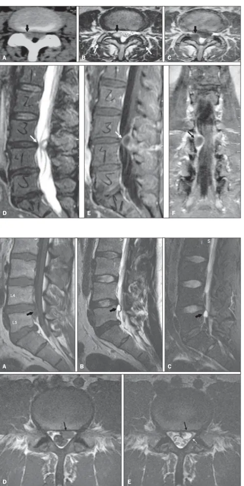
Figure 2. Interspinous bursitis associated with epi-dural cyst. Female, 53-year-old patient with intense, bilateral lumbar pain and claudication. Sagittal MRI demonstrates thickening and fluid in the interspinous ligament at the L4/L5 level (white arrows), associ-ated with the epidural cystic mass (black arrows) with intermediate signal intensity on T1-weighted se-quence (A), hyperintense signal on T2-weighted sequence (B) and peripheral contrast-enhancement (C). On axial T2-weighted image (D) and contrast-enhancedT1-weighted images (E) an epidural cys-tic mass can be observed in the midline (arrows) with marked dural sac compression.
A B C
D E
Figure 1. Interspinous bursitis at the L4/L5 level with epidural cyst. Female, 42-year-old patient with sud-den perineal paresthesia. Sagittal, T1-weighted MRI (A), T2-weighted sequence (B) and contrast-en-hanced T1-weighted sequence with fat saturation (C). The interspinous ligament presents edema and contras-enhancement (white arrows). An epidural cystic mass is observed with intermediate signal intensity on T1-weighted sequence, hyperintense signal on T2-weighted sequence, and peripheral contrast enhancement (black arrows) with anterior displacement of the dural sac. Epidural cyst in the midline (D,E – arrows) characterized by hyperintense signal on T2-weighted (D) and peripheral contras-enhancement (E) determining marked dural sac compression. Interapophyseal arthrosis, ligamentum flavum hypertrophy, and concentric and diffuse discal protrusion at the L4/L5 level with consequential vertebral canal stenosis can also be observed.
A B C
D E
spine, the L4-L5 transition being the pref-erential site of these lesions. There is a subtle predominance of these lesions in women, rarely appearing before the age of 30(7).
Synovial cysts are associated with osteoarthrosis, spondylolisthesis and ab-normal displacement of interapophyseal
joints. These lesions result from a hernia-tion of the joint tissue through a defect of the capsule(7,8) and present a layer of syn-ovial cells at histologic(9).
Synovial cysts may be asymptomatic or, depending on their size and site, may cause symptoms of nervous compression, sciatic
pain, simulating disc herniation. At CT, synovial cysts are seen as hypoattenuating lesions adjacent to the joint facet, protrud-ing into the epidural space and determin-ing radicular and dural sac compression. In 30% of cases, peripheral calcification and intracystic gas can be observed(9). Figure 3. Inter- and supraspinous
bursitis. Female, 58-year-old patient with focal lumbar pain and pain at digital palpation. Sagittal, T1-wei-ghted MRI (A), FSE T2-weighted sequence (B) and FSE T2-weighted section with fat saturation (C). Thi-ckening of inter- and supraspinous ligaments at the L2/L3 level (arro-ws), with clear inflammatory signs only on the FSE T2-weighted
se-quence with fat saturation. A B C
Figure 4. Cyst of ligamentum flavum. Female, 55-year-old patient with signs of vertebral canal steno-sis. Sagittal MRI demonstrates a cystic mass (white arrow) with lobu-lated margins, anteriorly displacing the cauda equina roots (black ar-row), with intermediate signal inten-sity on T1-weighted sequence (A), hyperintense signal on T2-weighted sequences (B,C) and heteroge-neous gadolinium uptake (D) (black arrows). At the L2/L3 level, the in-terspinous ligament (C,D – white arrows) is normal, with no sign of bursitis, edema of contrast-en-hancement. MRI, axial T1-weighted (E), T2-weighted (F) and contrast-enhanced T1-weighted sequence with fat saturation (G). The cyst (ar-rows) is localized in the midline and determines a marked dural sac com-pression.
A B C D
E F G
Figure 5. Synovial cyst. Female, 65-year-old patient with right-sided sciatic pain. CT (A) demonstrates hypodense mass (black arrow) adjacent to the right interapophyseal joint at the L3/L4 level. Axial MRI demonstrates bilateral interapophyseal arthrosis (white arrows), with intra-articular fluid at right. A synovial cyst can be observed with hyperintense sig-nal on T2-weighted sequence (B), with contrast-enhancement on T1-weighted sequence (C). MRI, sagittal, T2-weighted sequence with fat saturation (D) demonstrates synovial cyst (arrow) with contrast-enhancement on T1-weighted sequence (E) and dural sac compression at left that is best observed on contrast-enhanced image at the coronal plane (F).
A B C
D E F
Figure 6. Discal cyst. Female, 31-year-old patient with left-sided lumbar sciatic pain. MRI, sagittal image demonstrates a cystic mass related to the disk at the L4/L5 level (arrows). Low signal intensity is observed on T1-weighted (A), hyperintense signal on T2-weighted sequence (B) and T2-weighted se-quence with fat saturation (C), with well defined margins. Hypohydration is observed in the L4/L5 disk, but herniation or protrusion is not character-ized. On axial T1-weighted (D) and T2-weighted (E) images the epidural cystic mass (black arrows) is ventrally compressing the dural sac and posteriorly displacing the left root at the L5 level (white arrow).
A B C
D E
ties should be considered as possible causes for myelopathy and radiculopathy(14). Other causes for vertebral canal stenosis or lum-bar sciatic pain, such as, for example, neu-rogenic tumors and disc herniation, should be ruled out. Magnetic resonance imaging is the main tool for the diagnosis of extra-dural cystic lesions(15).
REFERENCES
1. Evans A, Stoodley N, Halpin S. Magnetic reso-nance imaging of intraspinal cystic lesions: a pic-torial review. Curr Probl Diagn Radiol. 2002;31: 79–94.
2. Chen CKH, Yeh LR, Resnick D, et al. Intraspinal posterior epidural cysts associated with Baas-trup’s disease: report of 10 patients. AJR Am J Roentgenol. 2004;182:191–4.
3. Asamoto S, Jimbo H, Fukui Y, et al. Cyst of the ligamentum flavum: case report. Neurol Med Chir. 2005;45:653–6.
4. Baker JK, Hanson GW. Cyst of the ligamentum flavum. Spine. 1994;19:1092–4.
5. Vernet O, Fankhauser H, Schnyder P, et al. Cyst of the ligamentum flavum: report of six cases. Neurosurgery. 1991;29:277–83.
6. Mahallati H, Wallace CJ, Hunter KM, et al. MR imaging of a hemorrhagic and granulomatous cyst of the ligamentum flavum with pathologic corre-lation. AJNR Am J Neuroradiol. 1999;20:1166– 8.
7. Rosa ACF, Machado MM, Figueiredo MAJ, et al. Cistos sinoviais lombares. Radiol Bras. 2002;35: 299–302.
8. Hsu KY, Zucherman JF, Shea WJ, et al. Lumbar intraspinal synovial and ganglion cysts (facet cysts). Ten-year experience in evaluation and treatment. Spine. 1995;20:80–9.
9. Bureau NJ, Kaplan PA, Dussault RG. Lumbar facet joint synovial cyst: percutaneous treatment with steroid injections and distention – clinical and imaging follow-up in 12 patients. Radiology. 2001;221:179–85.
10. Hagen T, Daschner H, Lensch T. Juxta-facet cysts: magnetic resonance tomography diagnosis. Radiologe. 2001;41:1056–62.
11. Kono K, Nakamura H, Inoue Y, et al. Intraspinal extradural cysts communicating with adjacent herniated disks: imaging characteristics and pos-sible pathogenesis. AJNR Am J Neuroradiol. 1999;20:1373–7.
12. Toyama Y, Kamata N, Matsumoto M. Pathogen-esis and diagnostic title of intraspinal cyst com-municating with intervertebral disk in the lumbar spine. Rinsho Seikei Geka. 1997;32:393–400. 13. Gundry CR, Heithoff KB. Epidural hematoma of
the lumbar spine. 18 surgically confirmed cases. Radiology. 1993;187:427–31.
14. Shima Y, Rothman SL, Yasura K, et al. Degenera-tive intraspinal cyst of the cervical spine. case report and literature review. Spine. 2002;27:E18– 22.
15. Robinson Y, Reinke M, Haschtmann D, et al. Spi-nal extradural meningeal cyst with spiSpi-nal steno-sis. Spinal Cord. 2006;44:457–60.
diate signal intensity on T1-weighted se-quences, hyperintense signal on T2-weighted with a hypointense capsule. The signal intensity may be heterogeneous as a result of hemorrhages, calcifications and
spinal vacuum phenomena(6), which are
better visualized at CT(10). After gadolinium injection, peripheral contrast enhancement of the cyst and adjacent interapophyseal joint is observed(9).
Improvement of symptoms may be achieved by means of radioscopically or CT-guided intra-articular corticosteroid injection, postponing or even replacing the surgical procedure(7,9). Surgical decompres-sion is indicated in the presence of refrac-tory pain or other associated neurological symptoms.
DISCAL CYSTS
Discal cysts are extradural, well-defined, homogeneous masses adjacent to a herni-ated or non-herniherni-ated intervertebral disc. These lesions are characterized by hyperin-tense signal similar to liquor and peripheral contrast-enhancement. Communication with the intervertebral disc may be ob-served, particularly at CT-discography(11).
ment of discal cysts. According to the first one, the cyst would originate from an ex-tradural hematoma secondary to rupture of an epidural vein determined by mechanical irritation of a discal herniation(12). This theory is defended by its authors, consid-ering the presence of hematic content in four of the seven cases reported(12). Extra-dural hematomas have been reported in the literature(13) with findings similar to discal cysts although without a documented com-munication with adjacent intervertebral disc.
The other theory refers to focal degen-eration with softening of the collagen con-nective tissue and discal fluid leakage with formation of a pseudocapsule. According to some authors, these cysts can be errone-ously diagnose or even treated as epidural hematomas or synovial cysts. The treatment for this lesion is surgical excision(11).
CONCLUSION
Several types of cystic formations with different physiopathological mechanisms may affect the vertebral canal. The pres-ence of epidural cysts must be more fre-quent than suggested by the few reports
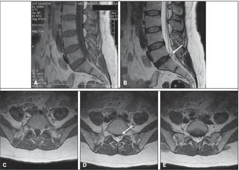
Figure 7. Discal cyst. Female, 42-year-old patient with left-sided lumbar sciatic pain. Sagittal MRI dem-onstrates a well-defined cystic mass at the L5/S1 level, with low signal intensity on T1-weighted sequence (A) and hyperintense signal on T2-weighted sequence (B). On axial T1-weighted (C) and T2-weighted sequences (D,E), the cystic mass (arrow) compresses the ventral face of the dural sac.
C D E