Creative Commons Non Commercial CC-BY-NC: This article is distributed under the terms of the Creative Commons Attribution-NonCommercial 3.0 License (http://www.creativecommons.org/licenses/by-nc/3.0/) which permits non-commercial use, reproduction and distribution of the work without further permission provided the original work is attributed as specified on the SAGE and Open Access pages (https://us.sagepub.com/en-us/nam/open-access-at-sage).
Introduction
Osteomyelitis (OM) is defined as an inflammatory con-dition of bone and bone marrow. In dogs and cats OM is most commonly caused by bacterial infection,1 but
mycotic and viral osteomyelitis have also been described.2 Osteomyelitis is often classified as either
haematogenous or post-traumatic in origin,3 and
frac-ture and subsequent surgical repair represent most of the cases of post-traumatic osteomyelitis reported in companion animals.4–6 Normal, healthy bone in an
immune-competent animal is highly resistant to infec-tion,2,5 but injury and infection of adjacent soft tissue
may induce a post-traumatic osteomyelitis subsequent to a bite or scratch.2,5,7 Haematogenous OM is
uncom-mon in dogs and cats and typically affects young animals.5,6 Bacteria lodge in the capillaries in the
meta-physeal area of the bone and, through inflammation and thrombus formation, create an ischaemic environ-ment that is conductive for further bacterial growth.5,8
Staphylococcal species are isolated in up to 74% of bone infections reported in small animals,5 and
infec-tions with a mixed bacterial flora, including aerobic and anaerobic combinations, are common.1,5,7,9 Osteomyelitis
may involve different bones in the body but most fre-quently occurs in long bones.1,6,10 Only a few cases of
scapular osteomyelitis have been reported in dogs,6,11
and to our knowledge, osteomyelitis in the scapula of a cat has not previously been reported.
Case description
A 12-week-old male intact domestic shorthair cat weigh-ing 1.1 kg was presented for a progressive lameness of the left thoracic limb first noticed by the owners a week prior to presentation. The cat had been introduced to the household with no signs of disease 2 weeks earlier. There
Abstract
Case summary A 12-week-old, male, domestic shorthair cat was presented with severe left thoracic limb lameness.
Investigation included physical examination, diagnostic imaging with radiography and CT, histopathology and microbiological culture. Physical examination revealed a large, firm mass on the left scapula. Radiography and CT showed a monostotic spherical expansile bone lesion in the infraspinatus fossa of the left scapula. The histopathological description was a central acute suppurative osteomyelitis with reactive fibrosis and new bone formation at the periphery. Aerobic and anaerobic cultures were negative and the underlying cause of the osteomyelitis could not be identified. The use of broad-spectrum antibiotics for 8 weeks proved effective with full clinical recovery and no signs of relapse during the follow-up time of 8 months.
Relevance and novel information This report describes the management and outcome of a rare case of osteomyelitis
with severe deformation of scapular bone morphology in an immature cat that was treated successfully with full recovery of limb function and restored integrity of the scapula.
Accepted: 16 August 2016
1 Surgery, Evidensia Strömsholm Referral Small Animal Hospital,
Strömsholm, Sweden
2 Diagnostic Imaging, Evidensia Strömsholm Referral Small Animal
Hospital, Strömsholm, Sweden
Corresponding author:
Sivert Viskjer DVM, Evidensia Strömsholm Referral Small Animal Hospital, Djursjukhusvägen 11, 734 94 Strömsholm, Sweden
was one additional cat in the new household and both cats were strictly confined indoors with no physical con-tact with other animals.
Physical examination revealed a non-weight-bearing left thoracic limb lameness, mild muscle atrophy of the left thoracic limb and a large, firm swelling over the left scapula. No signs of skin wounds or trauma to the affected area were seen. The cat was otherwise bright, alert and responsive and no other abnormal clinical find-ings were noted.
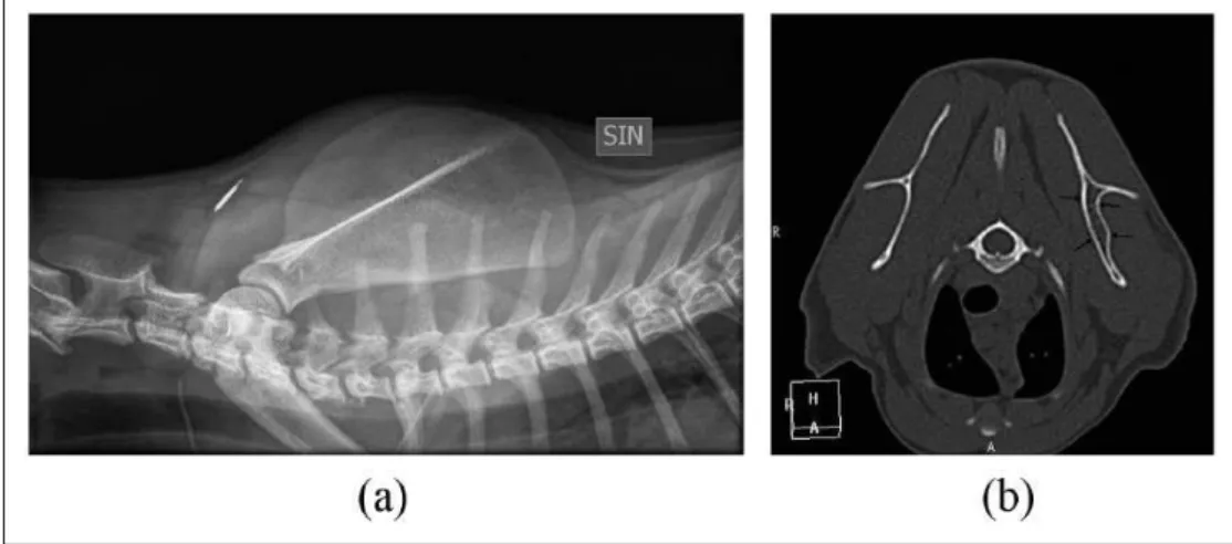
Initial radiographic examination showed a monosto-tic spherical expansile bone lesion in the infraspinatus fossa of the left scapula with a central heterogenous min-eral opacity surrounded by a zone of radiolucency and an outer rim of sclerotic and markedly thickened cortex and periosteal new bone with thin perpendicular radiat-ing brush border-like reactions. Swellradiat-ing of the sur-rounding soft tissue was noted (Figure 1a,b).
CT (Philips Brilliance [40-slice]; Philips Medical Systems) was performed on the thoracic limbs. The expansile lesion had a central core of irregular mineral density with a heterogeneous lucent centre. The perios-teum was thickened with a brush border-like reaction and there was marked surrounding soft tissue swelling (Figure 1c). CT of the abdomen and thorax, including lung parenchyma, was unremarkable.
The differential diagnoses for the monostotic expan-sile bone lesion were an aggressive disease process such as bacterial osteomyelitis with sequestrum formation, fungal osteomyelitis or bone abscess. Bone cyst or neo-plasia, such as osteochondroma or osteoclastoma, were considered less likely.
It was decided to collect samples from the scapular lesion for histopathological and microbiological analy-ses. The cat was premedicated with medetomidine (Domitor; Orion Pharma Animal Health) 0.08 mg/kg SC, meloxicam (Metacam; Boehringer Ingelheim Vetmedica) 0.2 mg/kg SC and methadone (Metadon Recip; Meda) 0.3 mg/kg. Ketamine (Ketaminol; Intervet) 5 mg/kg was administered intramuscularly to maintain a dissociative anaesthesia. Oxygen was delivered by flow-by during the procedure. With the cat positioned in right lateral recumbency a skin inci-sion was made centrally over the left scapula parallel to the scapular spine. Blunt dissection through subcu-taneous tissue, the aponeurosis of the deltoid muscle and the infraspinatus muscle gave access to the scapu-lar lesion. Wedge biopsies were taken with a number 11 scalpel blade from both the mineralised capsule and the softer inner core. Fresh material was placed on a sterile surgical sponge during surgery, then placed in a sterile plastic container and submitted for microbio-logical culture in the hospital’s internal laboratory. Material fixed in 10% buffered neutral formalin was submitted for histopathological examination. The sur-gical site was lavaged with saline and closed routinely with 1.5 metric polyglactin 910 and nylon, and the cat recovered from anaesthesia. The cat received meloxi-cam 0.05 mg/kg administered orally q24h for 6 days after surgery.
Aerobic and anaerobic microbiological culture did not reveal bacterial growth.
The histopathological description of the lesion was a central acute suppurative osteomyelitis (Figure 2) with
Figure 1 (a) Mediolateral projection of the left scapula showing the expansile lesion (black arrows) with adjacent soft tissue
ography and CT scan were performed which showed only mild expansion of the left infraspinatus fossa with near-normal thickness of the periosteum and no detecta-ble periosteal reaction (Figure 4a,b). The previous central heterogeneous mineral opacity was no longer present. A telephone interview with the owner was made 8 months after completion of the treatment; the cat was described by the owner as a completely normal healthy cat with no signs of lameness or scapular swelling.
Discussion
This report describes the diagnosis, treatment and out-come of a severe deforming OM affecting the left scapula of an immature, male, domestic shorthair cat. This is the first known case of scapular OM presented to our clinic.
Figure 2 Light microscopic section of a decalcified biopsy
from left scapula. Cell-rich inflammation with central eosinophilic acellular necrotic bone is seen. The cellular infiltrate comprises lymphocytes, plasma cells, macrophages and neutrophilic granulocytes, intermingled with nuclear debris. Haematoxylin and eosin
Figure 3 (a) Mediolateral projection of the left scapula at first follow-up 3 weeks after initiation of antibiotic treatment. The
The most common localisation of OM in small ani-mals is within the long bones of the appendicular skele-ton both for haematogenous and post-traumatic infections.1,6,10 Other skeletal involvement such as
verte-bral infections and discospondylitis are most often caused by a haematogenous spread from elsewhere in the body,12 whereas OM of the jaw in cats is often
associ-ated with periodontal disease and caused by a broad range of bacteria, most of which are normal oral flora.13
There are only a few cases of scapular OM described in the veterinary literature, all of which are reported in dogs.6,11 This also reflects the situation in people, where
only a few rare cases have been described in relatively young individuals,14 mostly haematogenous in origin,
but post-traumatic cases have also been reported.15
The pathogenesis of the scapular OM in this cat could not be determined and both haematogenous as well as post-traumatic origins are possible. There was no evi-dence of trauma to the skin over the affected area at the initial clinical examination, but it is possible that the cat previously had a small wound or abscess that was not noticed by the owner, and by the time of presentation it had completely healed. There are reports where there is a delay of weeks between a primary soft tissue lesion and a subsequent osteomyelitis of underlying bone.7
Skin trauma could, in this case, potentially have occurred when the mother carried the kitten by the scruff, or from a bite or a scratch caused by littermates.
Despite culture for both aerobic and anaerobic microbes, no causative agent could be detected. There are other reports of OM where the aetiological agent could not be established but where the animals also responded well to antibiotic treatment and were successfully treated with antibiotics.8,16 Previous investigations on sample
tech-niques and specimen handling have revealed the extreme importance of correct procedures to ensure optimal
laboratory results.2,17,18 In our case, it took approximately
15 mins before the specimen arrived in the laboratory for culture, and the specimen was, unfortunately, exposed to room air during this time. This suboptimal handling of the specimen may be a reason for the negative result of the anaerobic culturing.1,19 According to some authors, a
sus-picion of anaerobic infection should be raised in the case of a negative bacteriological culture.3,5
Samples from the lesion were not cultured for fungi even though the osteomyelitis potentially could be of mycotic origin. In dogs and cats primary mycotic osteo-myelitis are considered rare, it is most often haematoge-nous and the animals usually show signs of systemic disease.5 There was no histological evidence of fungal
organisms within the biopsies. This cat did not show any signs of fever, lethargy or systemic disease, only local pain and lameness. The fact that treatment with antibiotics was highly effective also makes a mycotic aetiology less likely. Additional testing, with in situ hybridisation or PCR, may have provided further information regarding the under-lying cause for the lesion seen. However, dehydration and decalcification could have altered the results according to correspondence with the laboratory involved.
For economic reasons, the owner declined urine analy-sis and blood analyses for complete blood counts, bio-chemistry, feline leukaemia virus/feline immunodeficiency virus and culture. An elevated white blood cell count together with an increased C-reactive protein could have supported an underlying infectious cause, despite the neg-ative culture result. Other tests may have revealed signs of altered bone metabolism, immunodeficiency and/or infectious disease. However, the diagnostic value of blood tests when evaluating a localised OM is debated,20 and the
authors do not believe that the results of blood tests and urine analysis would have changed the treatment plan or the outcome in this case.
Figure 4 (a) Twenty-week follow-up mediolateral projection of the left scapula. The lesion is no longer detectable. A microchip
Conlict of interest The authors declared no potential con-flicts of interest with respect to the research, authorship, and/ or publication of this article.
References
1 Muir P and Johnson KA. Anaerobic bacteria isolated
from osteomyelitis in dogs and cats. Vet Surg 1992; 21: 463–466.
2 Johnson KA. Osteomyelitis in dogs and cats. J Am Vet Med Assoc 1994; 205: 1882–1887.
3 Houlton JEF and Vannini R. Complications of fracture
treatment, osteomyelitis. In: Johnson AL (ed). AO princi-ple of fracture management in the dog and cat. Davos: AO Publishing Davos, 2005, p 416.
4 Caywood DD. Osteomyelitis. Vet Clin North Am Small
Anim Pract 1983; 13: 43–53.
5 Bubenik LJ. Infections of the skeletal system. Vet Clin North Am Small Anim Pract 2005; 35: 1039–1109.
6 Caywood DD, Wallace LJ and Braden TD. Osteomyelitis in the dog: a review of 67 cases. J Am Vet Med Assoc 1978; 172: 943–946.
7 Johnson KA, Lomas GR and Wood AK. Osteomyelitis in
dogs and cats caused by anaerobic bacteria. Aust Vet J
1984; 61: 57–61.
osteomyelitis of the scapula in children. Orthop Traumatol Surg Res 2009; 95: 632–635.
15 Buckley SL, Alexander AH and Barrack RL. Scapular
osteomyelitis. An unusual complication following sub-acromial corticosteroid injection. Orthop Rev 1989; 18: 321–324.
16 Dunn JK, Dennis R and Houlton JEF. Successful treatment of two cases of metaphyseal osteomyelitis in the dog. J Small Anim Pract 1992; 33: 85–89.
17 Hindiyeh M, Acevedo V and Carroll KC. Comparison of three transport systems (Starplex StarSwab II, the new Copan Vi-Pak Amies Agar Gel collection and transport swabs, and BBL Port-A-Cul) for maintenance of anaerobic and fastidious aerobic organisms. J Clin Microbiol 2001; 39: 377–380.
18 Dow SW, Jones RL and Adney WS. Anaerobic
bacte-rial infections and response to treatment in dogs and cats: 36 cases (1983–1985). J Am Vet Med Assoc 1986; 189: 930–934.
19 Walker RD, Richardson DC, Bryant MJ, et al. Anaerobic bacteria associated with osteomyelitis in domestic ani-mals. J Am Vet Med Assoc 1983; 182: 814–816.