201
https://doi.org/10.1590/0004-282X20160183
IMAGES IN NEUROLOGY
Intracranial hypotension secondary to
spontaneous spinal cerebrospinal fluid leaks
Hipotensão intracraniana secundária a fístula liquórica espinhal espontânea
Antonio Eustáquio Silva Junior
1, Patricia Pavan
2, Mariana Mari Oshima
1, Tânia Marchiori de Oliveira
Cardoso
2, Fabiano Reis
1A 37-year-old woman presented with acute
ortho-static hypotension and diffuse headache. Brain magnetic
resonance imaging (MRI) revealed T2 hyperintense
bilat-eral subdural effusions, diffuse pachymeningeal
enhance-ment, slit ventricles and venous engorgement
compat-ible with spontaneous intracranial hypotension. Single
photon emission computed tomography with
comput-ed tomography (CT) and CT-cisternography showcomput-ed
a cerebrospinal fluid (CSF) leak at the left C1-C2
tran-sition. Spontaneous intracranial hypotension is a rare
cause of daily headache, which remains largely
underdi-agnosed
1,2, and current evidence indicates that this
con-dition is the result of a spontaneous CSF leak
1,2,3,4. This
case illustrates the importance of CT-cisternography in
the detection of CFS leak
1,2,3, allowing appropriate
diag-nosis and treatment.
1Universidade Estadual de Campinas, Faculdade de Ciências Médicas, Departamento de Radiologia, Campinas SP, Brasil;
2Universidade Estadual de Campinas, Faculdade de Ciências Médicas, Departamento de Neurologia, Campinas SP, Brasil.
Correspondence: Fabiano Reis; Faculdade de Ciências Médicas - UNICAMP, Departamento de Radiologia; Rua Tessália Vieira de Camargo, 126, Cidade Universitária Zeferino Vaz; 13083-887 Campinas SP, Brasil, E-mail: [email protected]
Conflict of interest: There is no conflict of interest to declare. Received 20 January 2016; Accepted 03 October 2016.
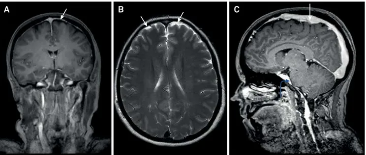
Figure 1.
Coronal T1 post contrast MRI demonstrates pachymeningeal enhancement (arrows); (B) axial T2 weighted MRI
demonstrates subdural effusions (arrows); (C) Sagittal T1-weighted post contrast demonstrates engorgement of the superior
sagittal sinus (straight arrow) and sagging of the brain (flattening of the pons and obliteration of prepontine cistern, curved arrow).
202
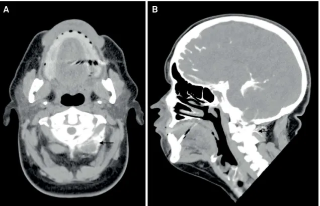
Arq Neuropsiquiatr 2017;75(3):201-202Figure 2.
Sagittal and axial sections of CT-cisternography (A) axial and (B) sagittal show a C1-C2 left cerebrospinal fluid leak, with
left fluid paravertebral collection (arrows).
A
B
References
1. Schievink WI. Spontaneous spinal cerebrospinal fluid leaks. Cephalalgia. 2008;28(12):1347-56. https://doi.org/10.1111/j.1468-2982.2008.01776.x
2. Purdy RA. Understanding and managing spontaneous
intracranial hypotension. Can J Neurol Sci. 2013;40(2):139-40. https://doi.org/10.1017/S0317167100013640
3. Mokri B. Spontaneous intracranial hypotension. Continuum
(Minneap Minn). 2015;21(4 Headache):1086-108.
4. Hoffmann J, Goadsby PJ. Update on intracranial hypertension