409
Radiol Bras. 2017 Nov/Dez;50(6):405–415Letters to the Editor
http://dx.doi.org/10.1590/0100-3984.2016.0081
Lucas de Pádua Gomes de Farias1, Igor Gomes Padilha1, Carla Jotta
Justo dos Santos2, Carol Pontes de Miranda Maranhão2, Christiana
Maia Nobre Rocha de Miranda2
1. Universidade Federal de Alagoas (UFAL), Maceió, AL, Brazil. 2. Clínica de Medicina Nuclear e Radiologia de Maceió (MedRadius), Maceió, AL, Brazil. Mailing address: Dra. Christiana Maia Nobre Rocha de Miranda. Clínica de Medicina Nuclear e Radiologia de Maceió (MedRadius). Rua Hugo Corrêa Paes, 104, Farol. Maceió, AL, Brazil, 57050-730. E-mail: maia.christiana@ gmail.com.
interlobular and intralobular septa, patients with GD can present with alveolar opacities, capillary plugging by Gaucher cells, and interstitial opacities, with a predominance of lymphatic distribu-tion, as well as respiratory infections(4–8). Other alterations de-scribed include pulmonary ibrosis, a miliary pattern and involve-ment of the hilar or mediastinal lymph nodes, as well as a reduc-tion in lung volume as a consequence of hepatosplenomegaly. Radiographic examinations can reveal an interstitial pattern and can show any changes in bone structures(3–7).
The diffuse pulmonary involvement seen in patients with GD indicates that it is a systemic disease. MDCT is an important tool for the initial evaluation and follow-up of these patients, and lung biopsy can be dispensed with when the tomography reveals interstitial opacities in an appropriate clinical and epidemiologi-cal context(6,7).
When there is no clinical suspicion of GD, a tomographic inding of the crazy-paving pattern makes the radiologic diagno-sis dificult(9). In such cases, the main differential diagnoses are alveolar proteinosis, pulmonary hemorrhage, pulmonary vasculi-tis, diffuse alveolar damage (acute respiratory distress syndrome), pulmonary edema, bronchioloalveolar carcinoma, Niemann-Pick disease, and radiation pneumonitis, as well as Pneumocystis, viral, lipoid, mycobacterial, interstitial, and eosinophilic pneumonia.
REFERENCES
1. Beutler E. Gaucher’s disease. N Engl J Med. 1991;325:1354–60.
2. Pastores GM, Hughes DA. Gaucher disease. 2000 Jul 27 [Updated 2015 Feb 26]. In: Adam MP, Ardinger HH, Pagon RA, et al. GeneReviews© [Internet]. Seattle (WA): University of Washington, Seattle; 1993-2017. [cited 2017 Oct 30]. Available from: www.ncbi.nlm.nih.gov/books/ NBK1269/.
3. Mendonça VF, Paula MTM, Fernandes C, et al. Manifestações esqueléti-cas da doença de Gaucher. Radiol Bras. 2001;34:151–4.
4. Wolson AH. Pulmonary indings in Gaucher’s disease. Am J Roentgenol Radium Ther Nucl Med. 1975;123:712–5.
5. Kerem E, Elstein D, Abrahamov A, et al. Pulmonary function abnormali-ties in type I Gaucher disease. Eur Respir J. 1996;9:340–5.
6. Aydin K, Karabulut N, Demirkazik F, et al. Pulmonary involvement in adult Gaucher’s disease: high resolution CT appearance. Br J Radiol. 1997;70:93–5.
7. Amir G, Ron N. Pulmonary pathology in Gaucher’s disease. Hum Pathol. 1999;30:666–70.
8. Yassa NA, Wilcox AG. High-resolution CT pulmonary indings in adults with Gaucher’s disease. Clin Imaging. 1998;22:339–42.
9. Müller CIS, D’Ippolito G, Rocha AJ. Tórax. Série Colégio Brasileiro de Radiologia e Diagnóstico por Imagem: Tórax. 1ª edição. Rio de Janeiro, RJ: Elsevier; 2010.
Figure 2. Axial (A), coronal (B), and sagittal (C) MDCT scans of the right lung showing diffuse, marked thickening of the interlobular and intralobular septa, ac-companied by ground-glass opacity of the lung parenchyma, characteristic of the crazy-paving pattern. Note also the irregularity with the pleural surface and the
thickening of the issures (arrowheads).
Retroperitoneal Ewing’s sarcoma/embryonal tumor: a rare differential diagnosis of back pain
Dear Editor,
A previous healthy 17-year-old female was referred to a rheumatology clinic due to a 6-month history of lower back pain. Her pain worsened at night and did not radiate. During that 6-month period, she had lost weight (5 kg). An initial evalu-ation produced normal cardiovascular and abdominal ind-ings. She had pain on lumbar spine palpation and pain when her sacroiliac joints were examined (Patrick’s test). Laboratory tests showed normal blood smear results and normal levels of inlammatory markers. While waiting for a magnetic resonance imaging (MRI) scan of her sacroiliac joint, she returned with signiicant worsening of her pain and weakness in her right leg. Examination showed grade 3 muscle strength and an absence
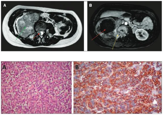
of the ipsilateral patellar relex. MRI revealed a right paraverte-bral mass, with intradural and foraminal components, showing a signal that was, in comparison with the muscle signal, pre-dominantly isointense (with a hyperintense component indicat-ing hemorrhage) on T1-weighted images and isointense (with a hyperintense necrotic component) on T2-weighted images (Fig-ure 1). Ultrasound-guided biopsy revealed an undifferentiated small round-cell morphology. Immunohistochemistry staining suggested a member of the Ewing’s sarcoma/embryonal tumor (ES/ET) family (Figure 2). The patient was submitted to chemo-therapy, which did not elicit an adequate response.
410
Radiol Bras. 2017 Nov/Dez;50(6):405–415 Letters to the EditorFabiano Reis1, Eduardo Macedo1, Marcondes Cavalcanti França Junior1,
Eliane Ingrid Amstalden1, Simone Appenzeller1
1. Universidade Estadual de Campinas (Unicamp), Campinas, SP, Brazil. Mail-ing address: Dr. Fabiano Reis. Unicamp – Radiologia. Rua Tessália Vieira de Camargo, 126, Cidade Universitária. Campinas, SP, Brazil, 13083-887. E-mail: [email protected].
The majority of ES/ETs are diagnosed during the irst two decades of life(1). The most common soft tissue sites are the chest wall, lower extremities, and pelvis/hip region. They are rarely found in the retroperitoneum, upper extremities, or internal organs(2). Patients often present with a painless mass or vague abdominal or chest pain, depending on tumor site(1). Muscle weakness can also be the predominant symptom(3).
There are certain red lags that should always be considered in the differential diagnosis of patients with focal neurological manifestations of myelopathy and radiculopathy. Although rare and having no speciic radiological indings, ES/ET should be suspected in young adults presenting with a large heterogeneous mass in the trunk, extremities, or soft tissues(4). In the case pre-sented here, an MRI inding of a large mass with isointense solid components on T1- and T2-weighted images, together with ne-crosis and hemorrhage, facilitated the diagnosis in this intrigu-ing case. In addition, ES of the retroperitoneum is dificult to differentiate from other tumors. The retroperitoneal tumors that can invade the neural foramen and vertebral canal are the following: ganglioneuroma and ganglioneuroblastoma; neuro-blastoma in younger patients (mean age, 22 months); leukemia (chloroma); and lymphoma. Invasion of the renal vein, inferior vena cava, and liver can be seen in ES, renal cell carcinoma, and adrenocortical carcinoma. Differentiation aspects that favor the diagnosis of ES are earlier age of presentation, absence of metastatic lymphadenopathy, and absence of calciications. ES
tends to be unilateral and does not cross midline(5). The deini-tive diagnosis can be made only by histopathological analysis.
REFERENCES
1. Javery O, Krajewski K, O’Regan K, et al. A to Z of extraskeletal Ewing sarcoma family of tumors in adults: imaging features of primary disease, metastatic patterns, and treatment responses. AJR Am J Roentgenol. 2011;197:W1015–22.
2. Ma Z, Brimo F, Zeizafoun N. Primary Ewing’s sarcoma/primitive neuro-ectodermal tumor (ES/PNET) of the penis: a report of an extraordinarily unusual site and a literature review of extraskeletal Ewing’s sarcoma. Int J Surg Pathol. 2013;21:63–7.
3. Sade R, Çakir M, Ogul H, et al. Primary extraosseous Ewing sarcoma of the lumbar spine presenting with left leg weakness.Spine J. 2015;15: 1488–9.
4. Holland MT, Flouty OE, Close LN, et al. A unique case of primary Ewing’s sarcoma of the cervical spine in a 53-year-old male: a case report and review of the literature. Case Rep Med. 2015;2015:402313. 5. Somarouthu BS, Shinagare AB, Rosenthal MH, et al. Multimodality
imaging features, metastatic pattern and clinical outcome in adult ex-traskeletal Ewing sarcoma: experience in 26 patients. Br J Radiol. 2014; 87:20140123.
http://dx.doi.org/10.1590/0100-3984.2015.0236
Figure 1. A: Axial T2-weighted MRI scan showing a huge heterogeneous retro-peritoneal mass (green arrow) with an isointense intradural component (red arrow). B: Axial gadolinium contrast-enhanced T1-weighted image showing a large mass, with intense peripheral enhancement and central necrosis (red arrow), extending through the neural fo-ramen (yellow arrow).
A
B
Ultrasound evaluation of diaphragmatic dysfunction
Dear Editor,
A 49-year-old male patient presented with a complaint of dyspnea when swimming, which he did regularly. The following were performed: chest X-ray, which showed elevation of the right hemidiaphragm; respiratory function tests, which revealed mild restrictive lung disease; and ultrasound of the diaphragm, which
demonstrated a signiicant reduction in the mobility of the right hemidiaphragm, although not to the point of paralysis.
Ultrasound of the diaphragm has been used mainly in pa-tients in intensive care. In such papa-tients, assessment of the dia-phragm by ultrasound can be used in order to predict successful weaning from mechanical ventilation(1), to inform decisions re-garding adjustments in mechanical ventilation parameters, and to investigate postoperative weakness/diaphragmatic paralysis(2), Figure 2. A: Hematoxylin-eosin staining
(original magniication, ×40) showing dense cellular proliferation in a diffuse or vaguely lobular pattern of uniform round cells, with scanty cytoplasm, ovoid
nuclei with ine chromatin and small nu